

 PDF
PDF ePub
ePub Citation
 Print
Print


Abstract
Study Design
A retrospective study of patients with contralateral recurrent lumbar disc herniation at the same level. Objectives : To analyze the risk factors of recurrence, clinical result and reoperative efficiency of contralateral recurrent lumbar disc herniation at the same level after primary discectomy compared with those after discectomy in primary lumbar disc herniation.
Summary of Literature Review
There have been many studies on recurrent disc herniation, but little investigation of risk factors and clinical result of contralateral recurrent lumbar disc herniation at the same level.
Materials and Methods
Ten cases who can investigate for 2 years among the patients who underwent reoperation for contralateral recurrent lumbar disc herniation at the same level after primary discectomy were selected as study group (groupⅠ) and thirty cases who underwent discectomy during the same study period were selected as control group (groupⅡ). A ge, gender, etiology and symptom of disc herniation, clinical improvement rate and amount of remove disc were recorded. Overall patient satisfaction, pain severity, functional outcome and work status were evaluated. Risk factors of recurrence were analyzed.
Results
Etiology was no different between both groups but showed the abrupt onset symptom in study group. Recurrence was more common in the case herniated posterolaterally and had severe degeneration change in lumbar disc before primary discectomy. The amount of bulging disc removed were average 1.5 cc in study group and 2.5 cc in control group. Recurrence was more in the cases removed smaller amount of bulging disc and remained the symptom of pain after primary discectomy. Clinical result show the same between both group after 2 years (p>0.05).
Go to : 
REFERENCES
1). Ahlgren BD, Vasavada A, Brower RS, Lydon C, Herkowitz HN, Panjabi MM. Anular incision technique on the strength and multidirectional flexibility of the healing in tervertebral disc. Spine. 1994; 19:948–54.
2). Bernard TN. Repeat lumbar spine surgery. Factors influencing outcome. Spine. 1993; 18:2196–200.
3). Boden SD, Davis DO, Dina TS, et al. Contrast enhanced MR imaging performed after successful lumbar disk surgery: Prospective study. Radiology. 1992; 182:59–64.
4). Brinckmann P, Grootenboer H. Change of disc height, radial disc bulge, and intradiscal pressure from discectomy. An in vitro investigation on human lumbar discs. Spine. 1991; 16:641–6.

5). Cervellini P, Curr D, Bernardi L, Volpin L, Benedetti A. Computed tomography after lumbar disc surgery: A comparison between symptomatic and asymp -tomatic patients. Acta Neurochir. 1988; 43:44–7.
6). Cinotti G, Roysam GS, Eisenstein SM, Postacchini F. Ipsilateral recurrent lumbar disc herniation. A prospective controlled study. J Bone Joint Surg 〔Br〕. 1998; 80:825–32.
7). Connolly ES. Surgery for recurrent lumbar disc herniation. Clin Neurosurg. 1992; 39:211–6.
8). Deutsch AL, Howard M, Dawson EG, Goldstein TB, Mink JH, Zeegen EH, Delamarter RB. Lum bar spine following successful surgical discectomy: Magnetic resonance imaging features and implications. Spine. 1993; 18:1054–60.
9). Ebeling U, Kalbarcyk H, Reulen HJ. Microsurgical reoperation following lumbar disc surgery. Timing, surgical findings, and outcome in 92 patients. J Neurosurg. 1989; 70:397–404.
10). Epstein JA, Lavine LS, Epstein BS. Recurrent herniation of the lumbar intervertebral disk. Clin orthop. 1967; 52:169–78.
11). Ethier DB, Cain JC, Yaszemski MJ, Glover JM, Kluznik RP, Pyka RE, Lauerman WC. The influence of anulotomy selection on disc competence. A radiographic, biomechanical, and histologic analysis. Spine. 1994; 19:2071–6.
12). Fandino J, Botana C, Viladrich A, Gomez-Bueno J. Reoperation after lumbar disc surgery: Results in 130 cases. Acta Neurochir. 1993; 122:102–4.
13). Goel VK, Nishiyama K, Weinstein JN, Liu YK. Mechanical properties of lumbar spinal motion segments as affected by partial disc removal. Spine. 1986; 11:1008–12.
14). Greenwood J, McGuire TH, Kimbell F. A study of the causes of failure in the herniated intervertebral disc operation: An analysis of sixty-seven reoperated cases. J neurosurg. 1952; 9:15–20.
15). Horton WC and Daftari. Which discs as visualised by magnetic resonance imaging is actually a source of pain? A correlation between magnetic resonance imaging and discography. spine. 1992; 17(6 Suppl):164–71.
16). Jackson RK. The longterm effects of wide laminectomy for lumbar disc excision: a review of 130 patients. J Bone Joint Surg(Br). 1971; 53-B:609–16.
17). Kim SS, Michelsen CB. Revision surgery for failed back surgery syndrome. Spine. 1992; 17:957–60.
18). Montaldi S, Fankhauser H, Schnyder P, de Tribo-let N. Computed tomography of the postoperative intervertebral disc and lumbar spinal canal: Investigation of twenty-five patients after successful operation for lumbar disc herniation. Neurosurgery. 1988; 22:1014–22.
19). O’ Sullivan MG, Connolly AE, Buckley TF. Rec u r -rent lumbar disc protrusion. Br J Neurosurg. 1990; 4:319–25.
20). Pappas CT, Harrington T, Sonntag VK. Outcome analysis in 654 surgically treated lumbar disc herniations. Neurosurgery. 1992; 30:862–866.
21). Roland M, Morris R. A study on the natural history of back pain: part 1, Development of reliable and sensitive measure of disability in low back pain. spine. 1983; 8:141–144.
22). Spengler DM. Lumbar discectomy. Results with limited disc excision and selective foraminotomy. Spine. 1982; 7:604–607.
23). Williams RW. Microlumbar discectomy: A conservative surgical approach to the virgin herniated lumbar disc. Spine. 1978; 3:175–182.
Go to :
Figures and Tables%
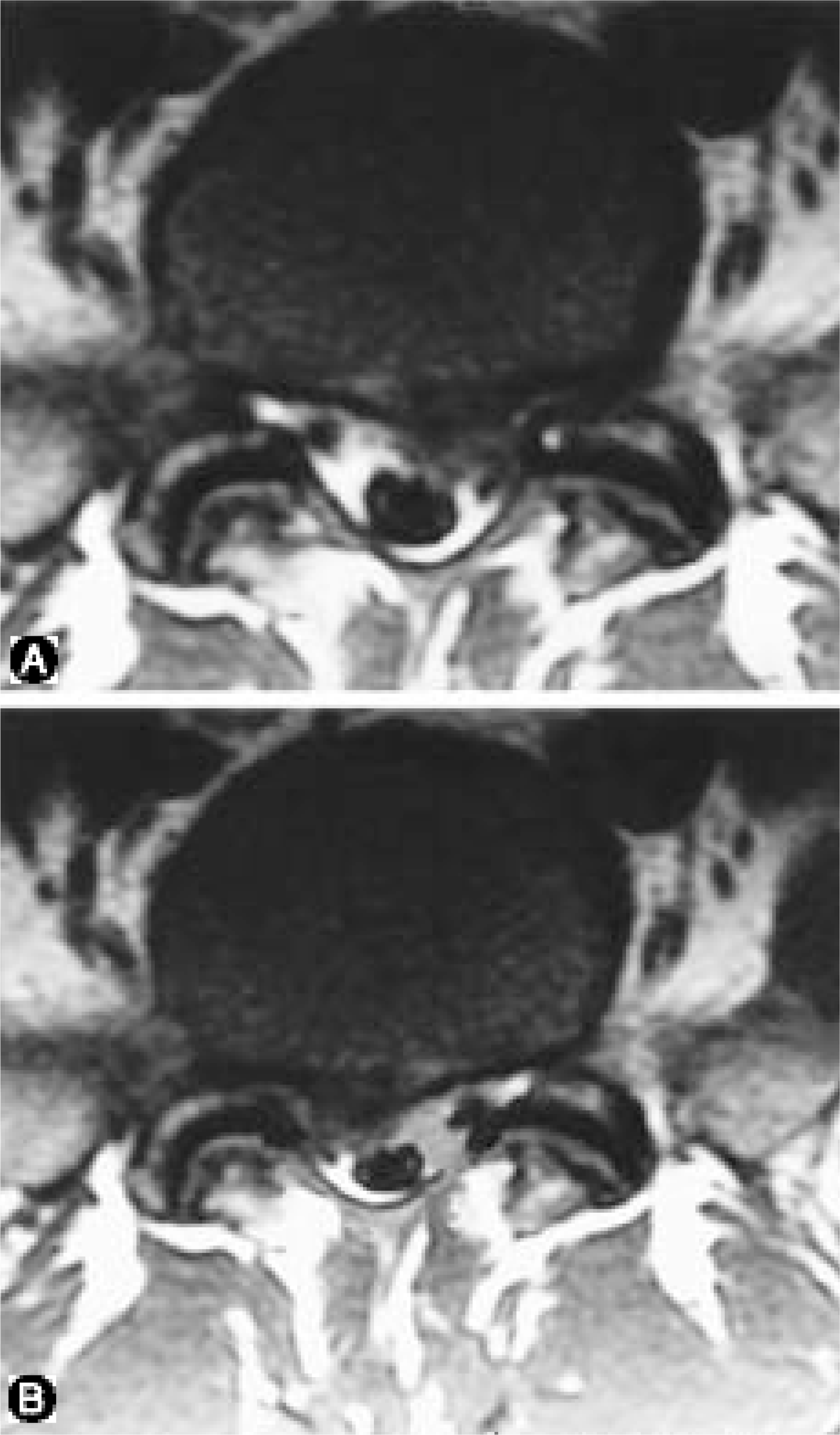 | Fig. 1.T2-weight axial MR image shows left-side extrusion of the L4-5 disc and the bottom MR image shows right-side extrusion of the recurrent disc at the same level. |
Table 1.
Symptom & Sign
 XML Download