

 PDF
PDF ePub
ePub Citation
 Print
Print


Abstract
Study Design
In vitro studies using human intervertebral disc for the localization of the type IV collagen.
Objective
1) To study the distribution pattern and immunoexpression of type 4 collagen in the intervertebral disc, 2) To study the function of type IV collagen in the intervertebral disc.
Summary of Back Ground
The correlations of degeneration changes and collagens in the dics have not been determined. The reports for type IV collagen were few. So far, the histologic analysis for the expression of type IV collagen in the intervertebral disc has not been done. There was no report to study the function of the type IV collagen in the intervertebral disc.
Methods
Fifty- four disc blocks obtained during anterior interbody fusion of the lumbar spine were used to observe the expression pattern of the type IV collagen with immunochemical stain. For the observation of the myxomatous degeneration in the intervertebral disc, the alcian blue stain with periodic acid- schilff was done. For the control group, 22 neonate intervertebral disc blocks were obtained at autopsy.
Results
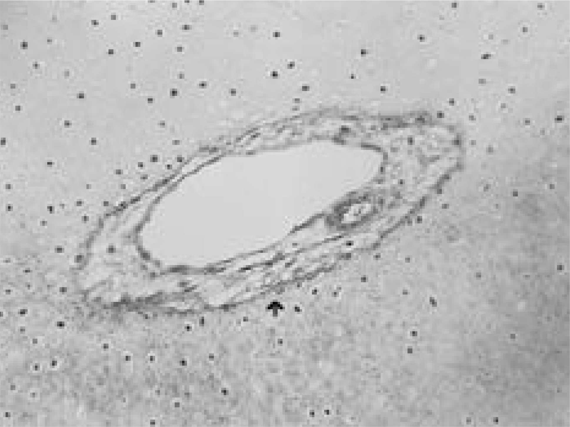
The immunoreactions for type IV collagen were associated blood vessels in the anulus fibrosus in the disc. There was no statistical significant difference of the type IV collagen expression between the control and disease groups. Myxomatous degenerations were observed as the irregular form in the degenerative intervertebral disc.
Go to : 
REFERENCES
1). Ahmed MU, Thorepe SR, Baynes JW. Identification of N (carboxymethyl)-lysine as a degradation product of fructose lysine in glycated protein. J Biol Chem. 261:4889–4894. 1986.
2). Beard HK, Ryvar R, Brown R, Muir H. Immunochemical localization of collagen types and proteoglycan in pig intervertebral discs. Immunology. 41:491–501. 1980.
3). Beard HK, Roberts S, O’ Brien JP. Immunofluores -cent staining for collagen and proteoglycan in normal and scoliotic intervertebral discs. J Bone Joint Surg. 63B:529–534. 1981.
4). Berkovitz B. Collagen crimping in the intra-articular disc and articular surfaces of the human temporo -mandibular joint. Arch Oral Biol. 45:749–756. 2000.
5). Brickley-Parsons D, Glimcher MJ. Is the chemistry of collagen in intervertebral discs an expression of Wolff's law? A study of the human lumbar spine. Spine. 9:148–163. 1984.

6). Eyre DR. Collagens of the disc. Chosh P, editor. ed.The Biology of the Intervertebral Disc. Boca Raton, Florida: CRC Press;p. 171–188. 1988.
7). Ghosh P, Bushelll GR, Taylor TFK, Akeson WH. Collagens, elastins and noncollagenous protein of the intervertebral disc. Clin Orthop. 129:124–132. 1977.
8). Herbert CM, Lindberg KA, Jayson MIV, et al. Changes in the collagen of human intervertebral discs during ageing and degenerative disc disease. J Mol Med. 1:79–91. 1975.
9). Kaapa E, Holm S, Han X, Takala T, Kovanen V, Vanharanta H. Collagens in the injured porcine intervertebral disc. J Orthop Res. 12:93–102. 1994.
10). Nerlich AG, Boos N, Wiest I, Aebi M. Immunolo -calization of major interstitial collagen types in human lumbar intervertebral discs of various ages. Vichows Archv. 432(1):67–76. 1998.
Go to :
Figures and Tables%
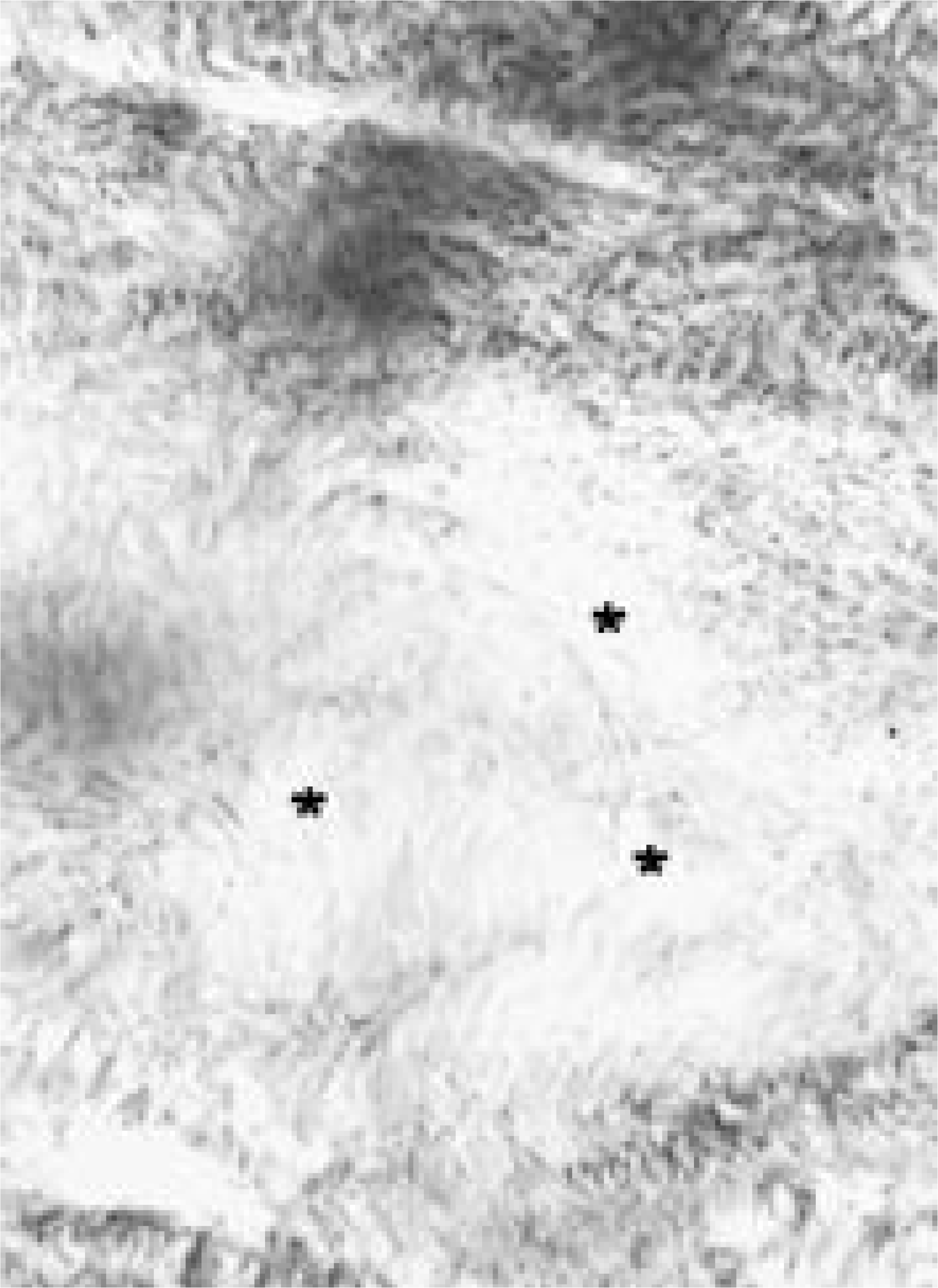 | Fig. 1.The myxomatous degenerations(*) in the degenerative intervertebral disc were observed in irregular form (×200). |
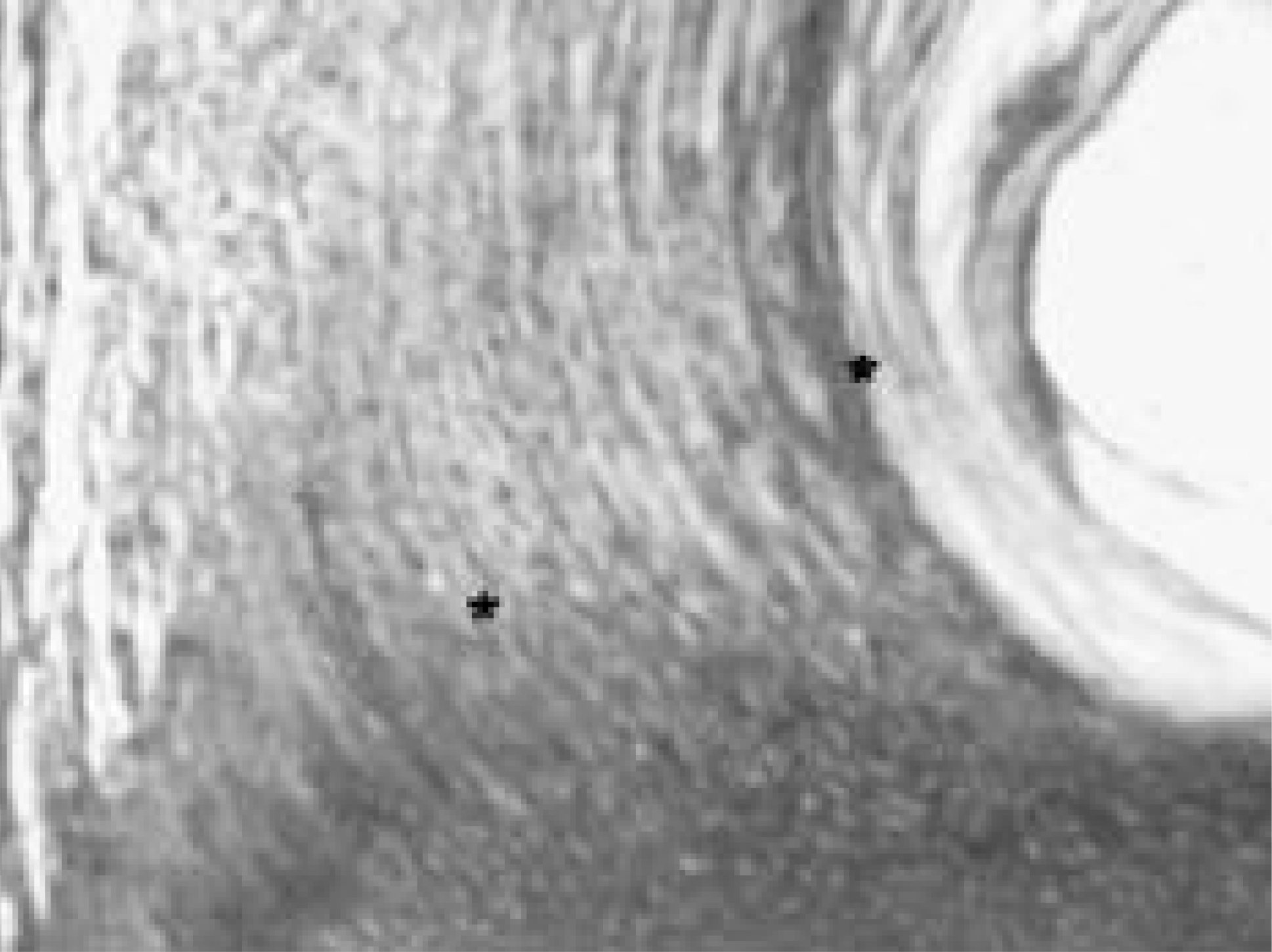 | Fig. 2.Extracellular matrix fibers were seen with regular form(*) in the neonate disc (× 200). |
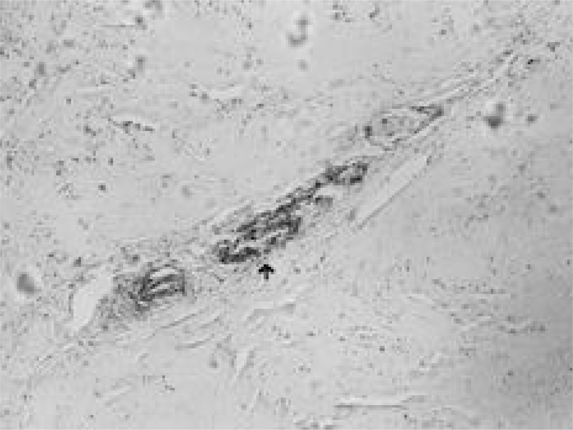 | Fig. 3.Blood vessels associated with the type IV collagen was abserved in anulus fibrosus (× 100). |
 XML Download