

 PDF
PDF ePub
ePub Citation
 Print
Print


Abstract
Materials and Methods
A 36- year-old woman had back pain and pain radiating to the left lateral abdomen. Straight leg raising was not limited. Plain roentgenograms show a small round radiolucent area in left L1 vertebral pedicle and expansile sclerotic margin in L1 vertebral body. T1- weighted MR images show the lesion displaying low signal intensity, T2- weighted images show high signal intensity, Gadolinium enhanced images show a necrotic area with low signal intensity in the lesion.
Results
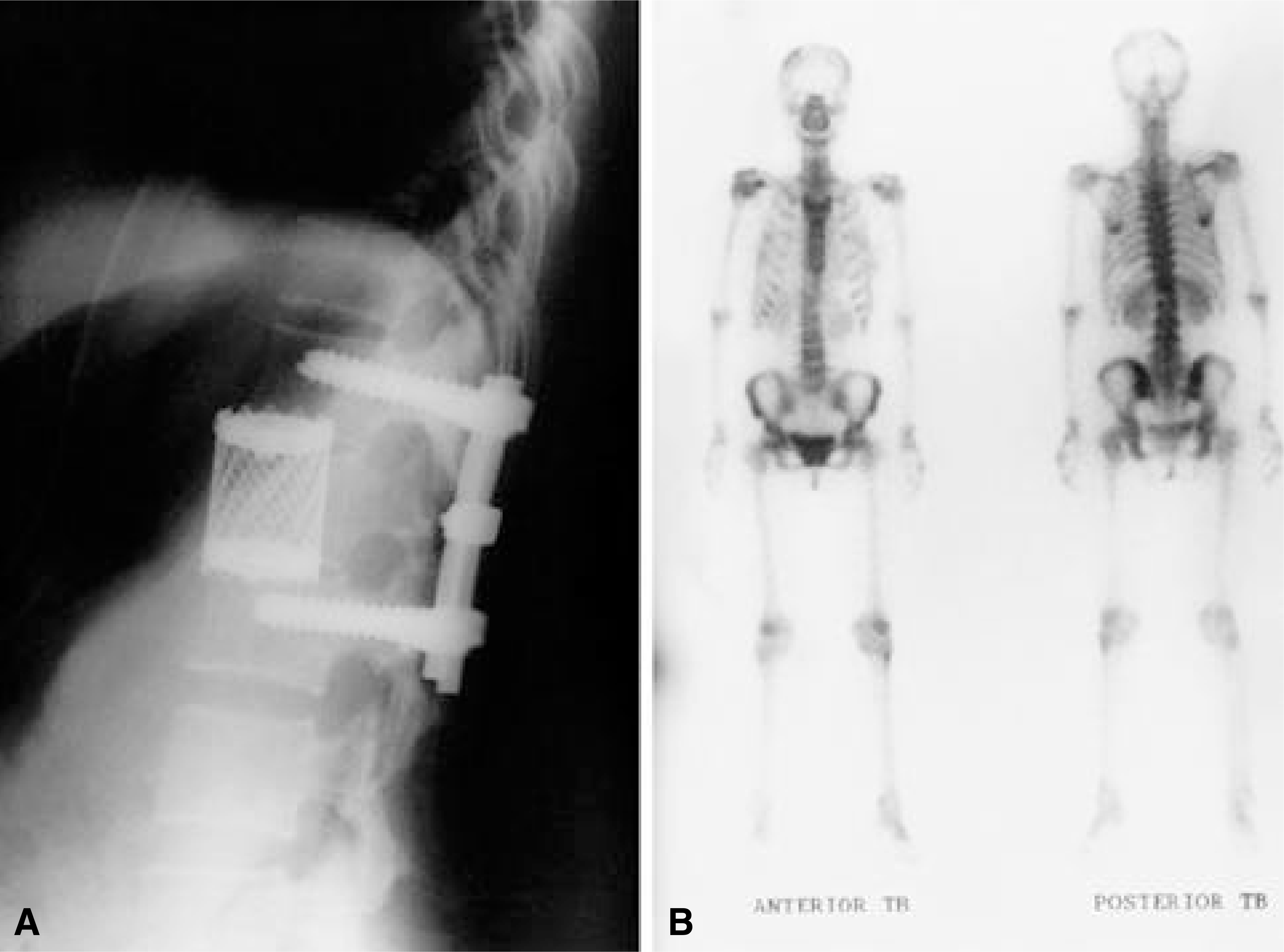
The mass of left L1 transverse process and pedicle was excised through posterior approach and pedicle screws were inserted T12 and L2 pedicle. L1vertebral body was excised through anterior approach and a titanium mesh was inserted. It was a ovoid mass, measured 2.3× 2× 1.5 cm in size and histologically diagnosed as chondroblastoma consisting of chondroid matrix and chondroblast. Soft tissue nodule shows chickenwire calcification. All the symptoms were relieved at 14 months follow- up and no evidence of recurrence on follow- up roentgenogram and bone scan. However, the patient had persistent lower back pain.
Go to : 
REFERENCES
1). 이상언, 안진환, 유명철, 김봉건 : 거대연골 아세포
종, 대한정형외과 학회지,. 14:125–128. 1979.
2). 이한구, 이상훈, 이춘기, 김희중, 이관희, 이영인,
진종수 : 골종양의 역학적 연구, 대한정형외과학회지,. 25:1–23. 1990.
3). Coleman SS. Benign chondroblastoma with recurrent soft tissue and intraarticular lesions, J Bone Joint Surg. 48-A:1554–1561. 1976.
4). Dahlin DC and Ivins JC. Benign Condroblastoma. A Study of 125 Cases. Cancer. 30:401–413. 1972.
5). Dahlin DC. Bone Tumors. 3rd ed.p. 43. Illinois: Charles C. Thomas Publisher;1978.
6). Ewing J. Neoplastic disease. A textbook on tumors. 3rd ed.p. 293. Philadelphia: WB Saunders;1928.
7). Fechner RE and Wilde HD. Chondroblastoma in the metaphysis of the femoral neck, J Bone Joint Surg. 56-A:413–420. 1976.
8). Hoeffel JC, Brasse F, Schmitt M, Plenat F, Vignaud JM, Czorny A and Marchal AL. About one case of vertebral chondroblastoma. Pediatr Radiolo. 17:392–396. 1987.

9). Huvos AG and Marcove RC. Chondroblastoma of Bone. Clin Orthop. 95:300–312. 1973.
10). Jaffe Hl and Lichtenstein L. Benign chondroblastoma of bone: A reinterpretation of the so-called calcifying or chondromatous giant cell tumor, Am J Pathol. 18:96–991. 1942.
11). Janusz Buraczewski, Janina Lysakowska and Witold Rudows ki. Chondroblastoma of the thoracic spine. J Bone Joint Surg. 39-B:705–710. 1957.
12). Kolodny A. Bone sarcoma: The primary malignant tumors of bone and the giant cell tumor, Surg Gynec Obset. 44(Suppliment):1927.
13). Pulm GE and Pugh DG. Roentgenologic aspects of benign chondroblastoma of bone, Am J Roentgenol. 79:584–591. 1958.
14). Salzer M, Salzer-Kuntschik M, Kretschmer G. Das Benign Chondroblasom, Arch. Orthop Unfallchir. 64:229–244. 1968.
15). Schajowicz Fritz and Gallardo Hector. Ep iphyseal Chondroblastoma of bone. A Clinicopathological Study of sixty-nine cases, J Bone Joint Surg. 52-B:205–226. 1970.
Go to :
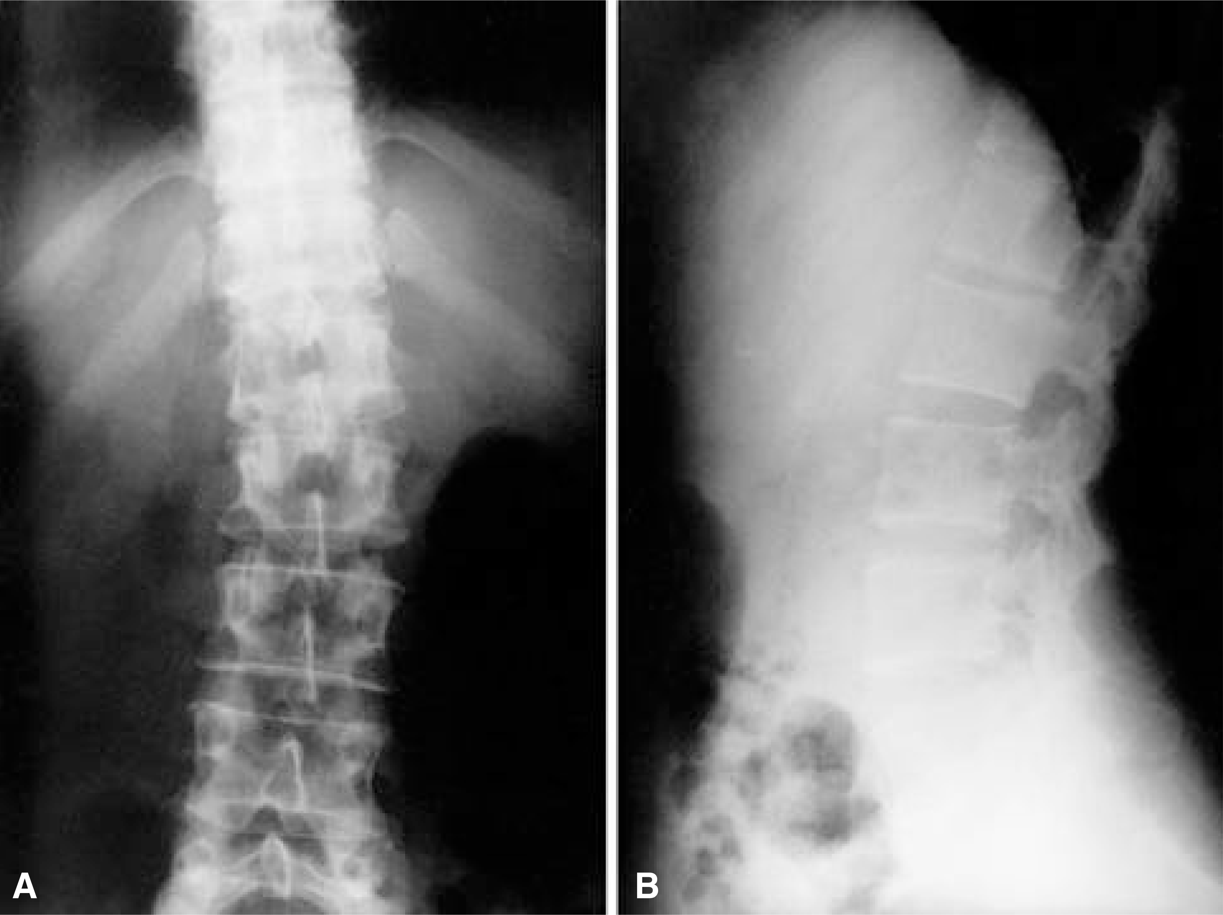 | Fig. 1-A.Preoperative lumbar AP radiograph showing osteolytic lesion on left L1 pedicle. Fig. 1-B. Preoperative lumbar lateral radiograph showing enlarged pedicle, osteolytic lesion on upper L1 body with sclerotic margin. |
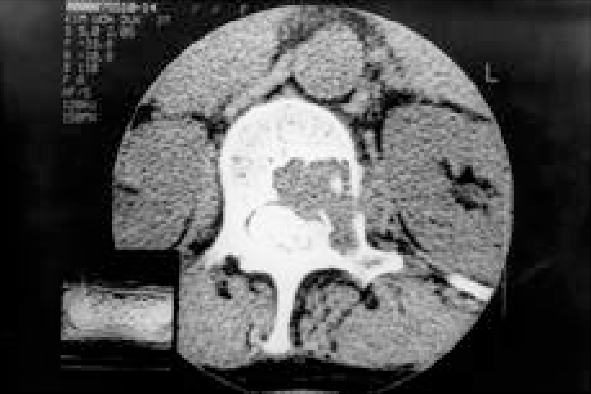 | Fig. 2.Preoperative CT scan of first lumbar vertebra shows osteolytic lesion on L1 body and left pedicle. Soft tissue and bone fragment was surrounded by sclerotic bone in osteolytic lesion. |
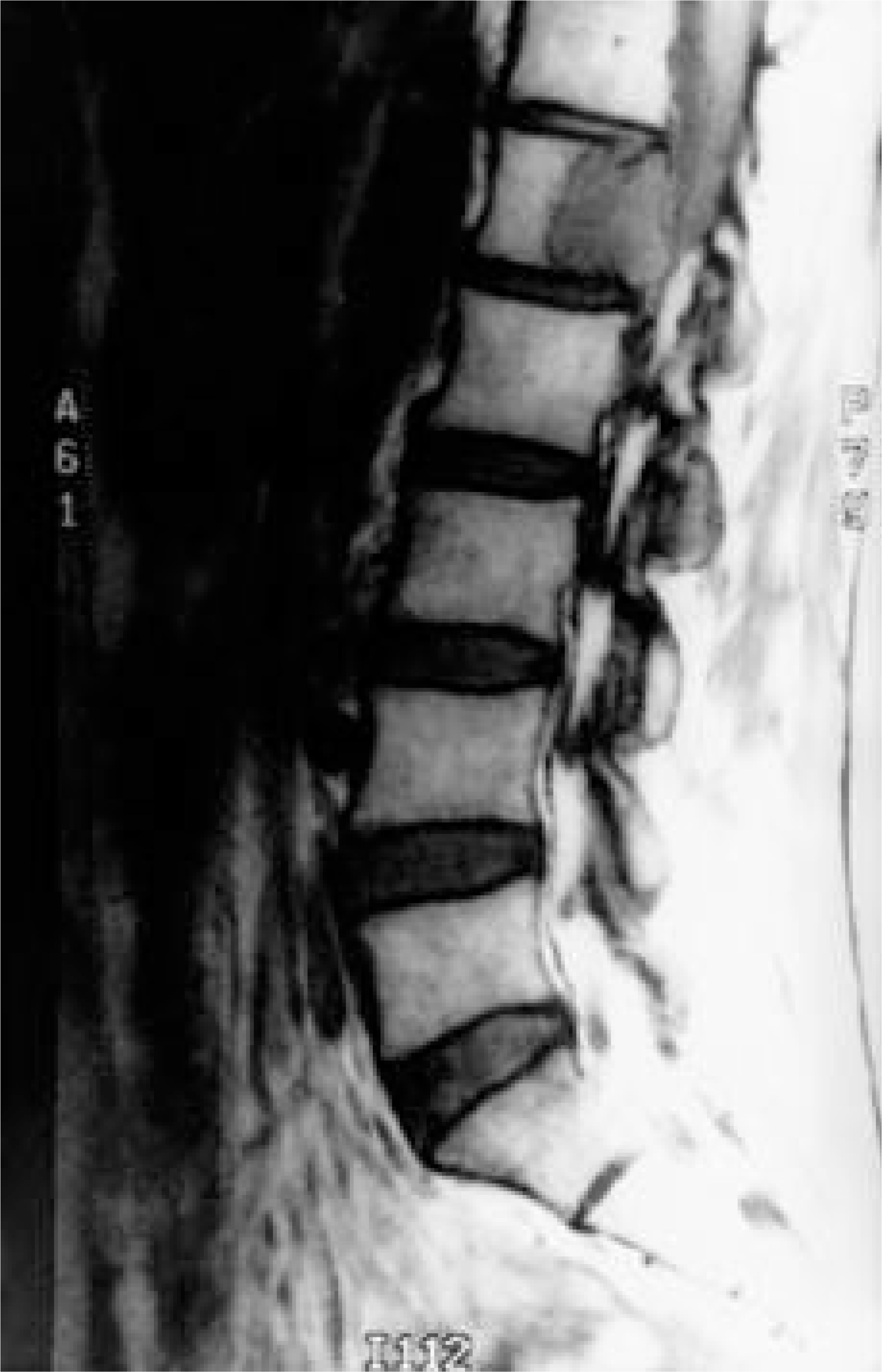 | Fig. 3.Preoperative T1-weighted sagittal sequence of MR images show well demarcated low signal intensity. |
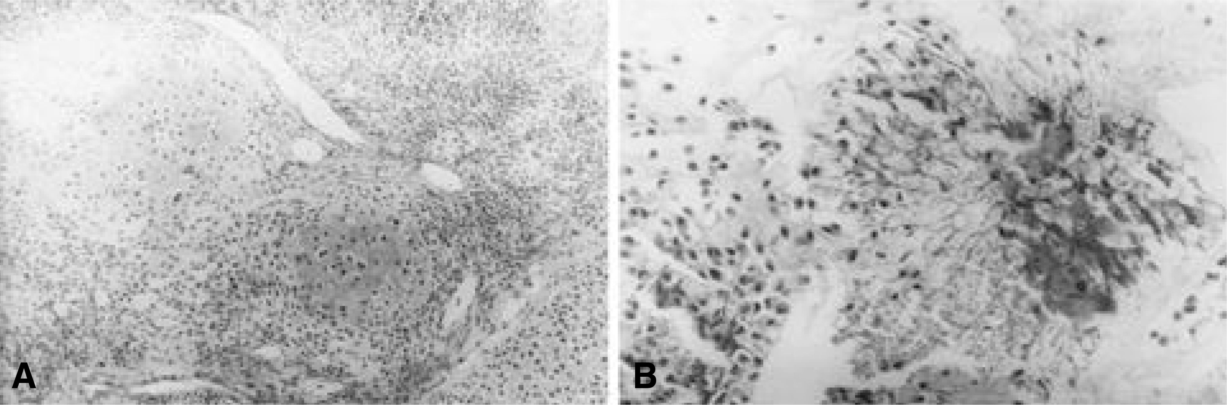 | Fig. 4-A.On histologic examination, small cuboidal tumor cells and areas of chondroid differentiation was observed (Hematoxylin and eosin stain, × 100). Fig. 4-B. Deposition of a thin layer of calcium around the tumor cells producing a characteristic chickenwire appearance (Hematoxylin and eosin stain, × 400). |
 XML Download