

 PDF
PDF ePub
ePub Citation
 Print
Print


Abstract
Objectives
To discuss a solitary bone cyst originating from the pedicle of the first lumbar vertebra.
Summary of Literature Review
The solitary bone cyst in the spinal colume is extremely rare. Since Dawson reported the first case in fourth cervical vertebral body, only 8 cases have been reported. The solitary bone cyst originating in the pedicle of the lumbar vertebra is the first reported case.
Materials and Methods
A 53- year-old female patient visited the emergency room for severe radiating pain to the left lower extremity. T2 weighted MRI image showed a solitary bone cyst in the left pedicle of the first lumbar vertebra.
Go to : 
REFERENCES
1). Aegerter EE and Kirkpatric JA Jr. Orthopedic Diseases. 3rd ed.Philadelphia, WB Saunders Co.: 491;1968.
2). Brodsky AE, Khalil M, and Vandeventer L. Unicameral bone cyst of a lumbar vertebra. J Bone Joint Surg. 68A:1283–1285. 1986.
3). Dawson EG, Mirra JM, Yuhl ET, and Lasser K. So litar y bone cyst of the cervical spine. Clin Orthop. 119:141–143. 1976.
4). Jaffe. HL and Lichtenstein L. Solitary unicameral bone cyst with emphasis on the roentgen picture., the pathologic appearance and the pathogenesis. Arch. Surg. 44:1004–1025. 1942.
5). Lee CC, Wei JD and How SW. Simple bone cyst in cervical vertebral spinous process and laminae: Report of a case. J Formos Med Assoc. 99:54–8. 2000.
6). Matsumoto K, Fuji S, Mochizuki T, and Hukuda S. Solitary bone cyst of a lumbar vertebra. Spine. 15:605–607. 1980.
7). Park CK, Cho KK, Lee SW, Jeon JS, Kang JK, and Choi CR. Simple bone cyst of the axis. Child's Nerv Syst. 13:171–174. 1997.

8). Kent K. Wu and Edwin R. Guise. Unicameral bone cyst of the spine, J Bone Joint Surg. 63A:324–326. 1981.
9). Sawai H, Nemoto F, Kushi M, and Hidaka S. A case report of solitary bone cyst in spinous process of lumbar vertebra. Kanto J Orthop Traumatol. 11:258–261. 1980.
10). Zenmyo M, Komiya S, Hamata T, and Inoue A. A solitary bone cyst in the spinous process of the cervical spine. Spine. 25:641–642. 2000.
Go to :
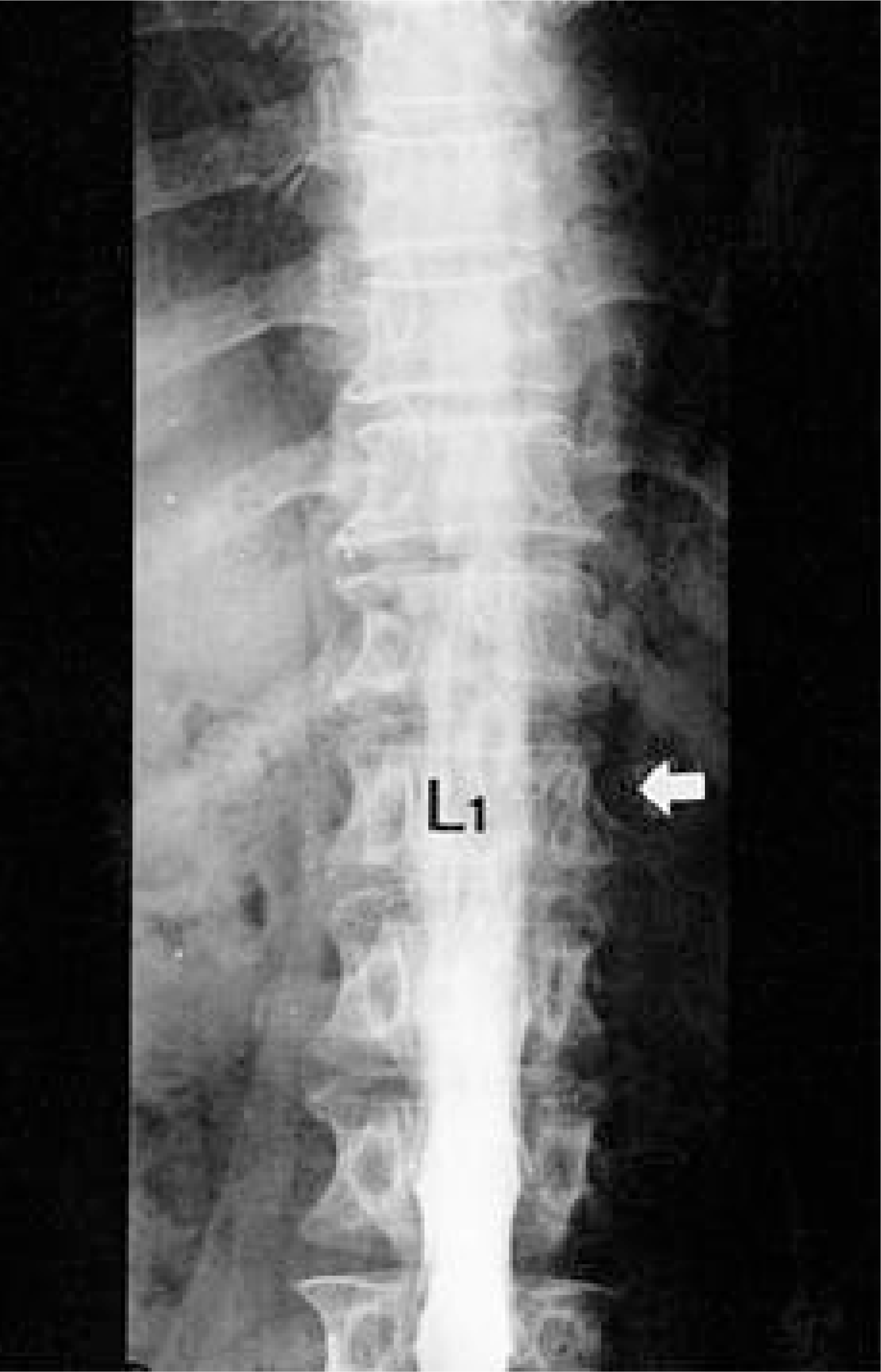 | Fig. 1.Myelogram of the patient showed expansile osteolytic lesion and cortical thinning at the left pedicle of the first lumbar vertebra. No evidence of thecal sac compression is observed. |
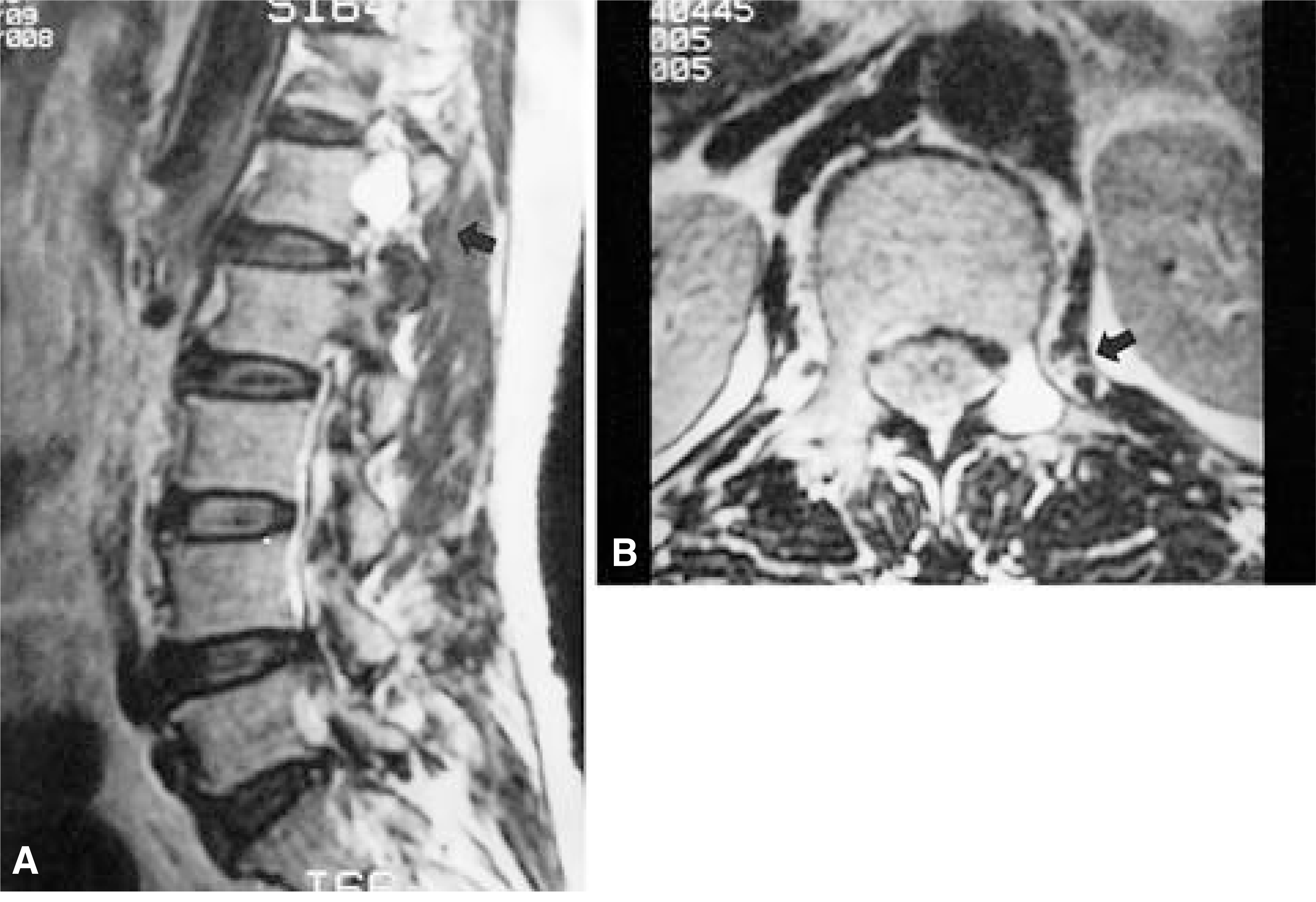 | Fig. 2-A.T2 weighted sagital MRI image showed cystic natured cavity at the left pedicle of the first lumbar vertebra. Fig. 2-B. T2 weighted axial MRI image showed the lesion is confined in the pedicle area without invasion into the spinal canal, but the medial wall of the pedicle shows cortical disruption. |
 XML Download