

 PDF
PDF ePub
ePub Citation
 Print
Print


Abstract
Study Design
We studied retrospectively the limbus vertebra by computed tomography or magnetic resonance image. Objectives : To analyze the clinical and radiologic characteristics of the limbus vertebra and to distinguish it from a fracture, infection or tumor.
Summary of Literature Review
The limbus vertebra is common. However, the clinical manifestations including the level, symptoms and radiologic characteristics of the limbus vertebra are not understood exactly in the literatures.
Materials and Methods
We presented 25 cases of the limbus vertebra that were confirmed by plain roentgenogram combined with computed tomography (CT) or magnetic resonance imaging (MRI). Of the 25 patients, 18 were males and 7 females.
Results
The levels of the limbus vertebra were L3 (2 cases), L4 (13 cases), and L5 (8 cases). There were two cases of 2 level involvement (L3/4 and L4/5). All cases showed the lower lumbar lesion and complained of the lower back pain. The accompa-nying diseases included 10 cases of herniated intervertebral discs, 2 cases of ankylosing spondylitis, 2 cases of spinal stenosis and one spondylolisthesis. Three patients were first diagnosed as tuberculous spondylitis and 2 patient as spine fracture on plain roentgenograms. But they can be confirmed by demonstrating the herniation of disc material between the anterosuperior bony fragment and the rest of the body in CT or MRI.
Conclusions
The CT or MRI could be great diagnostic modalities. The pathogenesis is thought to be the herniation of disc material into the vertebral body such as Schmorl's node and disc degeneration. Most limbus vertebra was found at the lower lumbar region and accompanied with disc bulging and degeneration. The correlation between the limbus vertebra and lower back pain is not certain.
Go to : 
REFERENCES
1). Begg AC. Nuclear herniations of the intervertebral disc: Their radiological manifestations and significance. J Bone Joint Surg. 36-B:180–193. 1954.
2). Brocher JEW. Spine. Schinz HR, Baensch WE, From-hold W, Glauner R, Uehlinger E, Wellauer J, editors. etc.Roentgen diagnosis. 2nd ed.New York: Grune and Stratton;p. 32–38. 1969.
3). Epstein JA, Lavin LS. Herniated lumbar disc in teen-age children, J Neurosurg. 21:1070. 1964.
4). Epstein NE. Lumbar surgery for 56 limbus fractures emphasizing noncalcified type III lesions. Spine. 17:1489–1496. 1992.

5). Ghelman B, Freiberger RH. The limbus vetebra: an anterior disc herniation demonstrated by discography. Am J Roentgenol. 127:854–855. 1976.
6). Goldman AB, Ghelman B, Doherty J. Posterior limbus vertebrae: a cause of radiating back pain in adolescents and young adults. Skeletal Radiol. 19:501–507. 1990.
7). Greene TL, Hensinger RN, Hunter LY. Back pain and vertebral changes stimulating Scheuermann's disease. J Pediatr Orthop. 5:1–7. 1985.
8). Heithoff KB, Gundry CR, Burton CV, Winter RB. Juvenile discogenic disease. Spine. 19:335–340. 1994.
9). Hellstadius A. A contribution to the question of the origin of anterior paradiscal defects and so-called persisting apo-physes in the vertebral bodies. Acta Orthop Scand. 18:377. 1949.
10). Henales V, Hervas JA, Lopez P, Martinez JM, Ramos R, Herrera M. Intervertebral disc herniations(limbus vertebrae) in pediatric patients: report of 15 cases. Pediatr Radiol. 23:608–610. 1993.
11). Laredo JD, Bard M, Chretien J, Kahn MF. Lumbar posteior marginal intra-oseous cartilaginous node. Skeletal Radiol. 15:201–208. 1986.
12). Lindblom K. Discography of dissecting transosseous ruptures of intervertebral discs in the lumbar region. Acta Radiol. 36:12. 1951.
13). Lippitt AB. Fracture of a vertebral body and endplate and disc protrusion causing subarachnoid block in a adolescent. Clin Orthop. 116:112–115. 1976.
14). Lowry JJ. Dislocated lumbar vertebral epiphysis in adolescent children. Report of 3 cases, J Neurosurg. 38:232. 1973.
15). Resnick D, Niwayama G. Diagnosis of bone and joint disorders. Philadelphia: WB Saunders;p. 1402–1404. 1981.
16). Schmorl G, Junghhanns H. The human spine in health and disease. 2nd ed.New York: Grune and Stratton;p. 2–14. 158-172. 1971.
17). Swischuk LE, John SD, Allbery S. Disc degenerative disease in childhood: Scheuermann's disease, Schmorl's nodes, and limbus vetebra: MRI findings in 12 patients. Pediatr Radiol. 28:334–338. 1998.
18). Takata K, Inoue SI, Takahashi K, Ohtsuka Y. Fracture of the posterior margin of a lumbar vertebral body. J Bone Joint Surg. 70-A:589–594. 1988.
19). Yagan R. CT diagnosis of limbus vertebra. J Comput Assist Tomogr. 8:148–151. 1984.
Go to :
Figures and Tables%
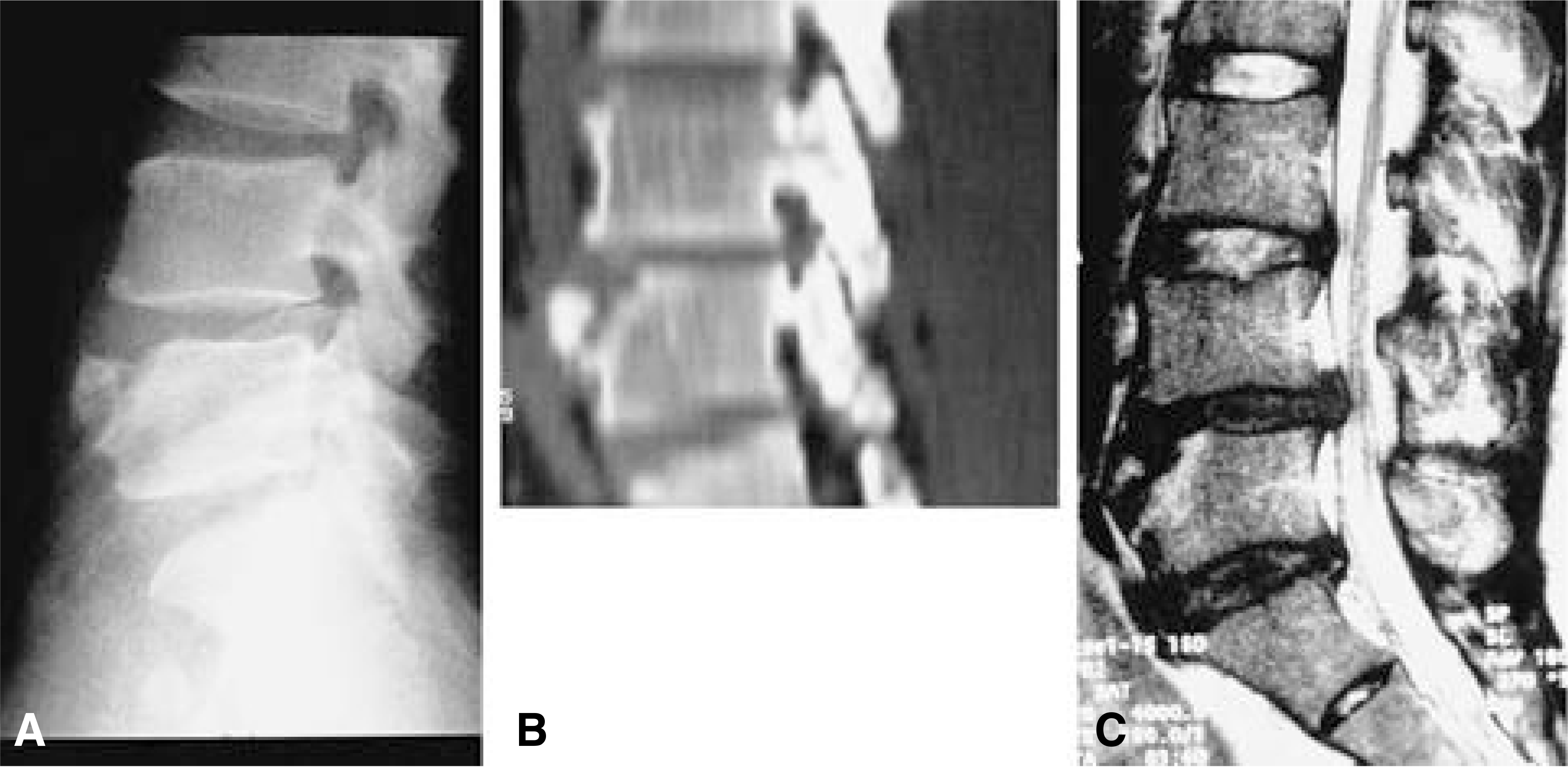 | Fig. 1-A.Lateral radiography of the lumbar spine. The bony defect with sclerotic contour at the anterosuperior margin of L4 body and a small detached triangular bony fragment was seen. Fig. 1-B. CT sagittal reformat view. The bony defect at the anterior edge of the vertebral body with irregular and increased density was seen. Fig. 1-C. Sagittal view of MRI showed that the erosion of the anterior part of the vertebral end plates, presence of an anterior bone fragment and herniation of disc material between this fragment and the vertebral body. |
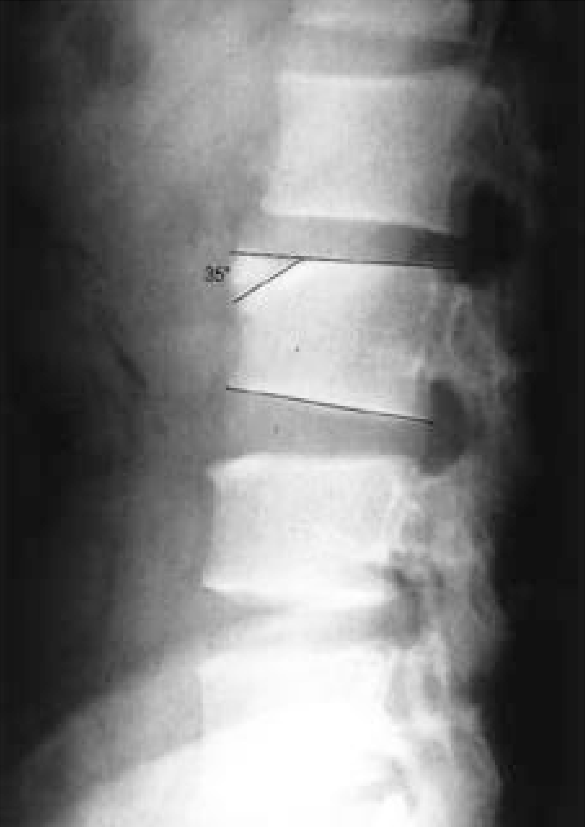 | Fig. 2.Lateral radiography of the lumbar spine. The length of detached bony fragment usually did not exceed 1/3 of endplate length and the angle between endplate and bony defect line was under 40 degrees. |
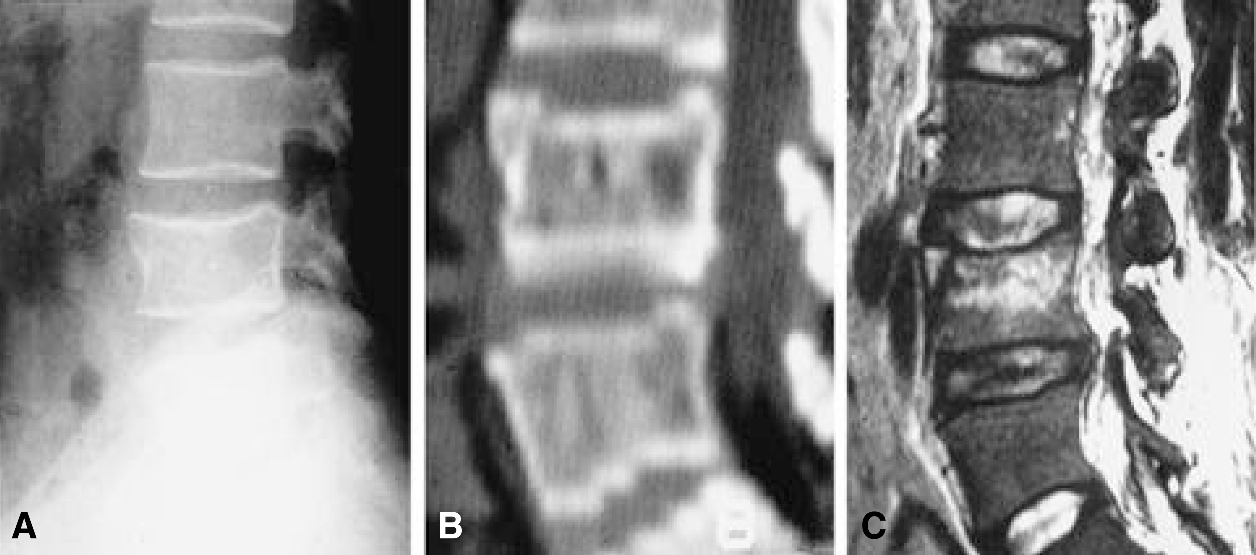 | Fig. 3-A.This case was differential diagnosis. The bony defect at the anterosuperior margin of the L4 body implies the vertebral body fracture. Fig. 3-B. CT sagittal reformat view. Fracture line is well visible. No sclerosis and no disc material is in the fracture site. Fig. 3-C. Sagittal view of MRI. The findings of no herniation of disc material between this fragment and the vertebral body implies vertebral body fracture. In addition, bony contusion of vertebral body is another evidence of vertebral body fracture. |
Table 1.
Age distribution
| M | F | |
|---|---|---|
| 5~10 | 1 | |
| 11~20 | 3 | 1 |
| 21~30 | 7 | |
| 31~40 | 4 | 1 |
| 41~50 | 2 | 2 |
| 51~60 | 3 | |
| 60~00 | 1 | |
| Total | 16 (64%) | 9 (36%) |
Table 2.
Initial diagnosis by plain radiographic examination
| Disease | No. of patients |
|---|---|
| Spinal Tuberculosis | 2 |
| Spine fracture | 3 |
| Limbus vertebra | 20 |
| Misdiagnosis rate | 5/25 = 20% |
 XML Download