

 PDF
PDF ePub
ePub Citation
 Print
Print


Abstract
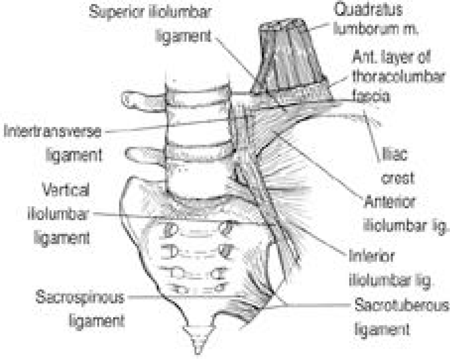
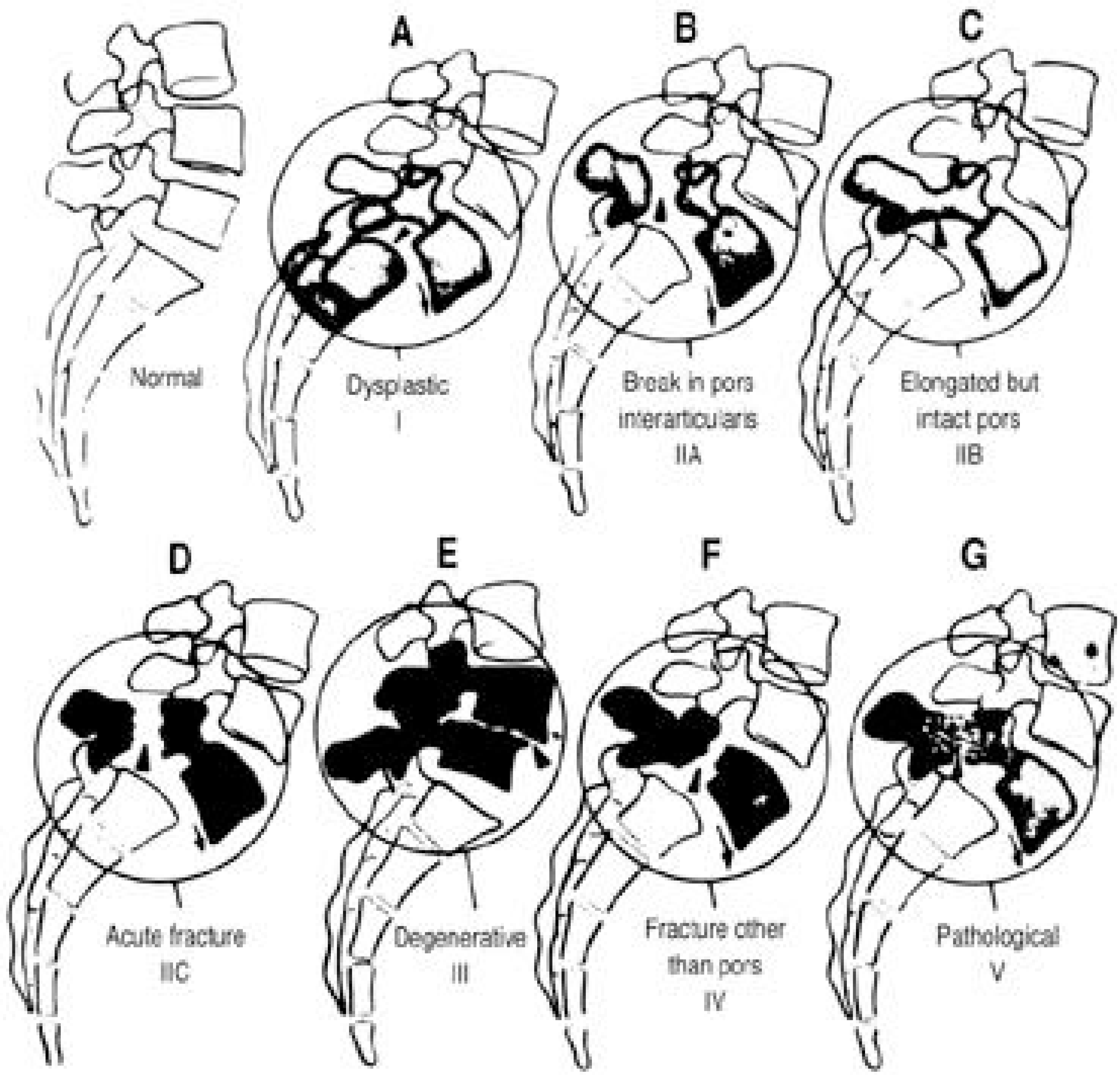
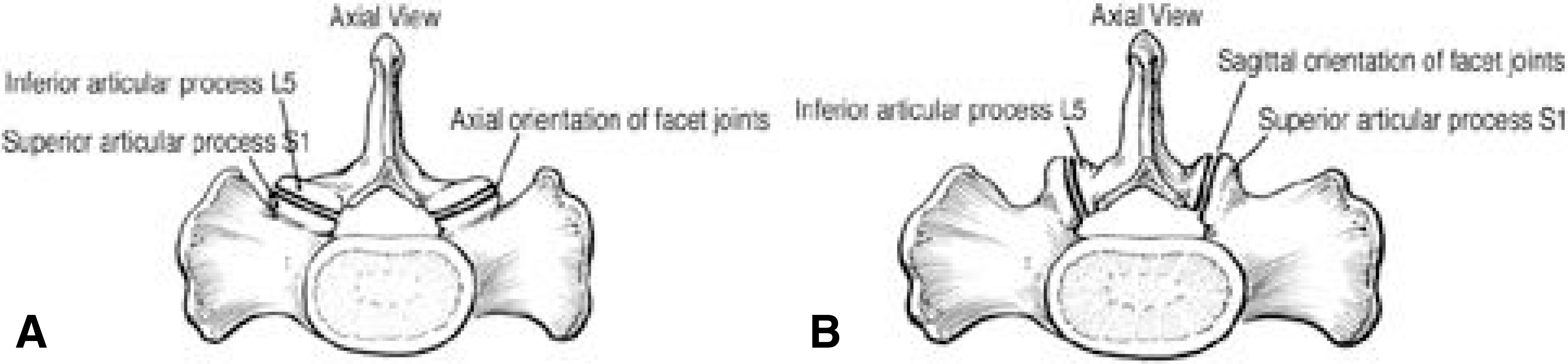
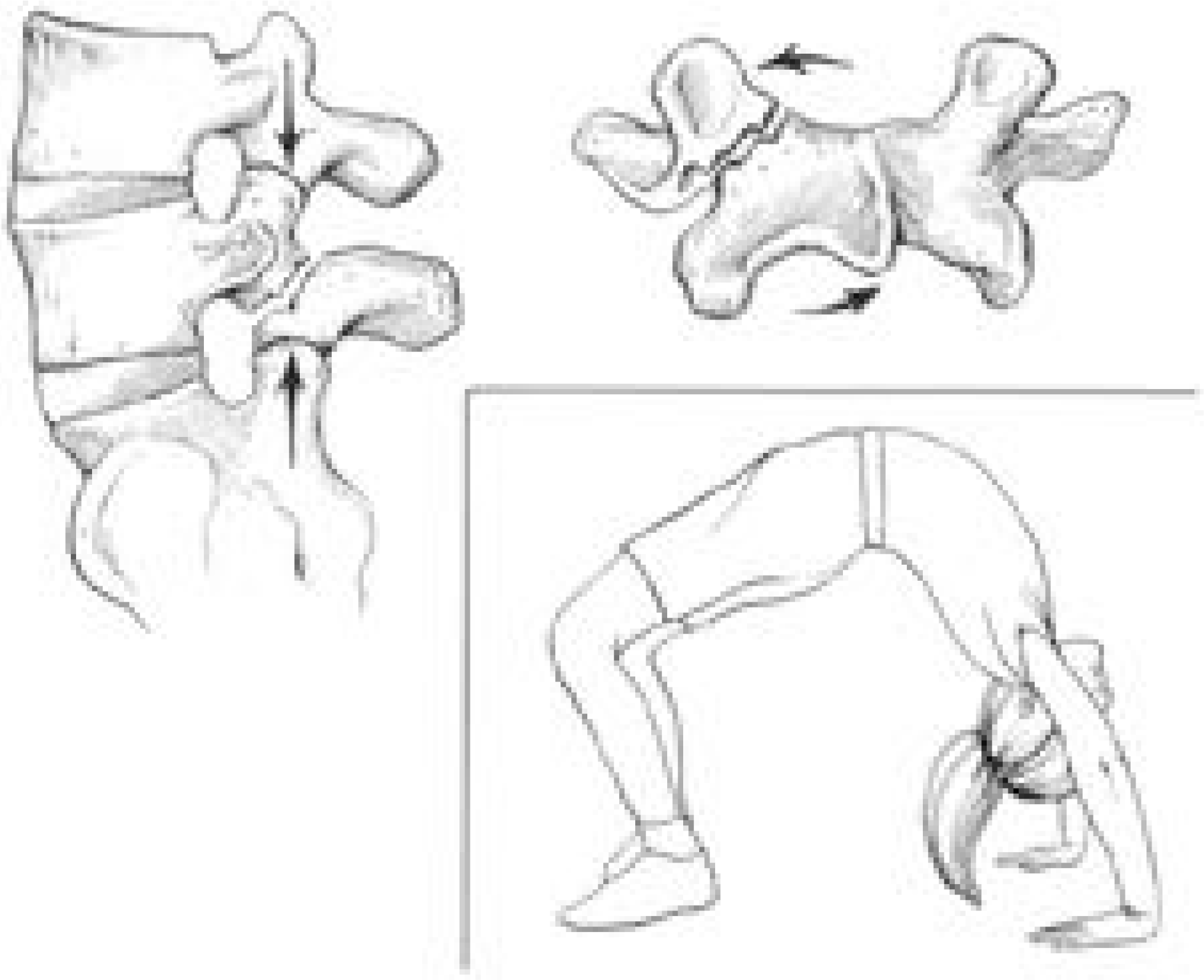
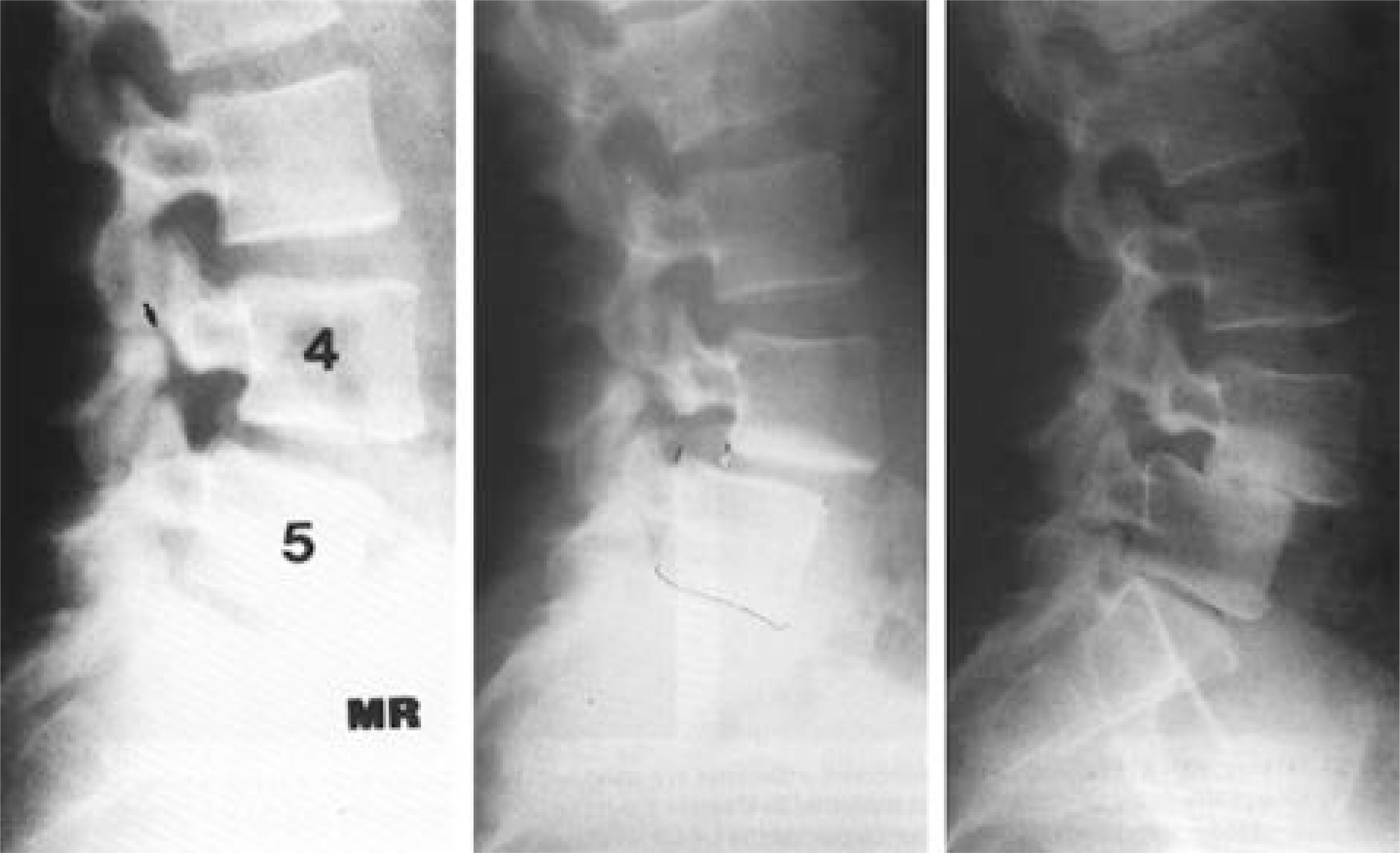
Spondylolisthesis is the slippage of all or part of one vertebra onto another. The term is derived from the Greek words spondylos and olisthesis. Wiltse, Macnab, and Newman combined their concepts in what remains the most widely accepted classification. Wiltse and Rothman in 1989 suggested a common congenital component in the etiology of dysplastic and isthmic types and added the postsurgical group to the original classification. The precise anatomy of the pars lesion is extremely important in understanding the pathogenesis. The pars interarticularis is the connecting link between pedicles, transverse processes, laminae, and the two articular facets acting as the pivot center. The L5- S1 articulation, being in the coronal plane, is more stable than the sagittal placement of the L4- 5 facet joint. The fifth lumbar vertebra is stabilized by a large L5 transverse process, which supports strong muscular and ligamentous(iliolumbar) attachment. A n increased lumbar lordosis increases the shear stress at the L4- 5 level. Both congenital and isthmic types are often associated with spina bifida of the L5 or S1 segments. There seems to be a definite sex and racial difference, with black women (1.1%) having the lowest prevalence and white men(6.4%) the highest. The increased prevalence is in A laskan natives and young sportsmen, ranging from 11% to 35%. Repetitive flexion, combined flexion- extension and both forcible hyperextension and rotation of the lumbar spine predispose to a pars stress fracture. Most of isthmic type develops during the first year of school, and by age 7 the prevalence is 4%. A further 1.4% of cases occur during adolescence, most between 11and 15 years of age. In patients under 25 years of age with low back pain and isthmic spondylolisthesis, this lesion is most probably the cause of the symptoms(18.9%). In patients older than 40 years, it is seldom the only cause of low back pain (5.2%). The radicular pain is found in 14% of patients with isthmic spondylolisthesis. Degenerative spondylolisthesis results from long standing intersegmental instability.10% of women over 60 years had a 1st or 2nd degree. It occurs 6 times more often at the L4- 5 level and 5 times more often in women, mostly in those older than 40 years. The patient may have back pain (5.6%) with or without leg pain and/or may have intermittent claudication (80%).
In traumatic spondylolisthesis, acute fractures of the pars interarticularis are rare and are always due to fracture of the other part of the posterior elements caused by severe trauma. In pathological spondylolisthesis, the bony strength is insuffient to resist forward motion of the proximal vertebra on the one below. In postsurgical spondylolisthesis, the most common etiology is extensive decompression with sacrifice of the facet joints.
Go to : 
REFERENCES
1). Apel DL, Lorenz M, Zindrick M. Symptomatic spondylolisthesis in adults: Four decades later. Spine. 14:345–348. 1989.
2). Borkow SE, Kleiger B. Spondylolisthesis in the new -born. A case report. Clin Orthop. 81:73–76. 1971.
3). Dandy DJ, Shannon MJ. Lumbosacral subluxation (Group 1 spondylolisthesis). J Bone Joint Surg. 53B:578–595. 1971.
4). Farfan HF. The pathological anatomy of degenerative spondylolisthesis: a cadaver study. Spine. 5:412–418. 1980.
5). Farfan HF, Osteria V, Lamy C. The mechanical etiology of spondylolysis and spondylolisthesis. Clin Orthop. 117:40–55. 1976.

6). Fredrickson BE, Baker D, McHolick WJ, et al. The natural history of spondylolysis and spondylolisthesis. J Bone Joint Surg. 66A:699–707. 1984.
7). Frennered AK, Danielson BI Nachemson AL, et al. Midterm followup of young patients fused in situ for spondylolisthesis. Spine. 16:409–416. 1991.
8). Grobler LJ, Rovertson PA, Novotny JE, Pope MH. Etiology of spondylolisthesis. Assessment of the role played by lumbar facet joint morphology. Spine. 18:80–91. 1993.
9). Harms J, Boehm H, Zielke K. Surgical treatment of spondylolisthesis. The Text Book of Spinal Surgery, vol 1. Philadelphia: PA, Lippincott;p. 585–592. 1991.
10). Harris I, Weinstein S. Longterm followup of spondylolisthesis. J Bone Joint Surg. 69A:960–969. 1987.
11). Hefti F, Brunazzi M, Morscher E. Natural course in spondylolysis and spondylolisthesis. Orthopaed. 23:220–227. 1994.
12). Hensinger RN. Spondylolysis and spondylolisthesis in children. Instr Course Lect, Am Acad Orthop Surg, St Louis CV Mosby. 32:132–151. 1983.
13). Henson J, McCall IW, O'Brien JP. Disc damage above a spondylolisthesis. Br J Radiol. 60:69–72. 1987.
14). Herbiniaux G. Traife'surdivers accouchements laborieux et sur les polypes de la matrice. Brussels: JL Boubers;1782.
15). Jackson DW, Wiltse LL, Circincione RJ. Spondylolysis in female gymnasts. Clin Orthop. 118:68. 1976.
16). Junghann H. Spondylolisthese, pseudospomdylolisthese und wirbelverschiebung nach hinten. Brun's Beitr Klin Chir. 151:376. 1931.
17). Kettlekamp DB, Wright GD. Spondylolysis in the Alaskan Eskimo. J Bone Joint Surg. 53A:563. 1971.
18). Kilian HF. Schilderungen neuer Beckenformen and ihres verhaltens in Leben. Mannheim: Verlag von Bassermann & mathey;1854.
19). Krenz J, Troup JDG. The structure of the pars interarticularis of the lower lumbar vertebrae and its relation to the etiology of spondylolysis, with a report of the healing fracture in the neural arch of a fourth lumbar vertebra. J Bone Joint Surg. 55B:735–741. 1973.
20). Lamble W. Beifrage zur geburtskunde und gynacko -logue. Von F W Scanzoni;1884.
21). Letts M, Smallman T, Afanasiev R, Gouw G. Fr ac tur e of the pars interarticular in adolescent athletes: a clinical biomechanical analysis. J Pediatr Orthop. 6:40–46. 1986.
22). Macnab I. Backache. 2nd ed. Baltimore: Williams and Wilkins;pp. p. 84–103. 1990.
23). Marchetti PF, Bartolozzi P. Spondylolisthesis: classification and etiopathogenesis. Progress in spinal pathology: Spondylolisthesis II. Bologna, Italy: Italian Scoliosis Research Group;p 35. 1986.
24). Marchetti PG and Bartolizzi. Spondylolisthesis: Morden trends in orthopaedic surgery. In Gaggi A. Bologna. 165:1982.
25). Matsunaga S, Ikiri K, Hayashi K. Non surgi cally managed patients with degenerative spondylolisthesis: a 10-to 18-year followup study. J Neurosurg (spine 2). 93:194–198. 2000.
26). Mimura M, Moriya H, Takahashi K, et al. Rotational instability in degenerative spondylolisthesis-A possible mechanical etiology of the disease. Presented at the International Society for the Study of the Lumbar Spine Meeting. Marseilles, France, June. 1993.
27). Neugebauer FL. A new contribution to the history and etiology of spondylolisthesis. New Sydenham Soc. Select. Monogr. 121B:1–64. 1888.
28). Newman P, Stane K. The etiology of spondylolisthesis with a special investigation by KH stone. J Bone Joint Surg. 45B:39–59. 1963.
29). Roche MB, Rowe GG. Incidence of separate neural arch and coincident bone variations: a survey of 4,200 skeletons. Anat Rel. 109:233–252. 1951.
30). Rossi F. Spondylolysis, spondylolisthesis, and sports. J Sports Med Phys Finess. 18:317–400. 1978.
31). Rowe GG, Roche MB. The etiology of separate neural arches. J Bone Joint Surg. 35A:102. 1953.
32). Saraste H. a radiologic follow up of spondylolysis and spondylolisthesis. J Pediatr Othop. 7:631–638. 1987.
33). Saraste H, Bronstrom LA, Aparisi T. Prognostic radiologic aspects of spondylolisthesis. Acta Radiol.[Diagn] (Stockh). 25:427–432. 1984.
34). Seitsalo S, Osterman K, Hyvarinen H, et al. Progression of spondylolisthesis in children and adolescents. A longterm followup of 272 patients. Spine. 16:417–421. 1991.
35). Szypryt EP, Twining P, Mulholland RC, Worthing-ton BS. The prevalence of disc degeneration associated with neural arch defects of the lumbar spine assessed by magnetic resonance imaging. Spine. 14:977–981. 1989.
36). Wiltse L, Rothman S. Classification, diagnosis and natural history. Semin Spine Surg. 1:78–94. 1999.
37). Wiltse LL. Etiology of spondylolisthesis. J Bone Joint Surg. 44A:539–560. 1962.
38). Wiltse LL, Rothman SLG. Spondylolisthesis: Classification, diagnosis and natural history. Semin. Spine Surg. 1:78. 1989.
39). Wiltse LL, Newman PH, MacNab I. Classification of spondylolysis and spondylolisthesis. Clin orthop. 117:23. 1976.
40). Wiltse LL, Widell EH Jr, Jackson DW. F atigue fracture: the basic lesion in isthmic spondylolisthesis. J Bone Joint Surg. 57A:17–22. 1975.
41). Winter RB. Congenital kyphosis. J Bone Joint Surg. 55A:223–256. 1973.
42). Wynne-Davies R, Scott JH. Inheritance and spondylolisthesis: A radiographic family survey. J Bone Joint Surg, b. 1B:301–305. 1979.
Go to :

 XML Download