Abstract
The patient of lumbar disc herniation complains low back pain and sciatica. The intervertebral disc was degenerated by aging, the lifetime incidence of low back pain ranges from 80 to 90%, as determined by epidemiologic studies, whereas the incidence of sciatica is only 2~40%. This article was made with review of the natural history, clinical manifestations and physical examinations of lumbar disc herniation. In conclusion, a careful history and physical examination remain the key to accurate diagnosis of the cause of low back pain and /or sciatica, and very helpful adjuncts to make a diagnosis of lumbar disc herniations.
REFERENCES
1). Andersson GB, Svensson H, Oden A. The intensity of work recovery in low back pain. Spine. 8:880–884. 1983.

2). Berqquist-Ulman M, Larsson U. Acute low back pain in industry, A controlled prospective study with special reference to therapy and confounding factors. Acta Orthop Scand. 170(Suppl):1–117. 1977.
3). Biering-Sorensen F. Physical measurements as risk indi -cators for low back trouble over a one-year period. Spine. 9:106–119. 1984.
4). Biering-Sorenson F, Thomson C. Medical, social and occupational history as risk factors for low back trouble in a general population. Spine. 11:170. 1986.
5). Bigos SJ, Battie MC, Spengler SM, et al. A prospective study of work perceptions and psychosocial factors affecting the report os back injury. Spine. 16:1–6. 1991.
7). Bodguk N, Tynan W, Wilson AS. The nerve supply to the human lumbar intervertebral discs. J Anat. 132:39–56. 1981.
8). Bodguk N, Wilson AS, Tynan W. The Human lumbar dorsal rami. J Anat. 134:383–397. 1982.
9). Boumphrey FRS, Bell GR, Modic M, et al. Computed tomography scanning after chymopapain injection for herniated nucleus pulposus. A prospective study. Clin Orthop. 219:120–123. 1987.
10). Bradley KC. The anatomy of backache, Aust NZ: J Surg. 44:227–232. 1974.
11). Breig A, Troup JDG. Biomechanical considerations in the straght-leg-raising test. Cadaveric and clinical studies of the effects of medial hip rotation. Spine. 4:242–250. 1979.
12). Bundschuh CV, Modic MT, Ross JS at al. Epidural fib -risis and recurrent disk herniation in the lumbar spine: MR imaging assessment. AJR Am J. Roentgenol. 150:923. 1988.
13). Dehlin O, Hedenrund B, Horal J. Back symptoms in nursing aids in a geriatic hospital. Scand J Rehabil. 8:47–53. 1976.
14). Deyo RA, Tsui- Wu YJ. Descriptive epidemiology of low back pain and its related medical mare in the United States. Spine. 12:264–268. 1987.
16). Dixon ASJ. Diagnosis of low back pain-sorting the com -plainers. Jayson M, editor. The Lumbar Spine and Back Pain. New York: Grune & Stratton;p. 77–92. 1976.
17). Fahrni WH. Observations on straight leg-raising with special reference to nerve root adhesion. Can J Surg. 9:44–48. 1966.
18). Fry J. Back pain and Soft tissue Rheumatism. Advisoty Services. Clinical and General Ltd.;1972.
19). Frymoyer JW, Pope MH, Clements JH, Wilder DG, MacPherson B. Ashikaga T. Rick factors in low back pain. Bone Joint Surg. 65A:213–218. 1983.
20). Gulliver I. Acute low back pain in industry. Acta Orthop. Scand. 129:6. 1970.
21). Hakelius A. A prognosis in sciatica. A clinical followup of surgical and non-surgical treatment. Acta Orthop Scand. 129(Suppl):1. 1970.
22). Hwan-mo Lee. Imaging diagnosis and classification of th Lumbar disc herniation. J of korean spine surgery;p. 276–282. 2000.
23). Heliovaara MD. Body height, obesity and risk of herniated lumbar intervertebral disc degeneration. Spine. 12:469–472. 1987.
24). Hirsch C, Jonsson B, Lewin T. Low back symptoms in aa Swedish female population. Clin Orthop. 63:171–176. 1969.
25). Hirsch C, Nachemson A. The reliability of lumbar disck surgery. Clin Orthop. 29:189–195. 1963.
26). Hoppenfeld S. Orthopedic Neurology. Philadephia: Lippincott;1977.
27). Horal J. The clinical appearance of low back disorders in the City of Gothenburg, Sweden. Acta Orthop Scand. 118(Suppl):8–73. 1969.
28). Hult L. Retroperitoneal disc fenestration in low back pain and sciatica: A preliminary report. Acta Orthop Scand. 20:342–348. 1951.
29). Hult L. Cervical, dorsal and lumbarspine syndromes. Acta Orthop Scasnd. 17:65–73. 1954.
30). Lassoner EM, Alho A, Karakarju EO, Paavilaimen T. Short term prognosis in sciatica. Ann. Chir Gynaecol. 66:47. 1977.
32). Malinsky J. The development of nerve terminations in the intervertebral discs of man. Acta Anat. 38:96–113. 1959.
34). Nachemson AL. The lumbar spine - an orthopedic challenge. spine. 1:59–71. 1976.
35). Nam-Hyun Kim, Hwan-mo Lee. Spinal Surgery(ed 1),1st, medical culture: 175-176. 1998.
36). ummi J. Iskiassyndroomaan leikkaushoidon indikaatiot. In Nummi J, Jarvinen(eds). Kipuselka ja sen hoito. Helsin -ki, Tyoterveyslaitoksen Julkaisuja, 33. 1974.
37). Ostgaard HC, Anderson GB, Karlsson K. Prevalence of back pain in pregnancy. Spine. 16:549–552. 1972.

38). Pearce J., Moll J. Conservative treatment and natural history of acute lumbar disc lesions. J. Neurosurg. Psychiatry. 30:13. 1967.
39). Roland M., Morris R. A study of the natural histpry of back pain. I. Development of reliable and sensitive measure of disability in low back pain. Spine. 8:141–144. 1983.
41). Rothman RH, Simeone FA, Bernini PM. Lumbar disc disease, Rothman and Simeone(eds): The Spine, vol 1 (ed 2). Philadelpia: Saunders;p. 508–645. 1982.
42). Rydevik BL, Brown MD, Lundborg G. Pathoana-tomy and pathophysiology of nerve root compression. Spine. 9:7–15. 1984.

43). Sangfort E. Lasegue's sign in patients with lumbar disc herniation. Acta Orthop. 42:459–460. 1971.
45). Spangfort EV. The lumbar disc herniation. A com-puter-acied analysis of 2, 054 operations. Acta Orthop Scand. 142:1–95. 1972.
46). Sugiura K, Yoshida T, Katoh S, et al. A study on tension signs in lumbar disc hernia. Int Orthop. 3:225–228. 1979.

47). Se-Il Suk. Orthopaedics(ed 5), new medical culture: 451-454. 1998.
48). Se-Il Suk. Spinal Surgery(ed 1), new medical culture: 190-202. 1997.
49). Weber H. Lumbar disc herniation. A prospective study of prognostic factors including a controlled trial. J Oslo City Hosp. 28:36. 1978.
50). Weber H. lumbar disc herniation: a controlled prospective study with ten years of observation. spine. 8:131. 1983.

51). Weinstein JN. Anatomy and neurophysiologic mechanisms of spinal pain. The adult spine. New York: Raven Press;p. 593–610. 1991.
52). White AWM. Low back pain in men receiving Work -men's compensation. Can Med Assoc J. 95:50–56. 1966.
53). Wood PH. Epidemiology of back pain. The lumbar spine and back pain. Kent: Pitman Medical publishing Company;p. 29–55. 1980.
Figures and Tables%
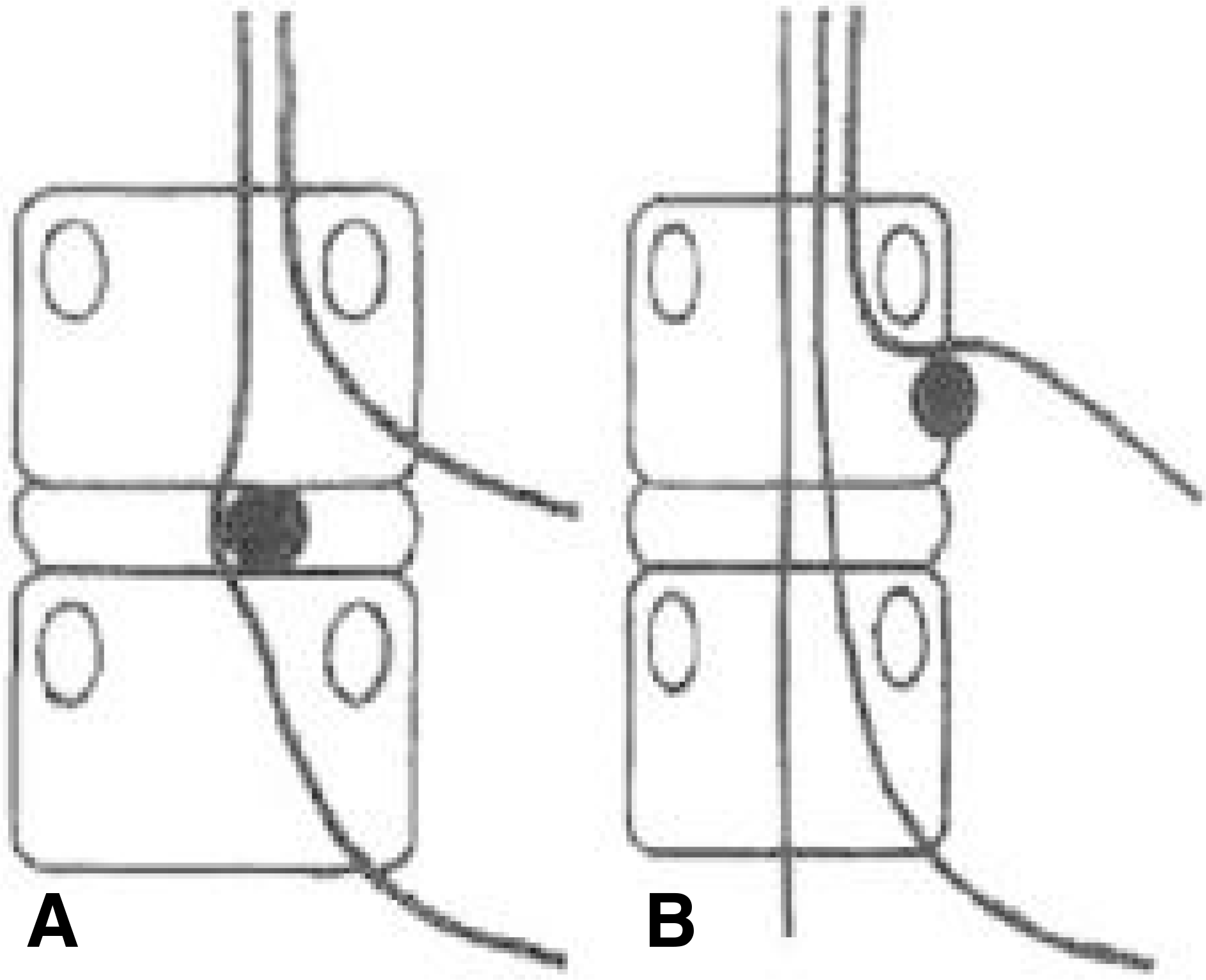
Fig. 1.
Root compression according to type of herniated disc Fig. 1-A. Posterior herniated disc Fig. 1-B. Extraforaminal herniated disc

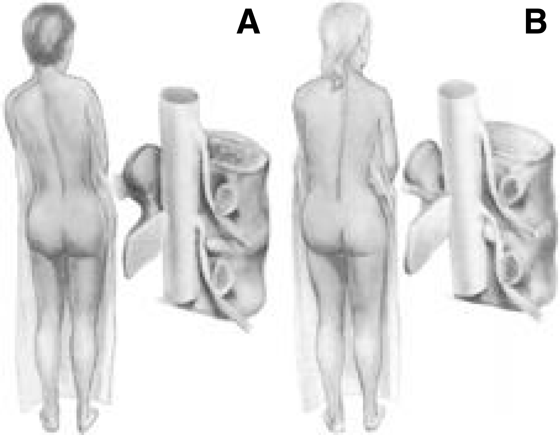
Fig. 2-A.
Herniated disc lateral to the nerve root. This usually produces a sciatic list away from the side of irritated nerve root. Fig. 2-B. Herniated disc medial to the nerve root and in an axillary position. This may produce a sciatic list toward the side of irritated nerve root.

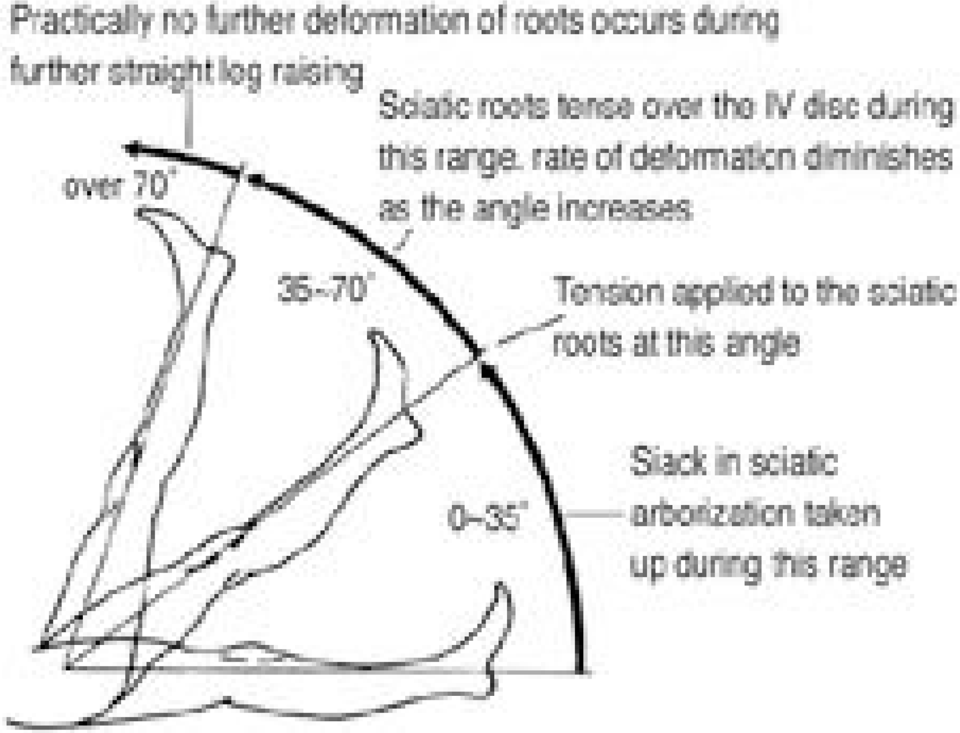
Fig. 3.
The dynamics of SLR : Observations on straight leg raising, with special reference to nerve root adhesion.

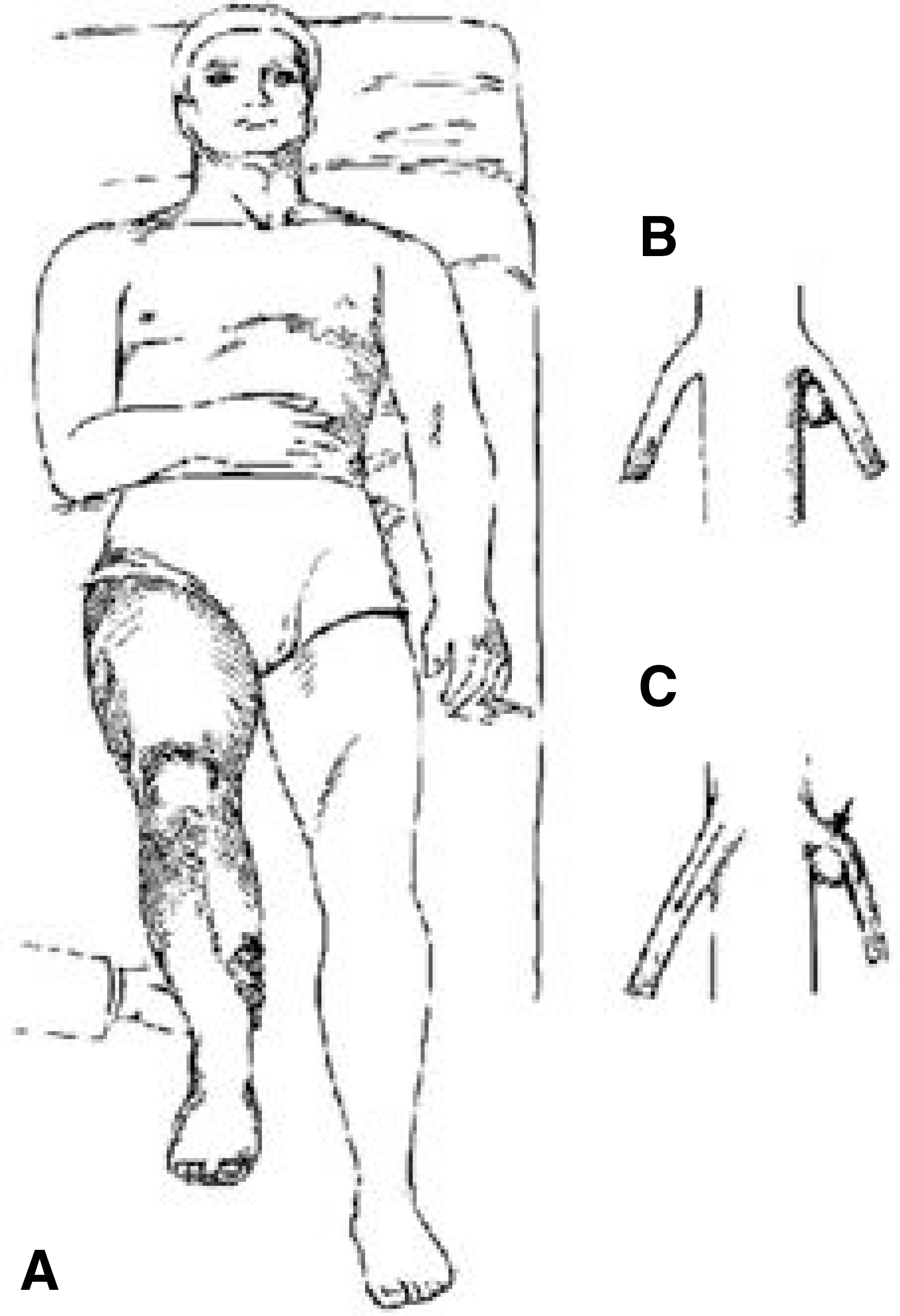
Fig. 4.
Movement of nerve roots when the leg on the opposite side is raised. Fig. 4-A. When the leg is raised on the unaffected side, the roots on the opposite side slide slightly downward and toward the midline. Fig. 4-B. In the presence of a disc lesion, particularly in an axillary locatin, this movement. Fig. 4-C. Increases the root tension.

Table 1.
neurologic test according to location of herniated disc
Table 2.
sensory test


 PDF
PDF ePub
ePub Citation
 Print
Print


 XML Download