

 PDF
PDF ePub
ePub Citation
 Print
Print


Abstract
Study Design
This case report presents a rare case of presacral giant schwannoma which originates from the S1 nerve root.
Objectives
To discuss a surgical approach for removal of presacral giant schwannoma and review the pertinent literatures.
Summary of Literature Review
Pre- sacral tumors are unusual neoplasms that cause approximately one in 40,000 hospital admissions. Schwannoma represents only a small fraction of the many types of tumors that may be present in this region. Less than 1% of all spinal schwannomas occur in the sacrum. The treatment of this lesion is complete removal, which is curative.
Materials and Methods
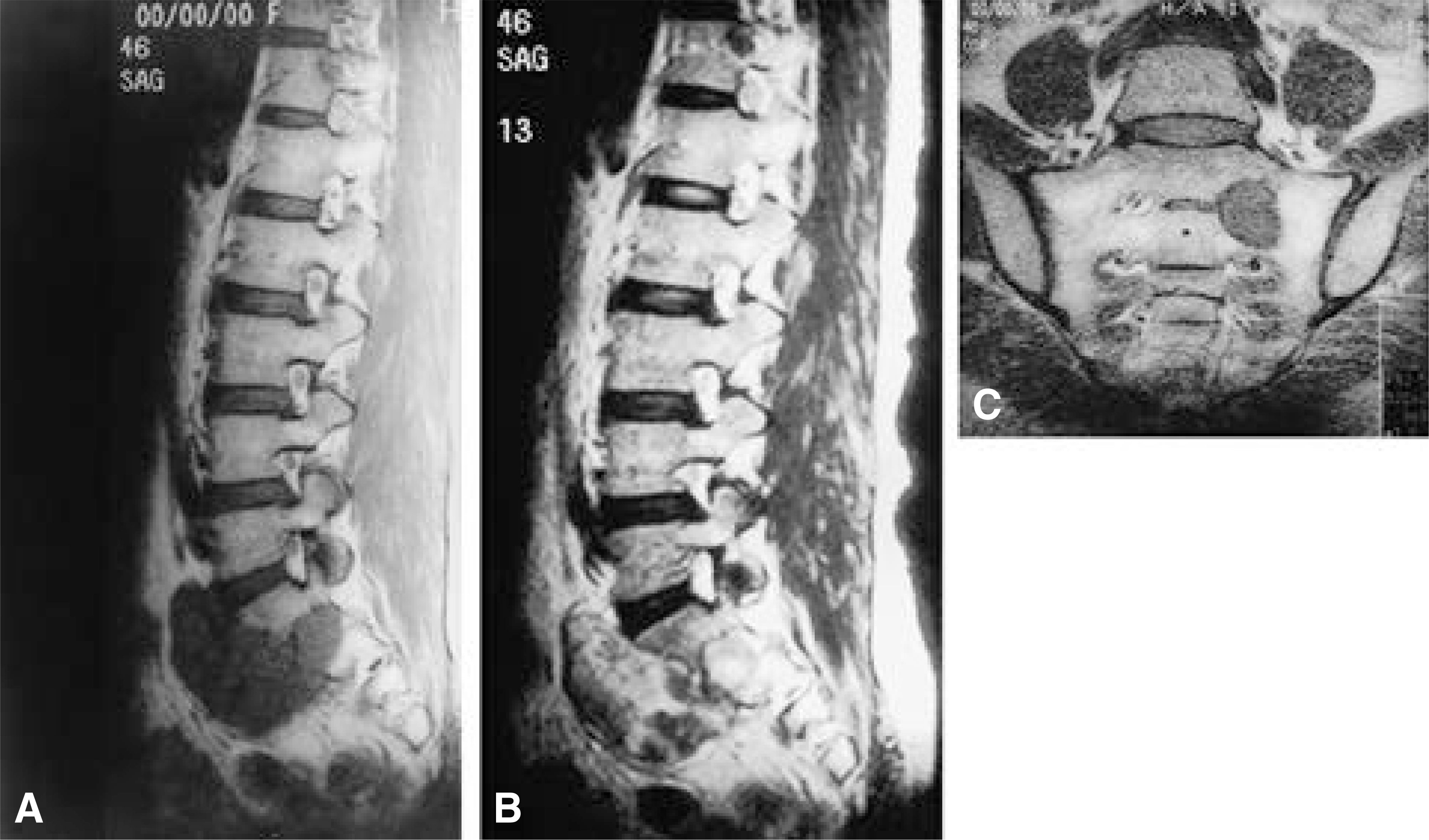
A 46- year- old woman developed gradual back pain and radiating pain on her left lower extremity for about a year. There was no noted improvement with the use of conservative treatment. T1- weighted sagittal MRI reveals a large homogeneous low- signal intensity mass on left presacral area and intrasacral extension of the tumor forming a dumbbell shaped mass.
Results
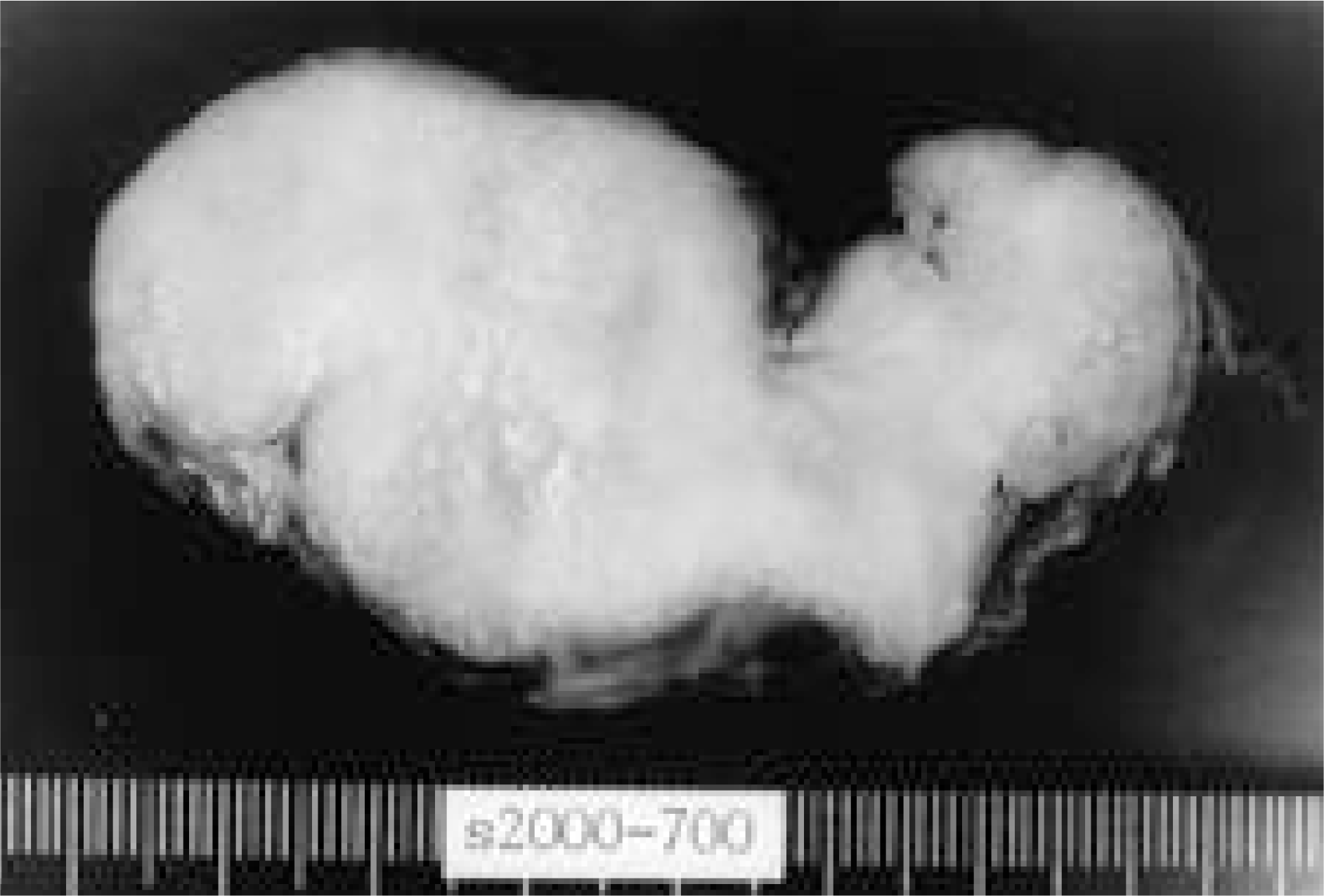
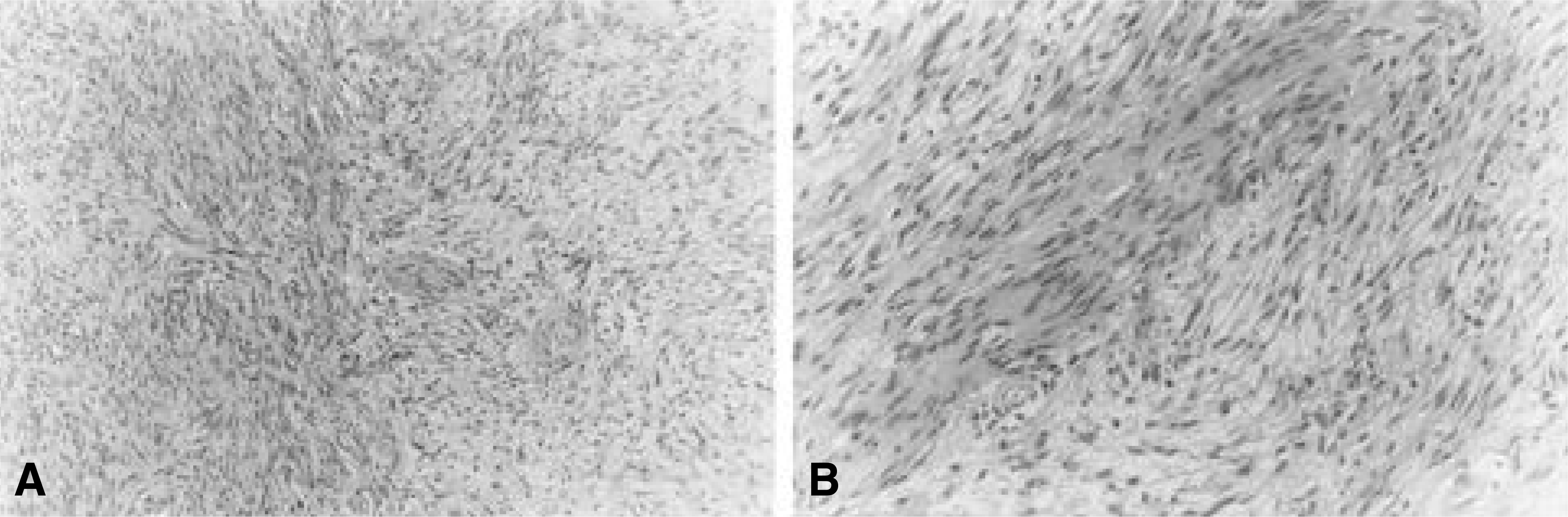
The tumor was completely removed by a combined anterior and posterior approach. The excised mass was cylindrical, measuring 8x4x3 cm in size, which had originated from the S1 nerve root. It was histologically diagnosed as benign schwannoma. Satisfactory result was obtained after the complete removal of the mass.
REFERENCES
1). Abernathey CD, Onofrio BM, Scheithauer B, Pairolero PC, Shives TC. Surgical management of giant sacral schwannomas. J Neurosurg. 65:286–295. 1986.

2). Acciarri N, Staffa G, Poppi M. Giant sacral schwannoma: removal by anterior, transabdominal approach. Br J Neurosurg. 10(5):489–492. Oct. 1996.
3). Das Gupta TK, Brasfield RD, Strong EW, Hajolu SI. Benign solitary schwannomas. Cancer. 24(2):355–366. Aug. 1969.
4). Gupta S, Sikora SS, Gupta R, Singh MK, Chatto-padhyay TK. Presacral neurilemoma(schwannoma): Report of a rare case. Jpn J Surg. 19(2):229–231. 1989.
5). Kim P, Ebersold MJ, Onofrio BM, Quast LM. Surgery of spinal nerve schwannoma. Risk of neurological deficit after resection of involved root. J Neurosurg. 71:810–814. 1989.
6). Madanes A, Ucci A, Mitchell GW Jr. Presacral neu-rilemmoma. A case report and literature review. J Reprod Med. 27(6):356–358. Jun. 1982.
7). Rengachary SS, O'Boynick P, Batnitzky S, Kepes JJ. Giant intrasacral schwannoma: Case report. Neurosurgery. 9(5):573–577. Nov. 1981.
Fig. 1-A.
T1-weighted sagittal MR image showing a large homogenous low signal intensity mass. The large tumor is located at the left presacral area and intrasacral extension of the tumor forming a dumbbell shaped mass. Fig. 1-B. T2-weighted sagittal MR image showing a large inhomogenous high signal intensity mass. Fig. 1-C. T1-weighted coronal MR image showing a well demarcated homogenous low signal intensity mass arising from S1 nerve root.
 XML Download