Abstract
Study design
Retrospective analysis of surgical treatment in patients with osteoporotic vertebral fracture associated myelopathy.
Objectives
To evaulate the clinical outcome of anterior decompression and fusion for osteoporotic vertebral fracture associated with myelopathy.
Summary of Literature Review
Major treatment of osteoporotic vertebral fracture were conservative methods. In patients with myelopathy, surgical treatment is recommanded.
Materials and Methods
From January 1995 to December 1998, twelve patients who had osteoporotic vertebral fracture associated with myelopathy and treated by operation were evaluated retrospectively. With simple roentgenography and dual energy absorptiometry, osteoporosis was evaluated. A nd with MRI and nerve conduction velocity test, we could diagnosed myelopathy. In ten patients, anterior approach was used, and in two patients, posterior approach was used.
Results
In all patients after operation, the neurologic symptoms according to the Frankel grading scale were improved over one grade and followup X - ray showed bone union finding unrelated to the site, shape, and severity of fracture. No significant complications such as increasing of kyphotic angle and metal loosening were existed in all cases.
Go to : 
REFERENCES
1). Barnett E, Nordin BEC. The radiological diagnosis of osteoporosis. A new approach. Clin Radiol. 11:166–174. 1960.

2). Burstein AH, Zika JH, Heiple KG, Klein LK. Contribution of collagen and mineral to the elastic plastic properties of bone. J Bone Joint Surg. 57-A:956–961. 1975.
3). Cameron JR, Sorenson JA. Measurement of bone and mineral in vivo. An improved method. Science. 142:230–232. 1963.
4). Cann CE, Genant HK, Ettinger B. Spinal mineral loss of oophorectomized women. Determination by quanti -tative computerized tomography. JAMA. 224:2056–2059. 1980.
5). Cohn SH, Ellis KJ, Wallach S. Absolute and relative deficit in total skeletal calcium and radial bone mineral in osteoporosis. J Nucl Med. 15:428–435. 1974.
6). Crilly RG, Horsman A, Marshall DH, Nordin BEC. Postmenopausal and corticosteroid induced osteoporosis. Front Horm Res. 5:53–54. 1978.
7). Eastell R, Cedel SL, Wahner HW, Riggs BL, Melton LJ III. Classification of vertebral fractures. J Bone Miner Res. 6:207–215. 1991.

8). Frankel HL, Hancock DO, Hyslopo , et al. The value of postural reduction in the initial management of closed injuries of the spine with paraplegia and tetraplegia. Paraplegia. 7:179–192. 1969.

9). Garnett ES, Kennet TJ, Kenyon DB. A photon scat-tering technique for the measurement of absolute bone density in man. Radiology. 106:209–212. 1973.

10). Genant HK, Bautovich GJ, Singh M. Bone seeking radionuclides. An vivo study of factors affecting skeletal uptake. Radiology. 113:373–382. 1974.
11). Iskrant AP. The etiology of hip fractures in females. Am J Public health. 58:485–490. 1968.
12). Jahng JS, Kang KS, Yang KH, Park HW, Lee SB. A clinical study on osteoporosis and back pain. J of Korean Orthop Assoc. 24:1210–1216. 1989.
13). Jensen PS, Orphanoudakis SC, Rauschkolb EN. Assessment of bone mass in the radius by computed tomography. A J R. 134:285–292. 1980.

14). Johnston CC Jr, Epstein S. Clinical, Biological, Radiographic, and Economic Features of Osteoporosis. Orthop Clin North America. 12:559–569. 1981; In vivo measurement of bone mass in the radius. Metabolism,. 17:1140–1142. 1968.
16). Jowsey J. Metabolic disease of bone. 1st ed.Philadelphia: WB Saunders Co;p. 96–114. 1977.
18). Lee DY, Choi IH, Lee CK, Kang SY, Roh SK. A comparison of bone mineral density in korean between normal population group and fracture risk group by photon absorptiometry. J of Korean Orthop Assoc. 23:945–953. 1988.
19). Mazess RB, Cameron JR. Bone mineral content in normal U.S. whites. Proceedings of the international con -ference on bone mineral measurement, DHEW Publication, 228-238. 1975.
20). Moon MS, Lee KS, Shin KS. Epidemiologic survey of the osteoporosis by simple spine roentgenograms in kore -ans(preliminary report). J of Korean Orthop Assoc. 24:571–581. 1989.
21). Nisson BE, Westlin NE. Bone and mineral content and fragility. Clin Orthop. 125:196–198. 1977.
23). Saville PD, Kharmosh O. Osteoporosis of rheumatoid arthritis. Influence of age, sex and corticosteroids. Arthritis Rheum. 10:423–430. 1967.

24). Verbiest H. Results of the surgical treatment of idiopathic developmental stenosis of the lumbar vertebral canal. J Bone Joint Surg. 59-A:181–189. 1977.
25). Wallach S. Osteoporosis in the female. Advances in obstetrics and gynecology. 1st ed.Batimore: Williams & Wilkins Co;p. 567–572. 1978.
Go to : 
Figures and Tables%
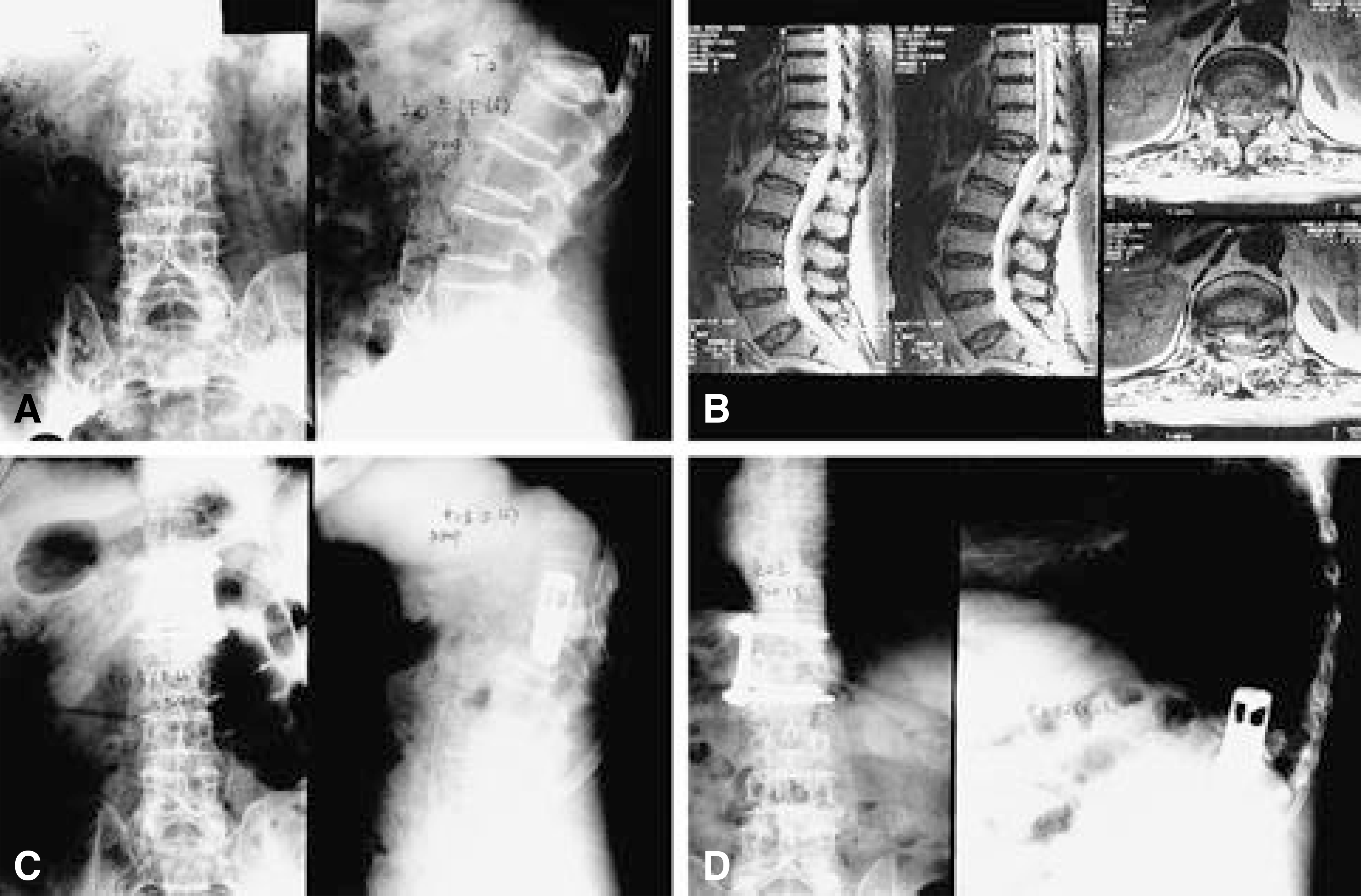
 | Fig. 1.66-year-old female. She was treated with T12 anterior decompression and T11-L1 anterior interbody fusion. Fig. 1-A. Preoperative X-ray shows severe compression fracture of T12 vertebral body. Fig. 1-B. Preoperative MRI shows compression of T12 spinal cord. Fig. 1-C. Immediate postoperative X-ray shows autogenous bone graft and plate and screws fixation state. Fig. 1-D. Postoperative one year X-ray shows well maintained state of plate and screws, and bone union of graft site. |
Table 1.
Results of preoperative evaluation and operation methods in osteoporotic vertebral fracture with myelopathy.
| Patient (Age/Sex) | Fracture site | Saville grade | Type of deformity | Spinal central score(Barnett) | Bone density* | Myelopathy (MRI†/NCV‡) | Kyphotic change(X-ray) | operation methods |
|---|---|---|---|---|---|---|---|---|
| 1(68/M) | T9 | III | Flat | 38 | 0.504 | +/+ | – | ant. decomp.§ |
| 2(70/F) | T10 | IV | Flat | 27 | 0.372 | +/+ | – | ant. decomp. |
| 3(58/F) | T12 | II | Biconcave | 42 | 0.497 | +/+ | – | ant. decomp. |
| 4(65/F) | T9 | III | Biconcave | 48 | 0.582 | +/+ | – | ant. decomp. |
| 5(64/F) | T9 | II | Flat | 32 | 0.571 | +/+ | – | ant. decomp. |
| 6(78/M) | T11 | II | Wedge | 63 | 0.729 | +/+ | + | post. decomp.|| |
| 7(72/F) | T1 | III | Flat | 41 | 0.425 | +/+ | – | ant. decomp. |
| 8(70/M) | T9 | IV | Flat | 37 | 0.397 | +/+ | – | ant. decomp. |
| 9(76/F) | L1 | IV | Biconcave | 50 | 0.492 | +/+ | + | post. decomp. |
| 10(66/F) | T12 | IV | Flat | 41 | 0.489 | +/+ | – | ant. decomp. |
| 11(69/M) | T12 | IV | Biconcave | 44 | 0.501 | +/+ | – | ant. decomp. |
| 12(66/F) | T10 | II | Biconcave | 50 | 0.577 | +/+ | – | ant. decomp. |
| mean 68.6 | 42.8 | 0.511 | ||||||
| Bone density* : bone density measured by dual energy absorptiometry | ||||||||
| MRI† : magnetic resonance imaging | ||||||||
| NCV‡ : nerve conduction velocity | ||||||||


 PDF
PDF ePub
ePub Citation
 Print
Print


 XML Download