Abstract
Study design
A nalysis was based on radiographic appearance of 57 cases of congenital scoliosis and associated anomaly
Purpose
The aim of the present study was to assess the incidence, morphology and the associated anomalies of the congenital spinal scoliosis.
Summary of Literature Review
Hemivertebra is the most common type of congenital scoliosis and urogenital, musculoskeletal and cardiac anomalies are strongly associated.
Materials and Methods
The authors analysed the morphology and the associated anomalies of 57 cases of congenital scoliosis from 1994 to 2000.
Re su l ts
It was more common in male(32 males and 25 females). The bony anomalies were classified as failure of formation(40cases, 70.2%), failure of segmentation(11cases, 19.3%) and mixed type(6cases, 10.5%). Of the failure of formation, there were 36 cases(63.2%) of hemivertebra, 2 cases of posterior quadrant vertebra and 2 cases of wedge vertebra. We found associated anomalies in 26 patients(45.6%). A ssociated cardiac anomalies were 2 dextrocardia, ventricular septal defect, atrial septal defect and patent ductus arteriosus. A ssociated musculoskeletal anomalies were 5 rib fusion, 2 developmental dysplastic hip, 3 Klippel-Feil syndrome, A chondroplasia, A rnold- Chiari malformation, spinal dysraphism with sacral hair patch, cleft palate with congenital anklyloglossia. A ssociated neurogenic anomalies were 2 cases of syringomyelia and 3 mental retardation. There were unilateral renal agenesis and undescended testicle in urogenital anomalies.
Go to : 
REFERENCES
1). Benard TN, Burke SW, Jhonston CE, Roberts JM. Congenital spinal deformities. A review of 47 cases. Orthopedics. 8:777–783. 1985.
2). Connor JM, Conner AN, Connor RAC, Tolmie JL, Yeung B, Goudie D. Genetic aspects of early child -hood scoliosis. Am J Med Genet. 27:419–424. 1987.
3). Day G, Upadhyay S, Ho E, Leong J, Ip M. Pulmonary function in congenital scoliosis. Spine. 19:1027–1031. 1994.
4). Drvaric DM, Ruderman RJ, Conrad RW, et al. Co ngen -ital scoliosis and urinary tract abnormalities: Are intravenous pyelograms necessary? J Pediatr Orthop. 7:441–443. 1987.
5). Hensinger RN, Lang JE, MacEwen GD. Klippel-Feil syndrome. a constellation of associated anomalies. J Bone Joint Surg. 56-A:1246–53. 1974.
6). Jaskwhich D, Ali RM, Patel TC, Green DW. Congenital scoliosis. Curr Opin Pediatr. 12:61–66. 2000.

7). McMaster MJ, David CV. Hemivertebra as a cause of scoliosis, A study of 104 patients. J Bone Joint Surg. 68-B:588–595. 1986.

8). McMaster MJ and Ohtsuka. The natural history of con -genital scoliosis: A study of two hunderd and fifty-one patients. J Bone Joint Surg. 64-A:1128–1147. 1982.
9). Peterson HA, Peterson LFA. Hemivertebra in iden -tical twins with dissimilar spinal columns. J Bone Joint Surg. 49-A:938–942. 1967.
10). Prahinski JR, Polly DW, McHale KA, Ellebogen RG. Occult intraspinal anomalies in congenital scoliosis. J Pediatr Orthop. 20:59–63. 2000.

11). Reckles LH, Peterson HA, Bianco AJ, Weidman WH. The association of scoliosis and congenital heart defects. J Bone Joint Surg. 57-A:449–455. 1975.

12). Shapiro F, Eyre D. Congenital scoliosis. A histopathologic study. Spine. 6:107–117. 1981.
13). Suk SI, Yoon GS, Bin SI. The brace treatment of congenital scoliosis. J of Korean Orthop Surg. 20:545–553. 1985.

14). Tanaka T, Uhtoff H. The pathogenesis of congenital vertebral malformations. Acta Orthop Scand. 52:413–425. 1981.
15). Winter R, Moe J, Lonstein J. The incidence of Klip -pel-Feil syndrome in patient with congenital scoliosis and kyphosis. Spine. 9:363–366. 1984.
16). Wynne-Davies R. Congenital vertebral anomalies: Etiology and relationship to spina bifida cystica. J Med Genet. 12:280–288. 1975.
Go to : 
Figures and Tables%
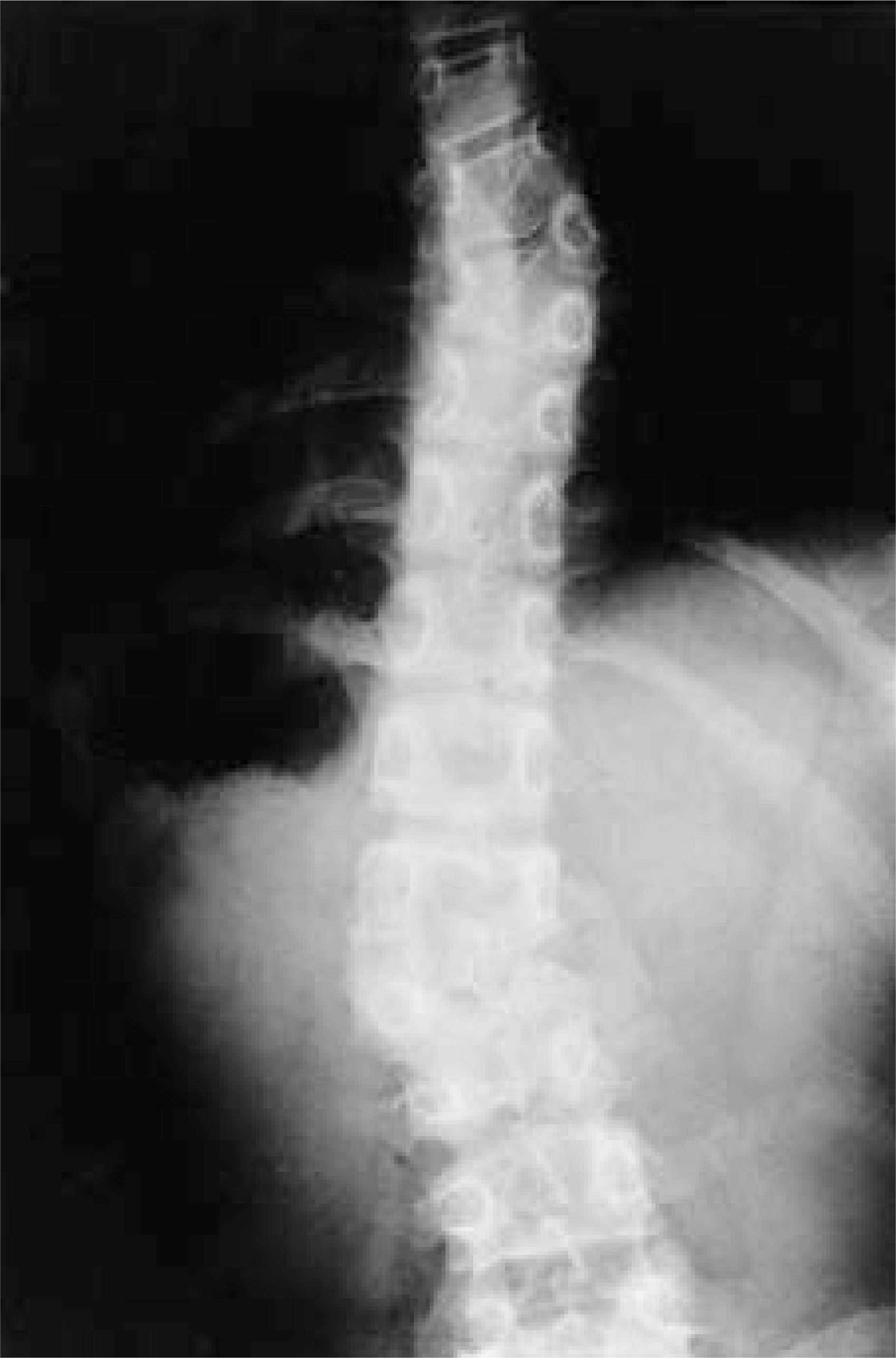
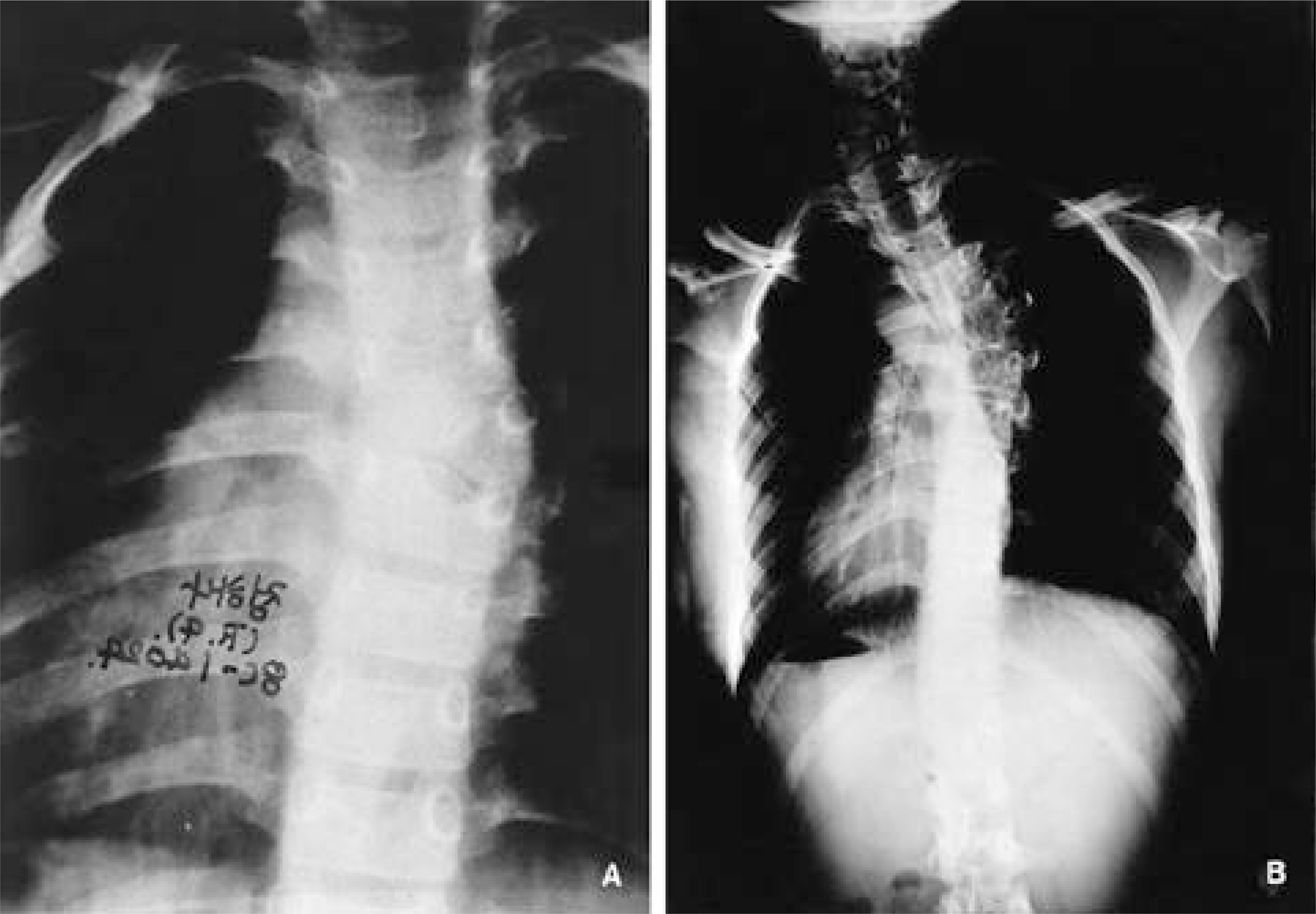 | Fig. 1-A.Plain radiograph in 1988 shows right T6 hemivertebra fully segmented. Fig. 1-B. 10 years later(1998), the Cobb's angle was increased to 39 degrees. |
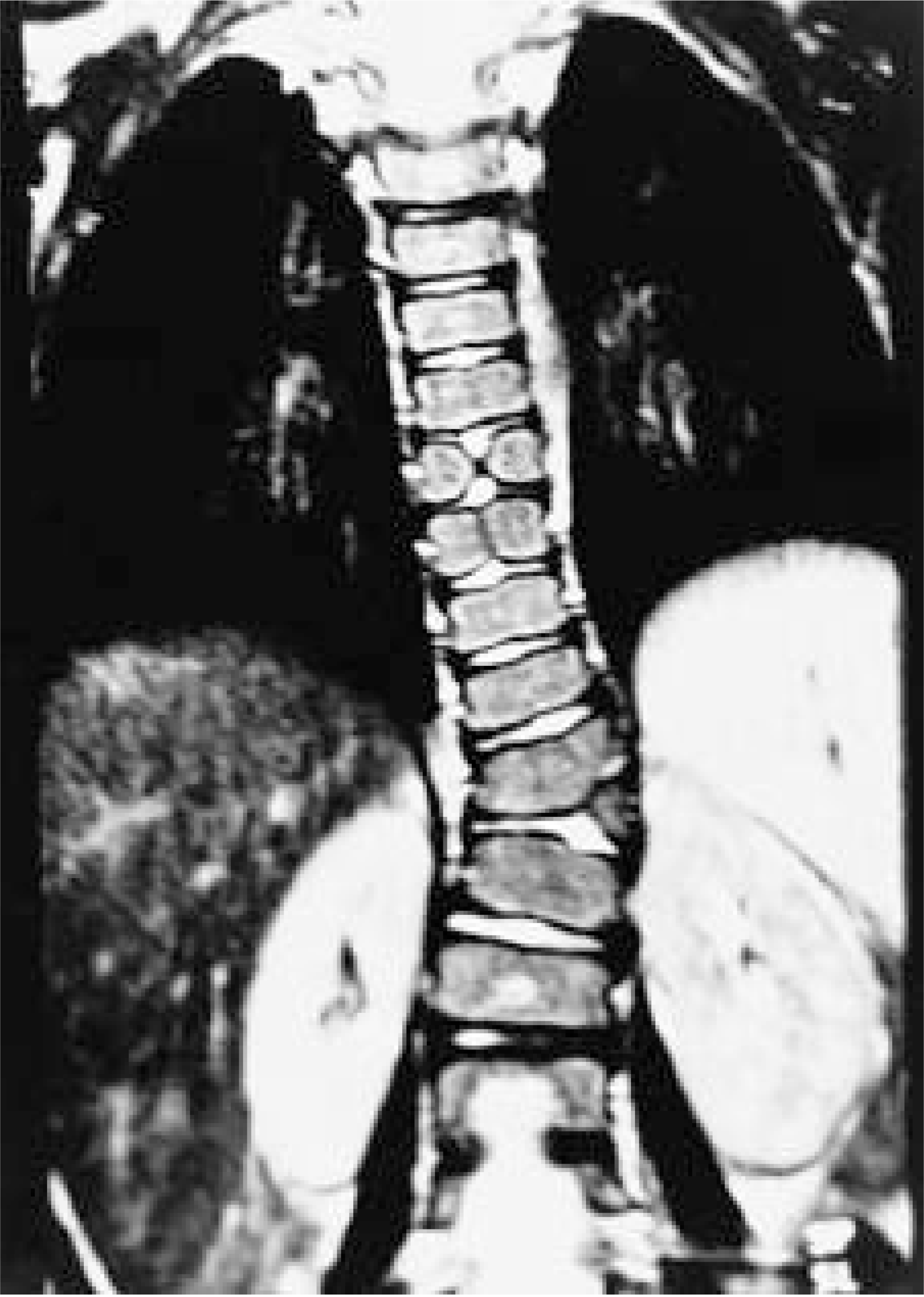
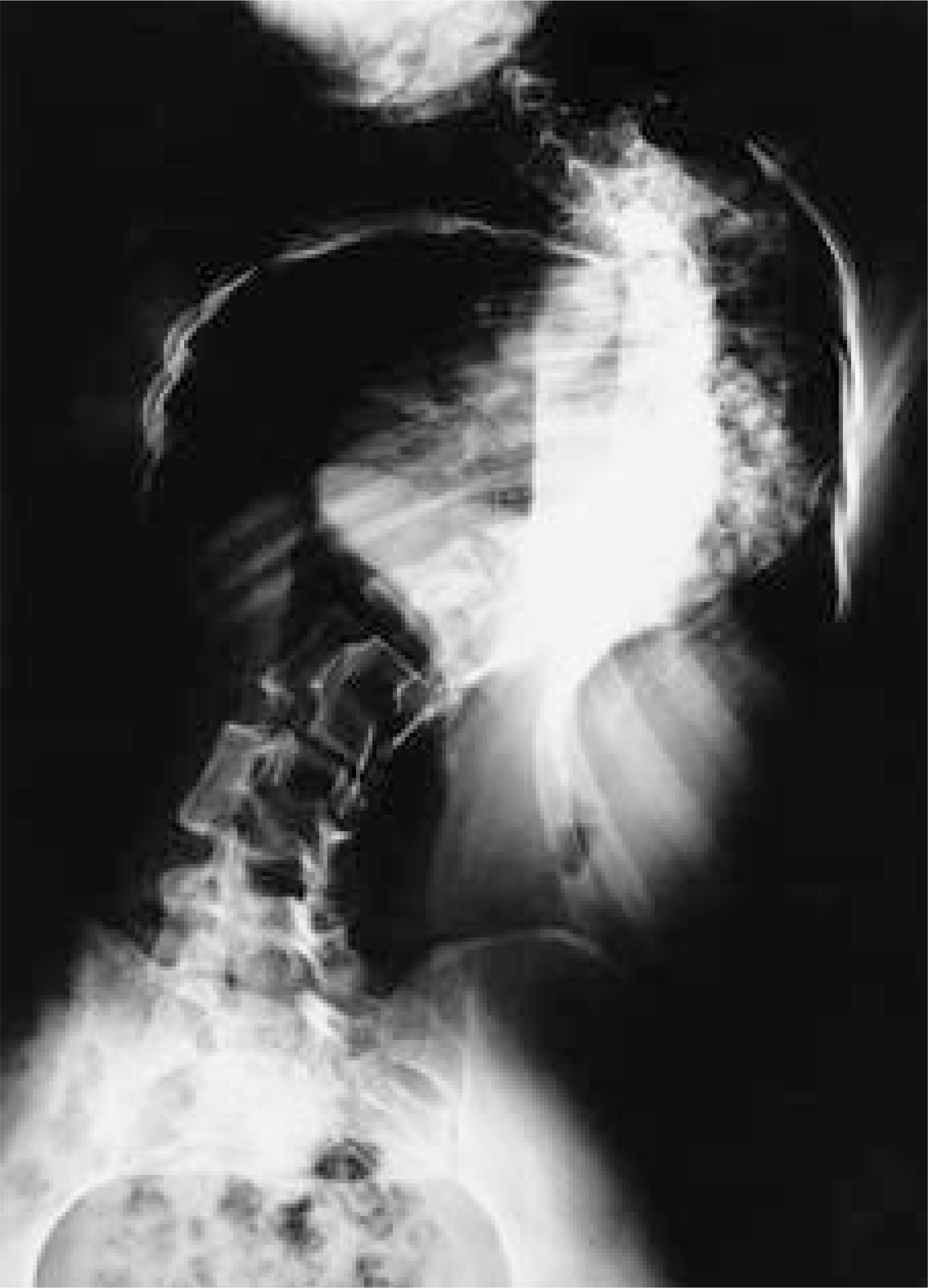 | Fig. 3.21-year-old-female with unilateral bar from T3-T11, the Cobb's angle was 122 degrees. |
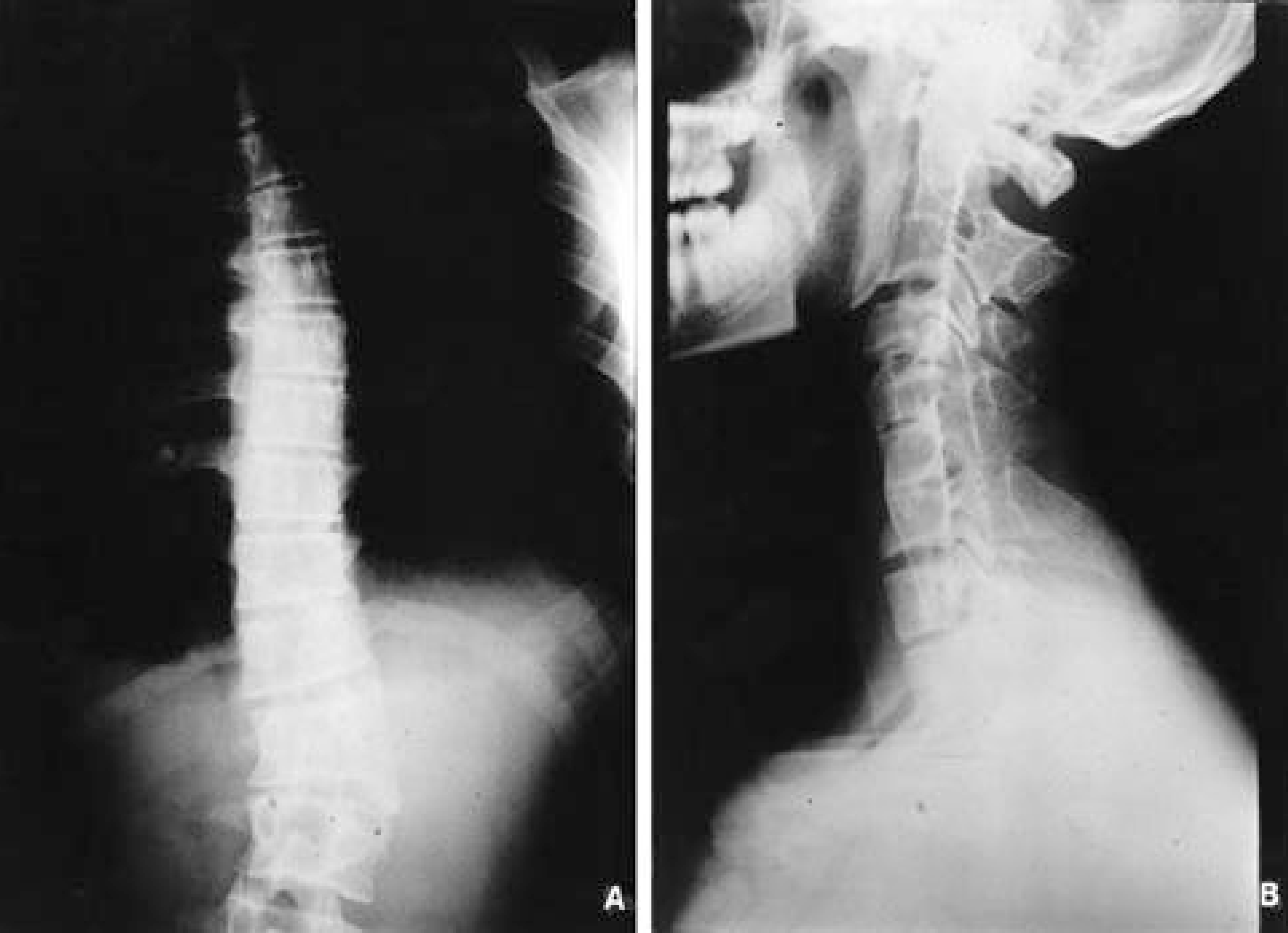 | Fig. 4-A.Whole spine anteriorposterior shows right L2 unincarcerated hemivertebra partially segmented. Fig. 4-B. Cervical spine lateral view shows fusion of C1-2 and C4-6 spine. |
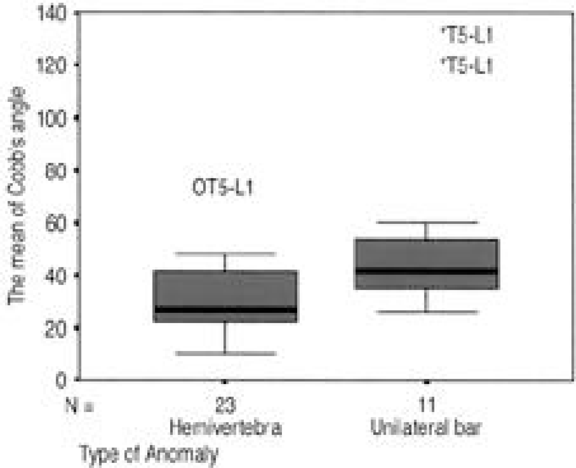 | Fig. 6.Distribution of Cobb's angle of hemivertebra and unilateral bar shows 32.1 degrees of the mean Cobb's angle in hemivertebra and 55.4 degrees in unilateral bar. |
Table 1.
Associated anomalies in 26 patients with congenital scoliosis.
Table 2.
Summary of vertebral anomalies
Table 3.
The mean of Cobb's angle of hemivertebra and unilateral bar
| Type of anomaly | location (No. of patient) | The mean of Cobb's angle |
|---|---|---|
| Hemivertebra | T∗1-T4(1) | 40 |
| (full segmented) | T5-L†1(8) | 38.1 |
| L2-L4(3) | 41 | |
| Hemivertebra | T1-4(1) | 39 |
| (semisegmented) | T5-L1(5) | 22.2 |
| L2-L4(5) | 24.4 | |
| Unilateral bar | T1-4(2) | 32.5 |
| T5-L1(7) | 66.3 | |
| L2-L4(2) | 40 |


 PDF
PDF ePub
ePub Citation
 Print
Print


 XML Download