Abstract
Purpose
To describe and analyze the clinical features of degenerative scoliosis so that we could guess the pathogenesis of the disease.
Materials and Methods
Forty- eight adults with degenerative scoliosis were reviewed. We evaluated the symptoms and physical findings. Simple radiographs of the lumbar spine and MRI films were investigated.
Results
All patients had low- back pain, and eleven of them had severe low- back pain. Forty- six patients(96%) had lower extremity symptoms, and 80% of them had severe symptoms. The mean curve was 18。 (range, 11o- 44。). The mean lordosis was 27。 (range, - 16。 - +45。). The frequency of significant degenerative change was highest in the low- lumbar region. Stenosis detected on MRI was present in the low- lumbar area in most cases and a limited number of cases revealed stenosis on the mid-lumbar area. The most frequent incidence of stenosis was at L4- 5.
Summary and Conclusion
The frequency of degenerative change was highest in the low- lumbar region and significant cases revealed degenerative change only in low- lumbar area. This may imply that degeneration and instability of low- lumbar area can cause secondary biomechanical compensation at above levels, resulting in scoliosis and degenerative change.
Go to : 
REFERENCES
1). Abei M. Correction of degenerative scoliosis of the lumbar spine-a preliminary report. Clin Orthop. 232:80–86. 1988.
3). Bridwell KH. Degenerative scoliosis. Bridwell KH and DeWald RL, editor. The textbook of spinal surgery. 2nd ed. Philadelphia: Lippincott-Raven;p. 777–795. 1997.
4). Epstein JA, Ebstein BS and Jones MD. Sym ptomatic lumbar scoliosis with degenerative changes in the elderly. Spine. 4:542–547. 1979.
5). Grubb SA and Lipscomb HJ. Diagnostic findings in painful adult scoliosis. Spine. 17:518–527. 1992.

6). Grubb SA, Lipscomb HJ and Suh PB. Results of surgical treatment of painful adult scoliosis. Spine. 19:1619–1627. 1994.

7). Jackson RP, Simmons EH and Stripinis D. Coronal and sagittal plane spinal deformities correlating with back pain and pulmonary function in adult idiopathic scoliosis. Spine. 14:1391–1397. 1989.

8). Marchesi DG and Aebi M. Pedicle fixation devices in the treatment of adult lumbar scoliosis. Spine. 17(8S):1619–1627. 1992.

9). Moon MS, Lee KS, Lim CI, Kim YB and Lee HS. A clinical study of degenerative lumbar scoliosis. Yonenobu K, editor. Lumbar fusion and stabilization. Springer-Verlag;p. 98–112. 1992.

10). Nash CL and Moe JH. A study of vertebral rotation. J Bone Joint Surg. 51A:223–229. 1969.
11). P rennou D, Marcelli C, H risson C and Simon L. Adult lumbar scoliosis- Epidemiologic aspects in a low back pain population. Spine. 19:123–128. 1994.
12). Pritchett JW and Bortel DT. Degenerative symptomatic lumbar scoliosis. Spine. 18:700–703. 1993.

13). Robin GC, Apan Y, Steinberg R, Makin M and Menczel J. Scoliosis in the elderly: A followup study. Spine. 7:355–359. 1982.
Go to : 
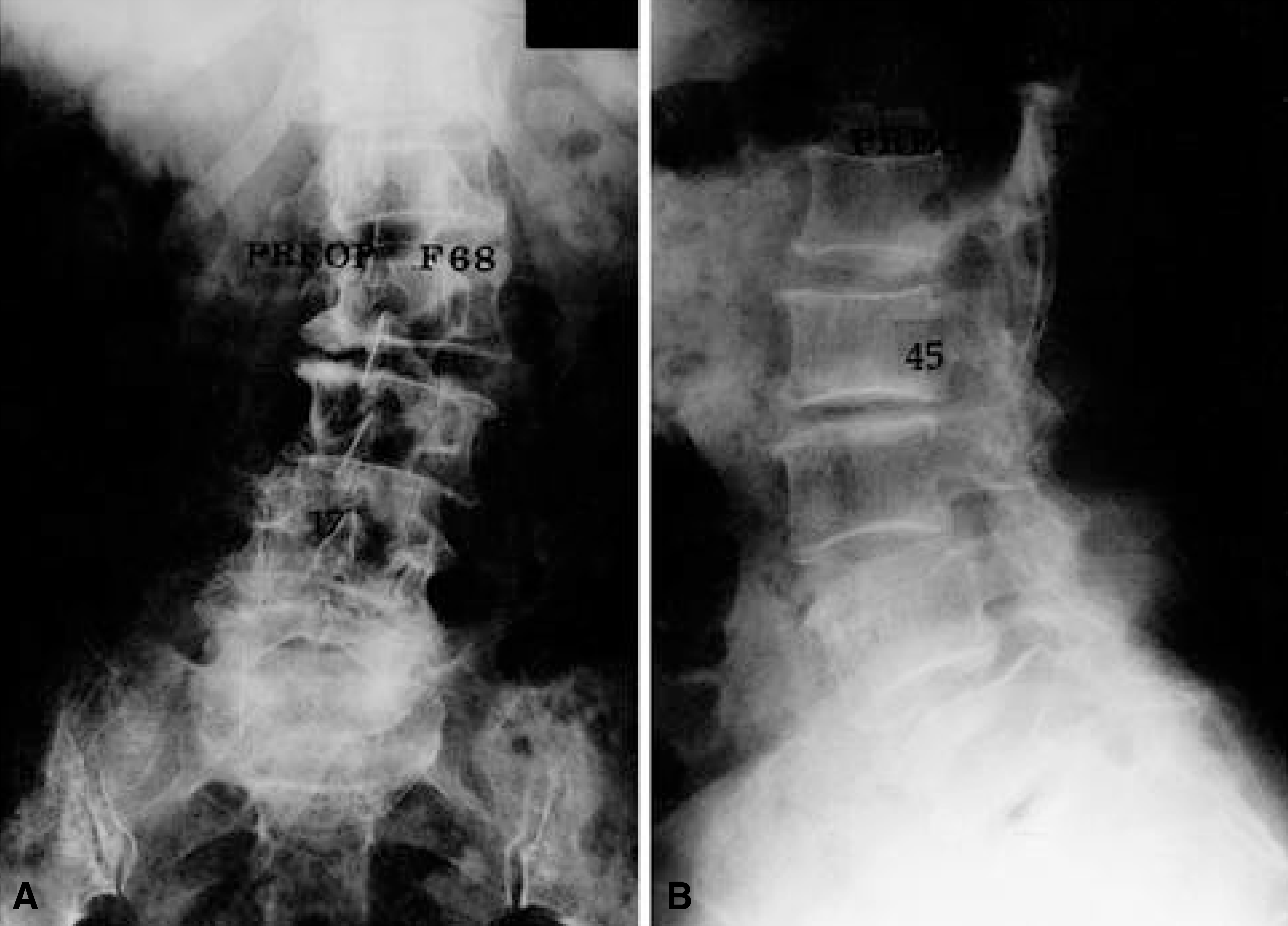
 | Fig. 1.These radiographs of a patient with scoliosis show typical pathologies of degenerative scoliosis including scoliosis, tilt, rotation of vertebral bodies, lateral subluxation and olisthesis. |
Table 1.
Symptoms and physical findings
Table 2.
Radiographic features of the curves


 PDF
PDF ePub
ePub Citation
 Print
Print


 XML Download