Abstract
Summary of literature Review
There were many proposed surgical treatments for lumbar degenerative kyphosis but the best treatment is still controversial.
Materials and Methods
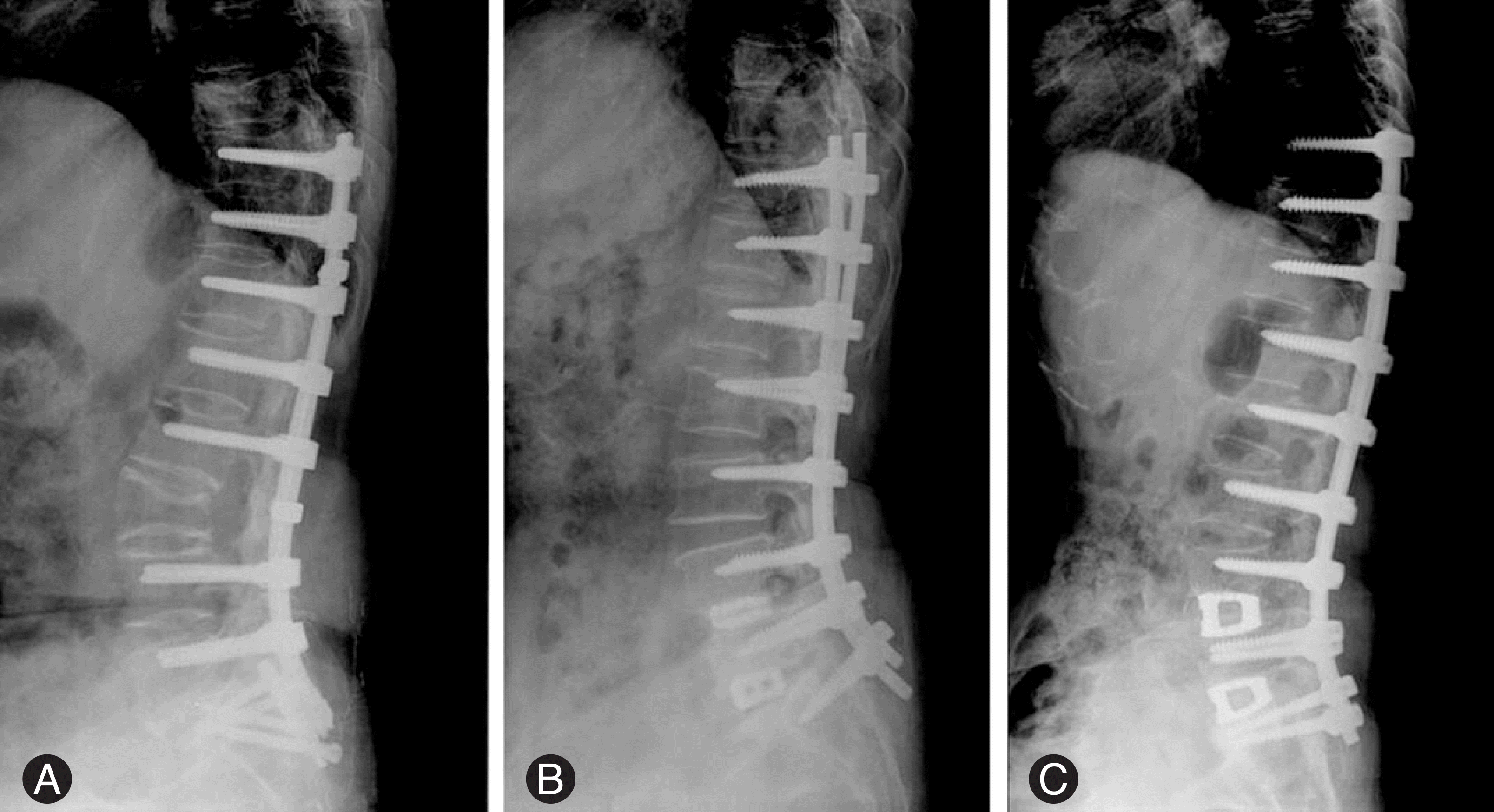
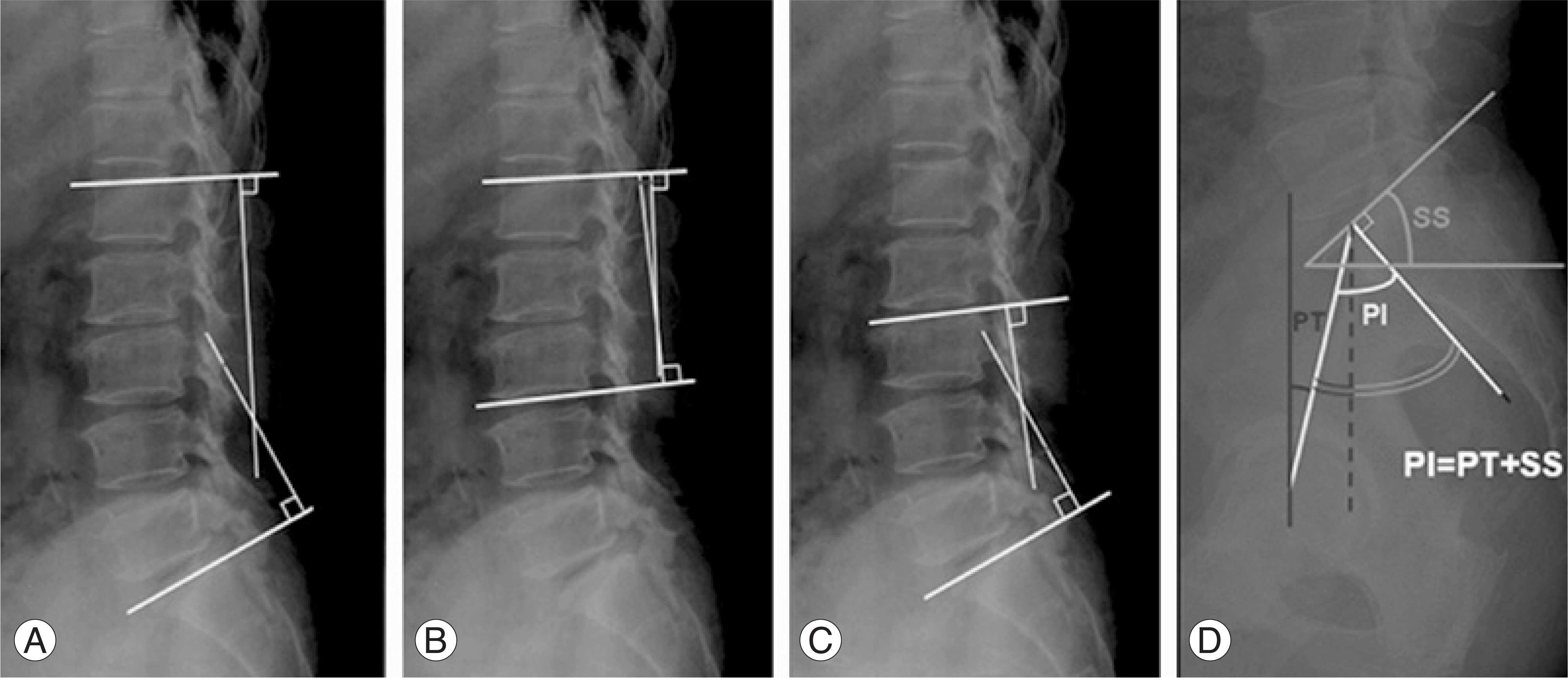
Thirty three patients (all female) had undergone surgery. The mean age at surgery was 61.2. The average follow-up period was 34.7 months. The patients were divided into three groups. Group A included 7 cases with a correction by a posterior osteotomy, Group B included 15 with a posterior correction without an osteotomy, and Group C included 11 with combined anterior-posterior surgery. The radiographic measurements of lumbar lordosis, upper lumbar lordosis, lower lumbar lordosis, and pelvic tilt were performed before surgery, after surgery, and at the final follow-up visit. The loss of correction, complication rates and the clinical results were also compared.
Results
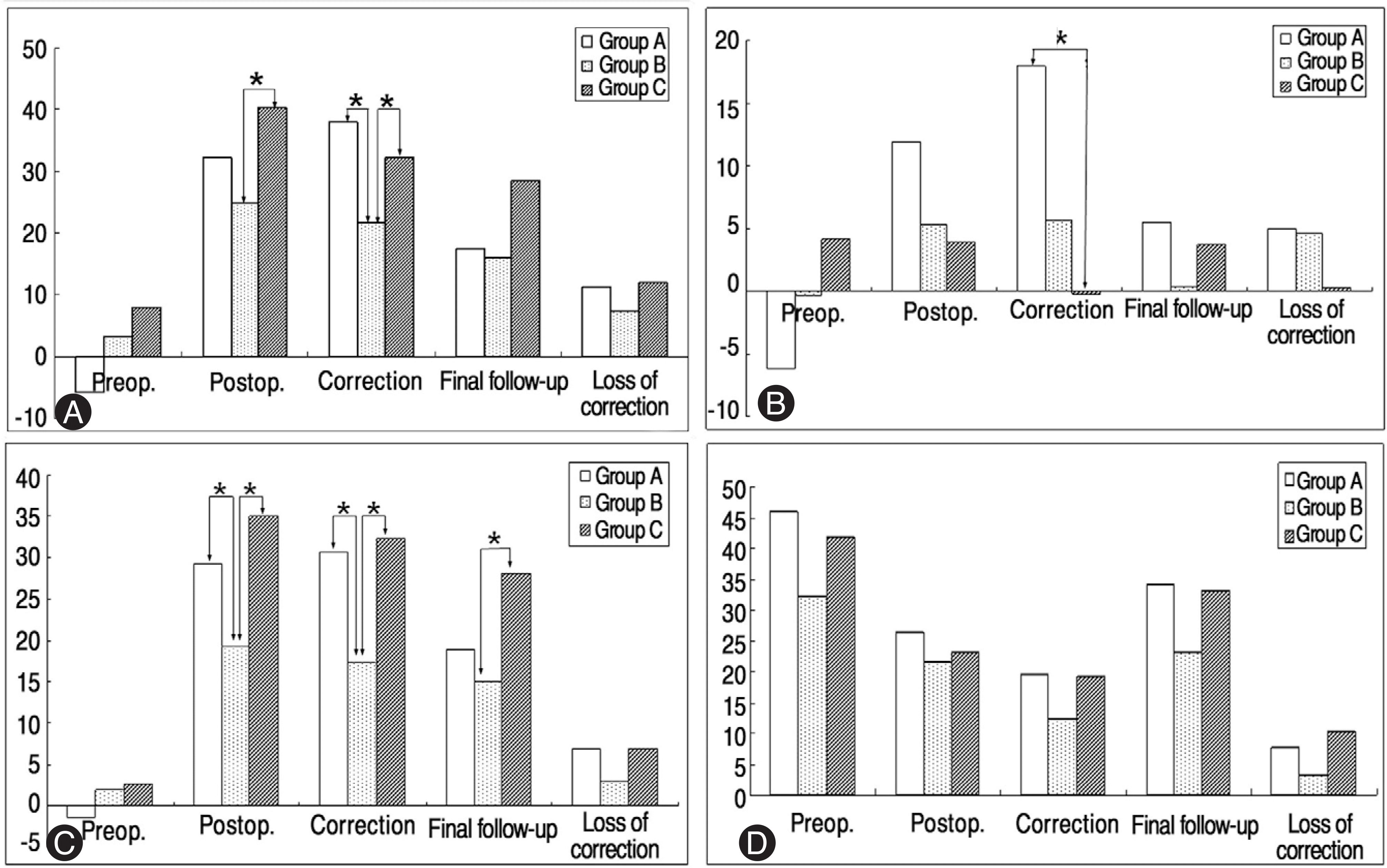
Postoperative correction of the lumbar and lower lumbar lordosis were significantly higher in group A and C than group B. The correction of upper lumbar lordosis was significantly higher in group A than group C. On the final follow-up, there was no significant difference in the loss of correction and clinical results between the three groups. The number of cases with complications in groups A, B and C was 4 (57%), 2 (13.3%) and 2 (18.2%), respectively. Two patients in group A required additional surgery.
Conclusions
Groups A and C were more effective than posterior-only correction. There was no significant difference in the clinical results between the three groups but complication rate was higher in Group A than the other groups. Combined anterior and posterior surgery can be a safe and effective method for correction.
Go to : 
REFERENCES
01). Lee CS., Kim YT., Kim EG. Clinical study of lumbar degenerative kyphosis. J Korean Spine Surg. 1997. 4:27–35.
02). Aota Y., Kumano KO., Hirabayashi S. Postfusion instability at the adjacent segments after rigid pedicle screw fixation for degenerative lumbar spinal disorders. J Spinal disord. 1995. 8:464–473.

03). Gelb DE., Lenke LG., Bridwell KH., Blanke K., McEnery KW. An analysis of sagittal spinal alignment in 100 asymptomatic middle and older aged volunteers. Spine. 1995. 12:1351–1358.

04). Takemitsu Y., Harada Y., Iwahara T., Miyamoto M., Mitatake Y. Lumbar degenerative kyphosis: Clinical, radiologic and epidemiological study. Spine. 1988. 13:1317–1326.
05). Doherty JH. Complications of fusion in lumbar scoliosis. Proceedings of the scoliosis research society. J Bone Joint Surg Am. 1973. 55:438–445.
06). Moe JH., Denis F. The iatrogenic loss of lumbar lordosis. Orthop Trans. 1977. 1:131.
07). Aaro S., Ohlen G. The effect of Harrington instrumentation on sagittal configuration and mobility of the spine in scoliosis. Spine. 1983. 8:570–575.
08). Casey MP., Asher MA., Jacobs RR., Orrick JM. The effect of Harrington rod contouring on lumbar lordosis. Spine. 1987. 12:750–753.

09). LaGrone MO. Loss of lumbar lordosis. A complication of spinal fusion for scoliosis. Orthop Clin North Am. 1988. 9:383–393.
10). LaGrone MO., Bradford DS., Moe JH., Lonstein JE., Winter RB., Ogilvie JW. Treatment of symptomatic flat-back after spinal fusion. J Bone Joint Surg Am. 1988. 70:569–580.

11). Swank S., Lonstein JE., Moe JH., Winter RB., Bradford DS. Surgical treatment of adult scoliosis. A review of two hundred and twenty-two cases. J Bone Joint Surg Am. 1981. 63:268–287.

12). Swank SM., Mauri TM., Brown JC. The lumbar lordosis below Harrington instrumentation for scoliosis. Spine. 1990. 15:181–186.

13). Van Dam BE., Bradford DS., Lonstein JE., Moe JH., Ogilvie JW., Winter RB. Adult idiopathic scoliosis treated by posterior spinal fusion and Harrington instrumentation. Spine. 1987. 12:32–36.

14). Kim EH., Han SK., Kim HJ. A clinical analysis of surgical treatment of lumbar degenerative kyphosis. J Korean Spine Surg. 2001. 8:210–218.

15). Nakai O., Yamaura I., Kurosa Y, et al. Posterior stabilization for lumbar degenerative kyphosis: In situ fusion in maximum extension on Hall's frame. Proc 5th Int Conf Lumbar Fusion and Stabilization,. Tokyo, Springer-Verlag;1993. p. 135–149.
16). Kim EH., Kim SW. Anterior and posterior surgical treatment with wedged cage(Syncage) in lumbar degenerative kyphosis. J of Korean Spine Surg. 2003. 10:240–247.
17). Suk SI., Kim JH., Kim WJ, et al. Treatment of fixed lumbosacral kyphosis by posterior vertebral column resection. J of Korean Spine Surg. 1998. 33:367–374.
Go to : 
 | Fig. 1.Radiographs of three operative groups (A) Posterior osteotomy, (B) Posterior surgery only, (C) Combined anterior and posterior surgery |
 | Fig. 2.Radiographic measurements (A) Lumbar lordosis angle, (B) Upper lumbar lordosis angle, (C) Lower lumbar lordosis angle, (D) Pelvic tilt angle (PT), pelvic incidence (PI), sacral slope (SS) |
 | Fig. 3.Changes of radiographic parameters (A) Lumbar lordosis angle, (B) Upper lumbar lordosis angle, (C) Lower lumbar lordosis angle, (D) Pelvic tilt angle. ∗p<0.05 by Kruskal-Wallis test |
Table 1.
Patients demographics and clinical data
Table 2.
Age and fused segments number of three operative methods
| Group A | Group B | Group C | Total | P value | |
|---|---|---|---|---|---|
| Number of cases | 7 | 15 | 11 | 33 | |
| Age (Yr) | 58.1 | 63.9 | 60 | 61.2 | 0.118 |
| F/U periods (Mo) | 53.9 | 34.2 | 34.7 | 39.2 | 0.097 |
| Fused segments | 5.4 | 4.9 | 4.7 | 4.9 | 0.825 |
Table 3.
Summary of radiographic measurements
Table 4.
Comparison of radiographic parameters according to clinical outcome


 PDF
PDF ePub
ePub Citation
 Print
Print


 XML Download