

 PDF
PDF ePub
ePub Citation
 Print
Print


Abstract
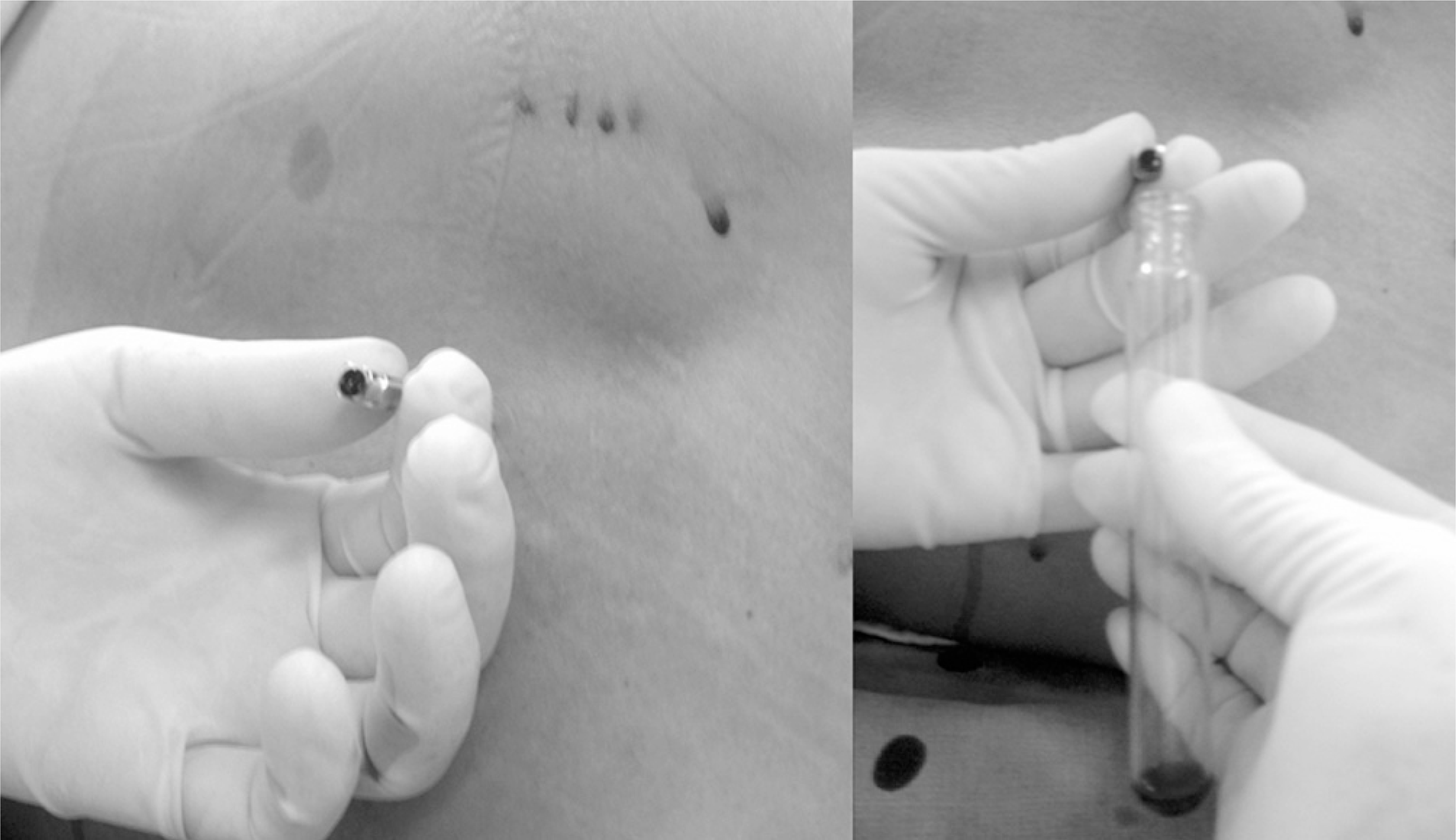
A 63 year-old female was brought to our hospital with severe lower back pain. She received antihypertensive drugs for 2 years but her blood pressure was normal upon arrival. She could not stand up or even walk. The MRI showed a subdural hematoma at the thoracolumbar region, which was extremely rare. The treatment applied was decompression through a spinal tap without surgery. After this, her pain subsided considerably. Two weeks later, MRI confirmed that there was no hematoma in the same region. She was discharged and has enjoyed her daily activities free of pain. A spontaneous subdural hematoma is an extremely rare disease. The condition was treated successfully in a conservative manner.
Go to : 
REFERENCES
01). Kyriakides AE., Lalam RK., El Masry WS. Acute spontaneous subdural spinal hematoma presenting as paraplegia: a rare case. Spine. 2007. 32:619–622.
02). Jimbo H., Asamoto S., Mitsuyama T., Hatayama K., Iwasaki Y., Fukui Y. Spinal chronic subdural hematoma in association with anticoagulant therapy: a case report and literature review. Spine. 2006. 31:184–167.
03). Schwerdtfeger K., Caspar W., Alloussi S., Storwitzki M., Loew F. Acute spinal subdural extramedullary hematoma: a nonsurgical approach for spinal cord decompression. Neurosurgery. 1990. 27:312–314.
04). Mashiko R., Noguchi S., Uemura K., Nagata T., Mastu-mura A. Lumbosacral subdural hematoma. Case report. Neurol Med Chir(Tokyo). 2006. 46:258–261.
05). Boukobza M., Haddar D., Boissonet M., Merrlant JJ. Spinal subdural haematoma: a study of three cases: Clin Radiol. 2001. 56:475–480.
06). Braun P., Nogues-Melendez P., Montoliu Forinas G., Martinez Martinez JC., Marcos Naranjo E. Spontaneous spinal subdural hematoma: MRI findings in two cases. Radiologia. 2007. 49:47–50.
07). Png MA. Spontaneous lumbar subdural hematoma-a case report. Singapore Med J. 1997. 38:76–78.
Go to :


 XML Download