Abstract
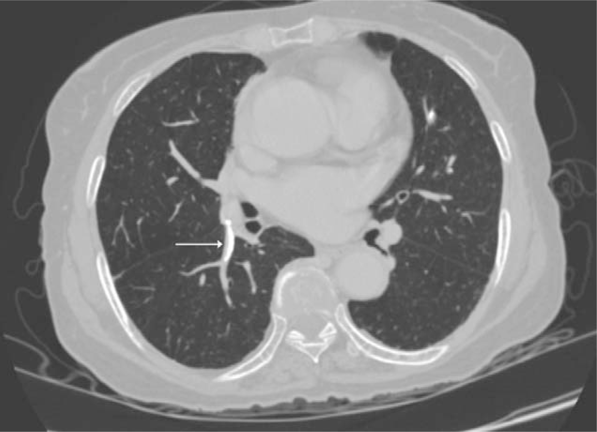
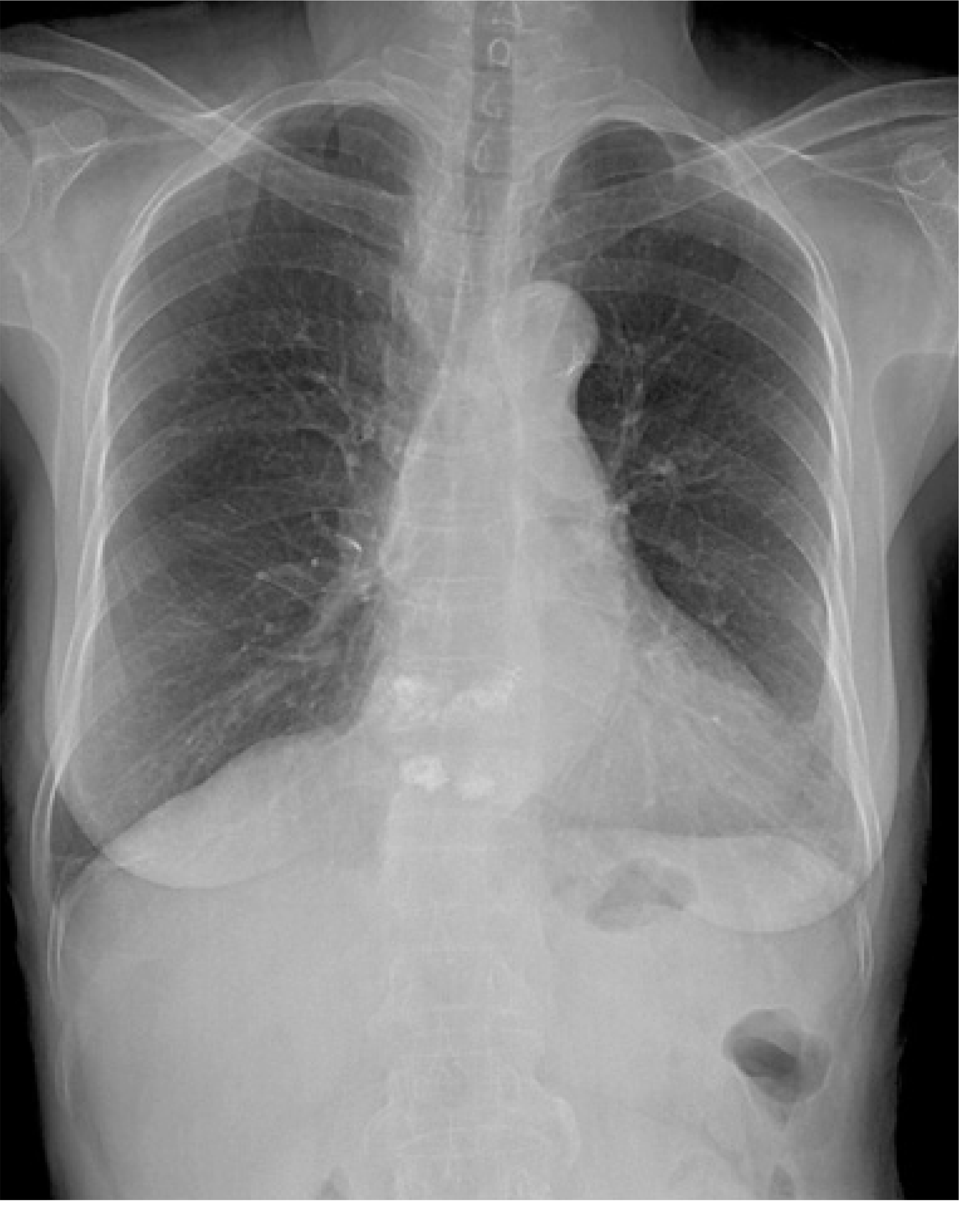
Percutaneous vertebroplasty for osteoporotic compression fractures or malignant osteolytic spinal tumors provides pain relief. A pulmonary embolism caused by polymethylmethacrylate migration after this procedure is rare and its major complication, pulmonary infarction, involves necrosis of the lung parenchyme, resulting from interference with the blood supply. We report a case of a large pulmonary embolus (diameter 2 cm) after cement vertebroplasty for an osteoporotic vertebral compression fracture and successful management with anticoagulation only.
Go to : 
REFERENCES
01). Orsini EC., Byrick RJ., Mullen JB., Kay JC., Waddell JP. Cardiopulmonary function and pulmonary microemboli during arthroplasty using cemented or non-cemented components. The role of intramedullary pressure. J Bone Joint Surg Am. 1987. 69:822–832.

02). Padovani B., Kasriel O., Brunner P., Peretti-Viton P. Pulmonary embolism caused by acrylic cement: a rare complication of percutaneous vertebroplasty. Am J Neuroradiol. 1999. 20:375–377.
03). Min SH., Kim MH., Park HG., Paik HD. A clinical analysis of 260 percutaneous vertebroplasty in the treatment of osteoporotic compression fracture. J Korean Fracture Soc. 2006. 19:357–362.

04). Moon SH., Kim DJ., Hwang CS., Lee SE., Park SW. A comparision of vertebroplasty versus conservative treatment in osteoporotic compression fractures. J Korean Fracture Soc. 2004. 17:374–379.
05). Phillips FM., Todd Wetzel F., Lieberman I., Campbell-Hupp M. An in vivo comparison of the potential for extravertebral cement leak after vertebroplasty and kyphoplasty. Spine. 2002. 27:2173–2179.

06). Jang JS., Lee SH., Jung SK. Pulmonary embolism of polymethylmethacrylate after percutaneous vertebroplasty. Spine. 2002. 27:416–418.

07). Coventry MB., Beckenbaugh RD., Nolan DR., Ilstrup DM. 2,012 total hip arthroplasties. A study of postoperative course and early complications. J Bone Joint Surg Am. 1974. 56:273–284.
Go to : 
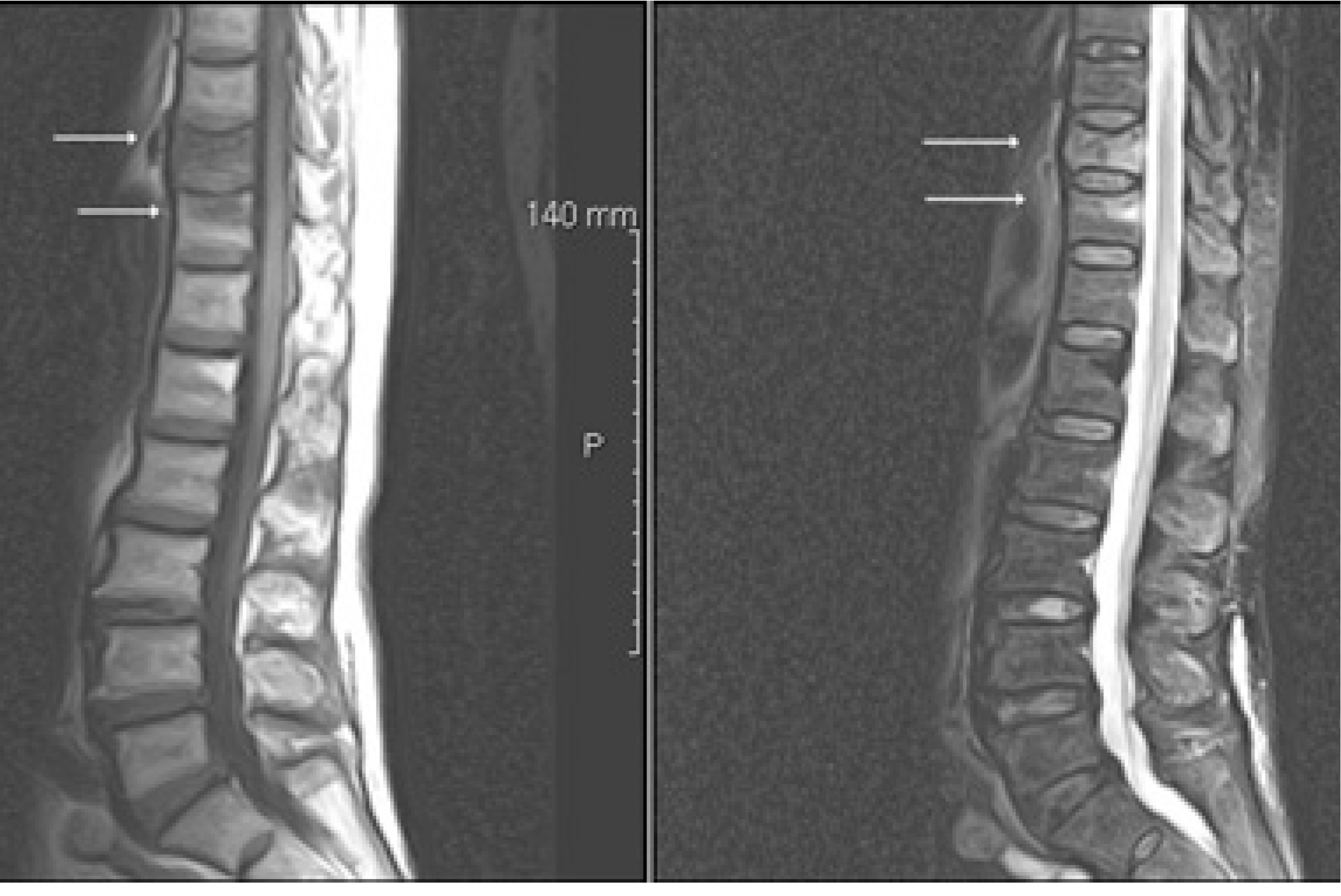 | Fig. 2.Sagittal images of thoracolumbar spine MR show signal change in T10 and T11 vertebral bodies (arrows). |
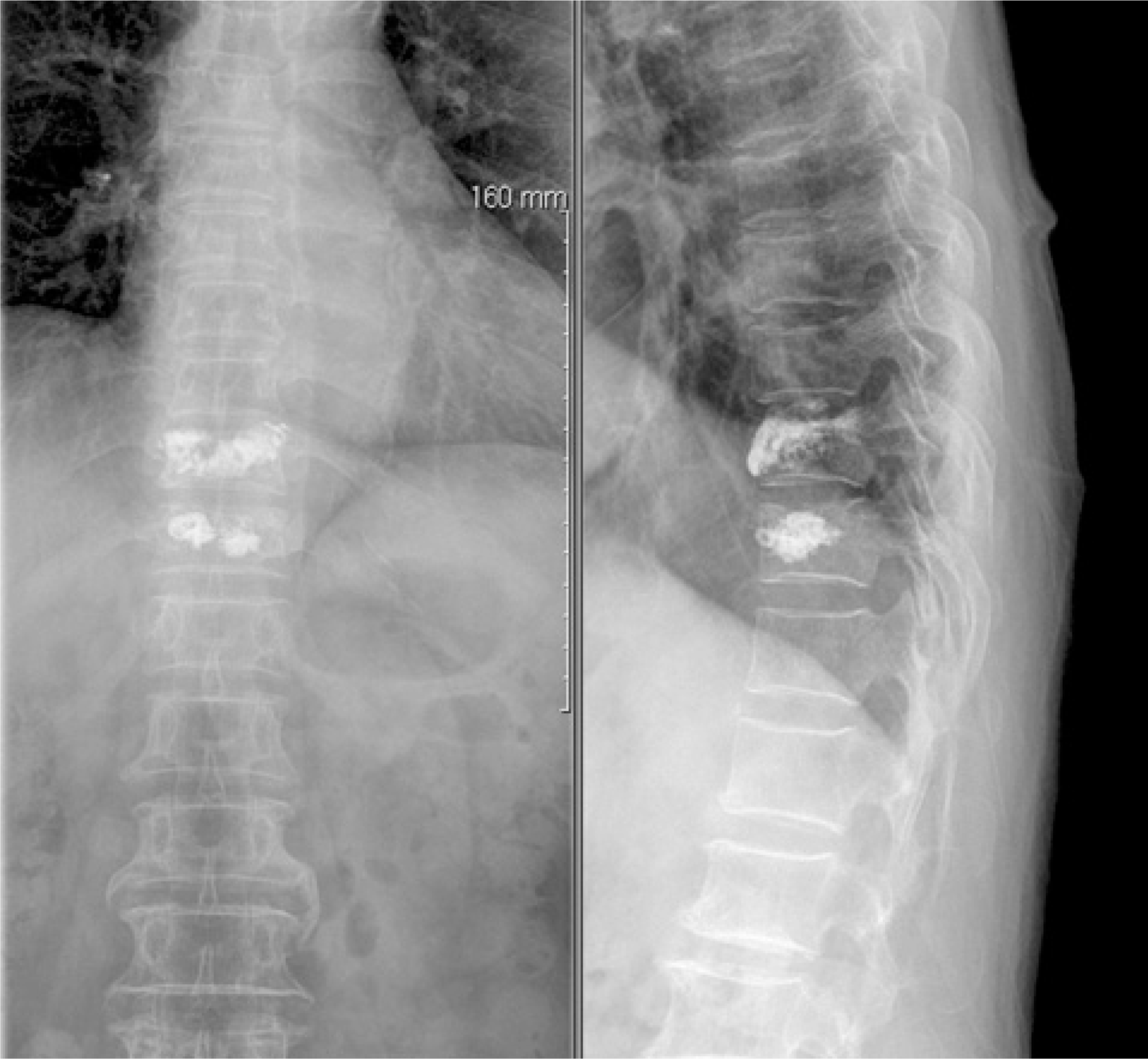 | Fig. 3.In postoperative radiographs of T10 and T11 vertebroplasty, there were no evidence of cement leakage out of vertebral bodies. |
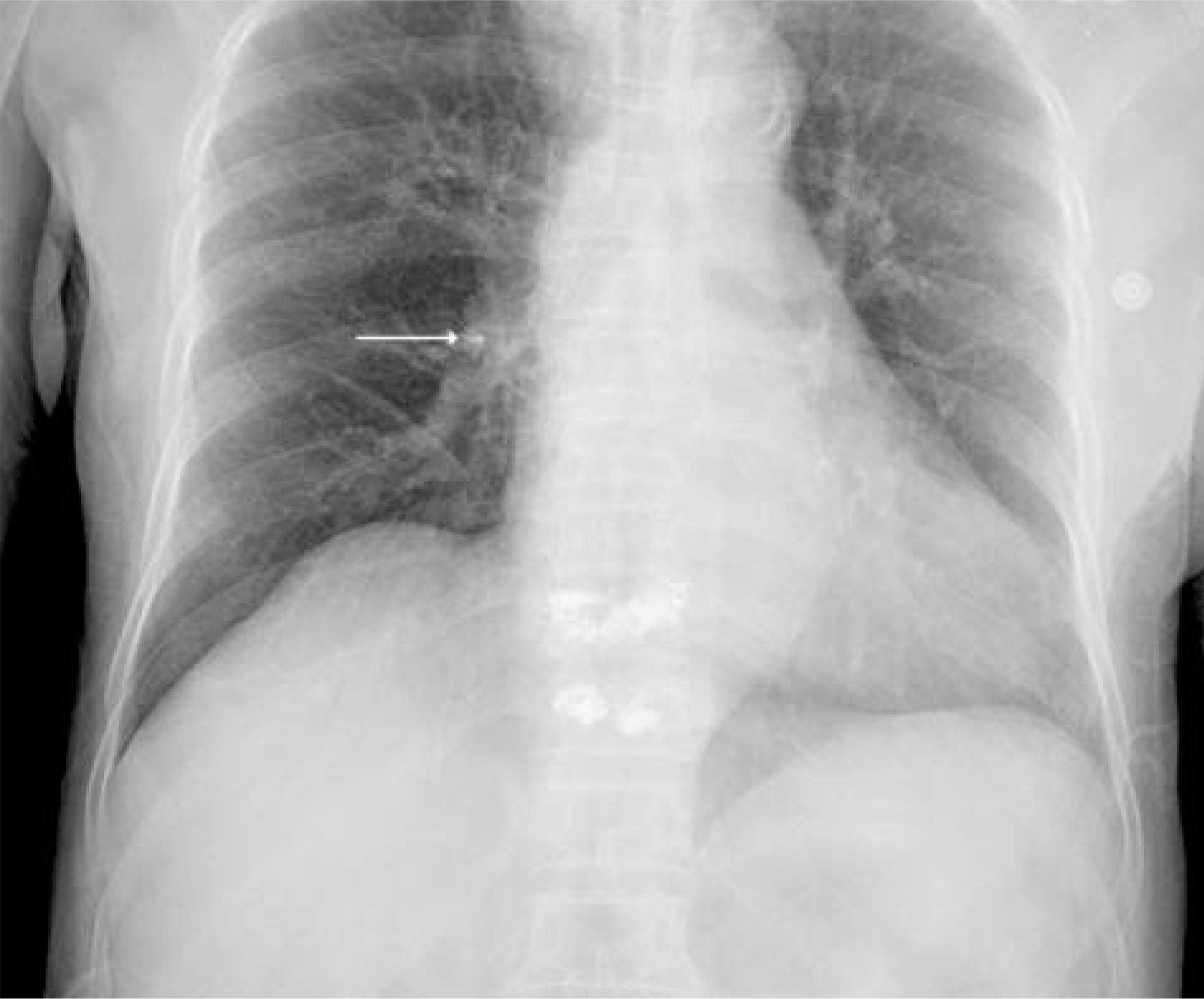 | Fig. 4.An immediate postoperative chest radiograph shows high density cement embolus in right lung hilar area (arrow). |


 PDF
PDF ePub
ePub Citation
 Print
Print


 XML Download