

 PDF
PDF ePub
ePub Citation
 Print
Print


Abstract
Survival after traumatic atlanto-occipital dislocation is rare. Severe persistent neurological deficits are common in the survivors, but early resuscitation and the use of the newer diagnostic techniques have contributed to improved outcomes. We present here the case of a 42 year old man with traumatic atlanto-occipital dislocation combined with a dens fracture, and the patient obtained good clinical results after we applied a Halo-vest and performed posterior fusion.
REFERENCES
01). Dibenedetto T., Lee CK. Traumatic atlanto-occipital instability. A case report with follow-up and a new diagnostic technique. Spine. 1990. 15:595–597.

02). Chang H., Park JB., Kim SK., Choi WS., Chun SK. Traumatic atlanto-occipital rotatory posterior dislocation combined with atlanto-axial rotatory subluxation, Spine. 1998. 5:326–332.
03). Blackwood NJ. Atlanto-occipital dislocation. A case of fracture of the atlas and axis, and forward dislocation of the occiput on the spinal column, life being maintained for thirty-four hours and forty minutes by artificial respiration, during which a laminectomy was performed upon the third cervical vertebra. Ann Surg. 1908. 47:654–658.
04). Traynelis VC., Marano GD., Dunker R., Kaufman HH. Traumatic atlanto-occipital dislocation. Case report. J Neurosurg. 1986. 65:863–870.
05). Alker GJ Jr., Oh YS., Leslie EV. High cervical spine and craniocervical junction injuries in fatal traffic accidents: A radiological study, Orthop Clin North Am. 1978. 9:1003–1010.
06). Bucholz RW., Burkhead WZ. The pathological anatomy of fatal atlanto-occipital dislocations, J Bone Joint Surg Am. 1979. 61:248–250.
07). Power B., Miller MD., Kramer RS., Matrinez S., Gehweil-er JA. Traumatic anterior atlanto-occipital dislocation, Neurosurgery. 1979. 4:12–17.
08). Harris JH Jr., Carson GC., Wagner LK., Kerr N. Radiologic diagnosis of traumatic occipitovertebral dissociation: 2. Comparison of three methods of detecting occipitovertebral relationships on lateral radiographs of supine subjects. Am J Roentgenol. 1994. 162:887–892.
09). Wholey MH., Bruwer AJ., Baker HL Jr. The lateral roentgenogram of the neck; with comments on the atlantoodontoid-basion relationship. Radiology. 1958. 71:350–356.
10). McCullen GM., Garfin SR. Cervical spine internal fixation using screw and screw-plate constructs. Spine. 2000. 25:643–652.
Fig. 1.
Radiologic measurements in occipito-cervical dislocation. A. Basion-axial interval: the distance between the posterior axial line and a parallel line drawn through the basion. B. Basion-dental interval: the distance from the caudad end of the basion to the rostral tip of the dens axis. C. Power's ratio (BC/OA): the quotient derived from the distance of the basion (B) to the posterior arch of C1(C), and the posterior aspect of the anterior arch of the atlas (A) to the opisthion (O).
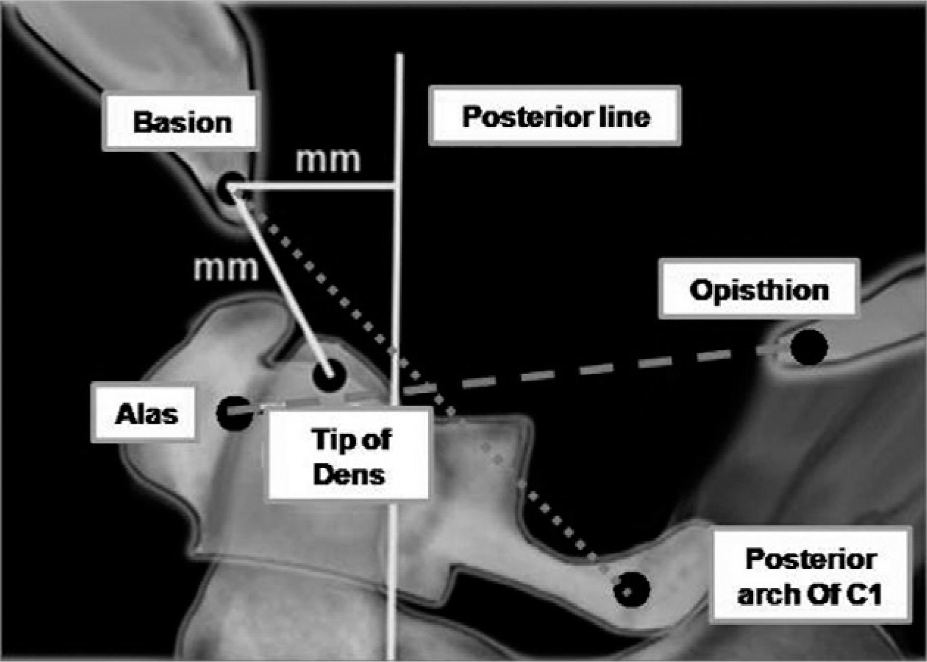
Fig. 2.
Initial lateral cervical spine radiograph and CT scan suggests atlanto-occipital dislocation. (A) Initial lateral roentgenographic view. (B, C) Axial CT scans. (D) Wackenheim's line. (E) Basion-axial and basion-dental interval. (F) Power's ratio.
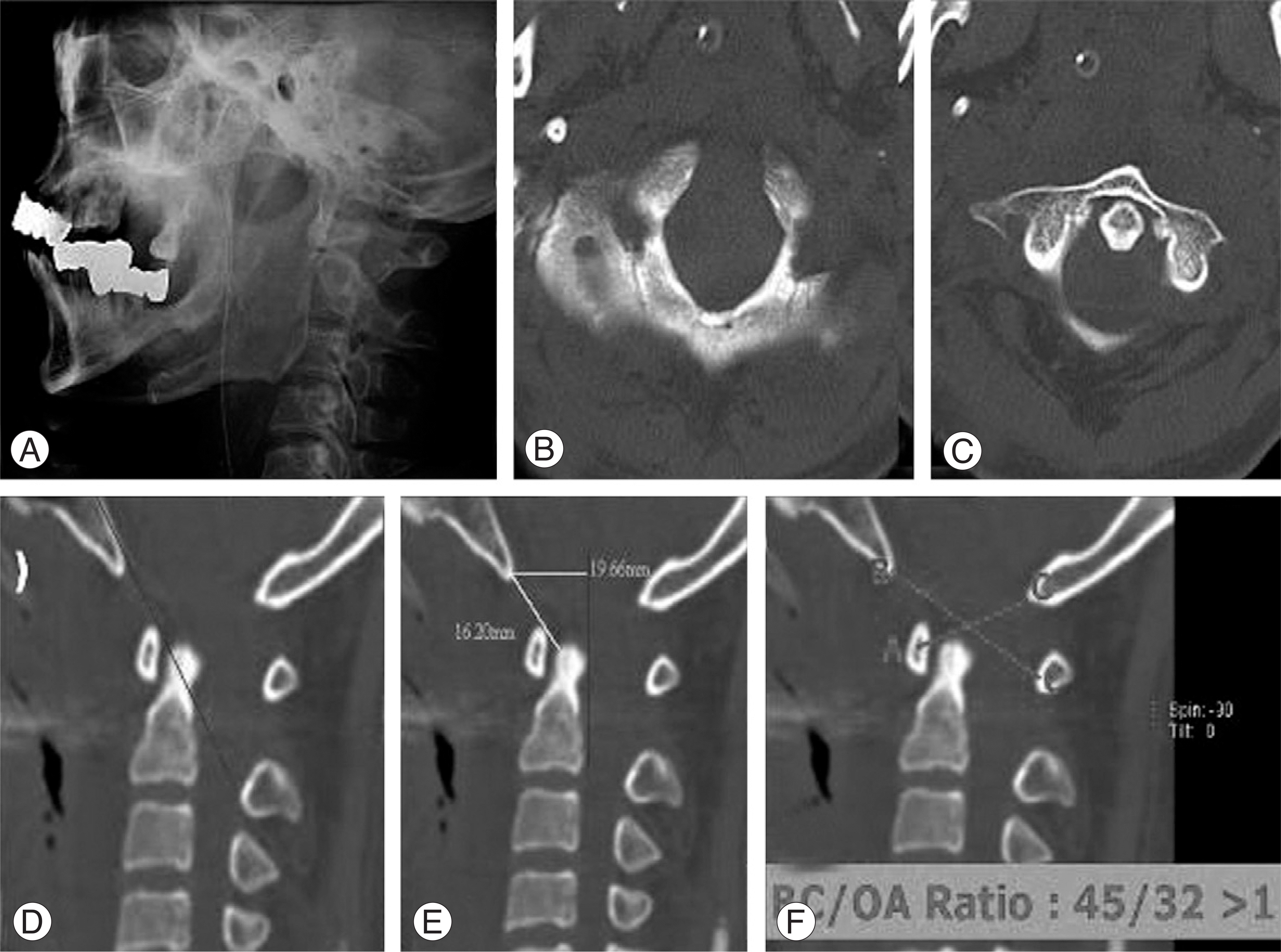
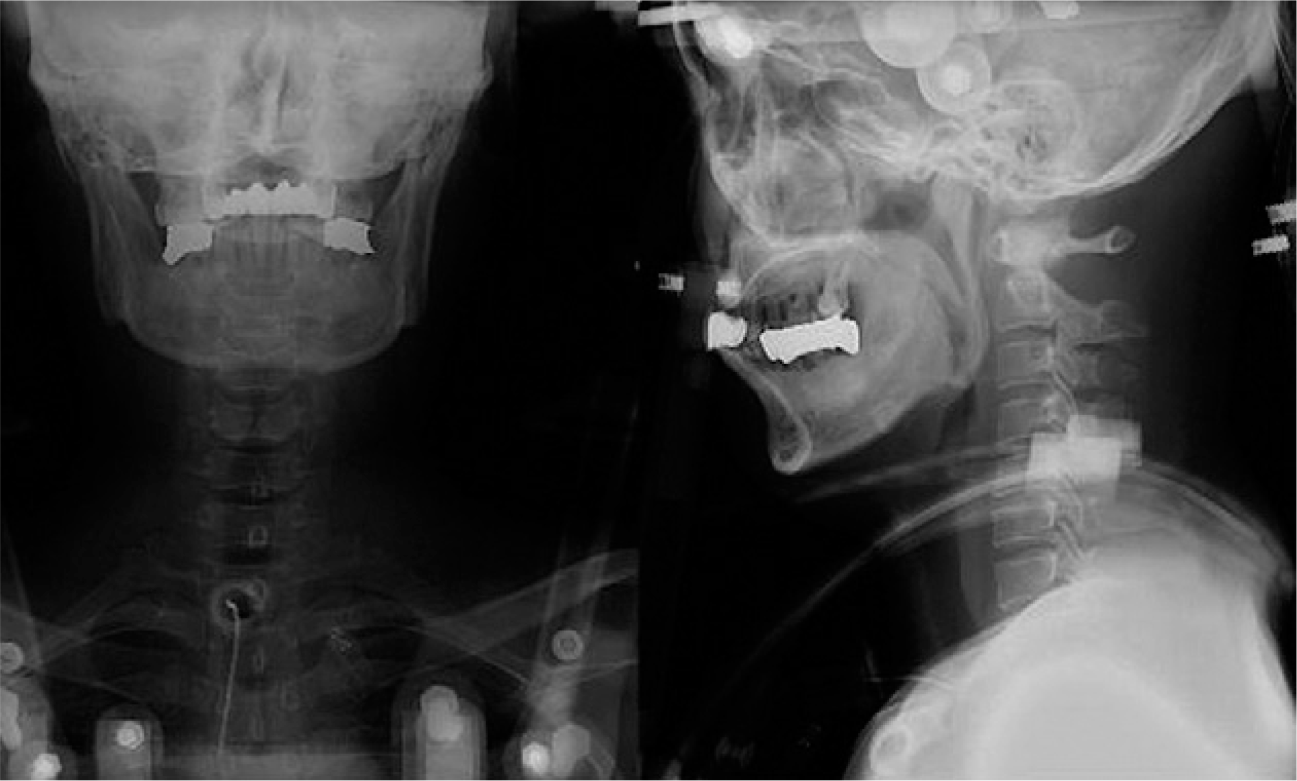
Fig. 5.
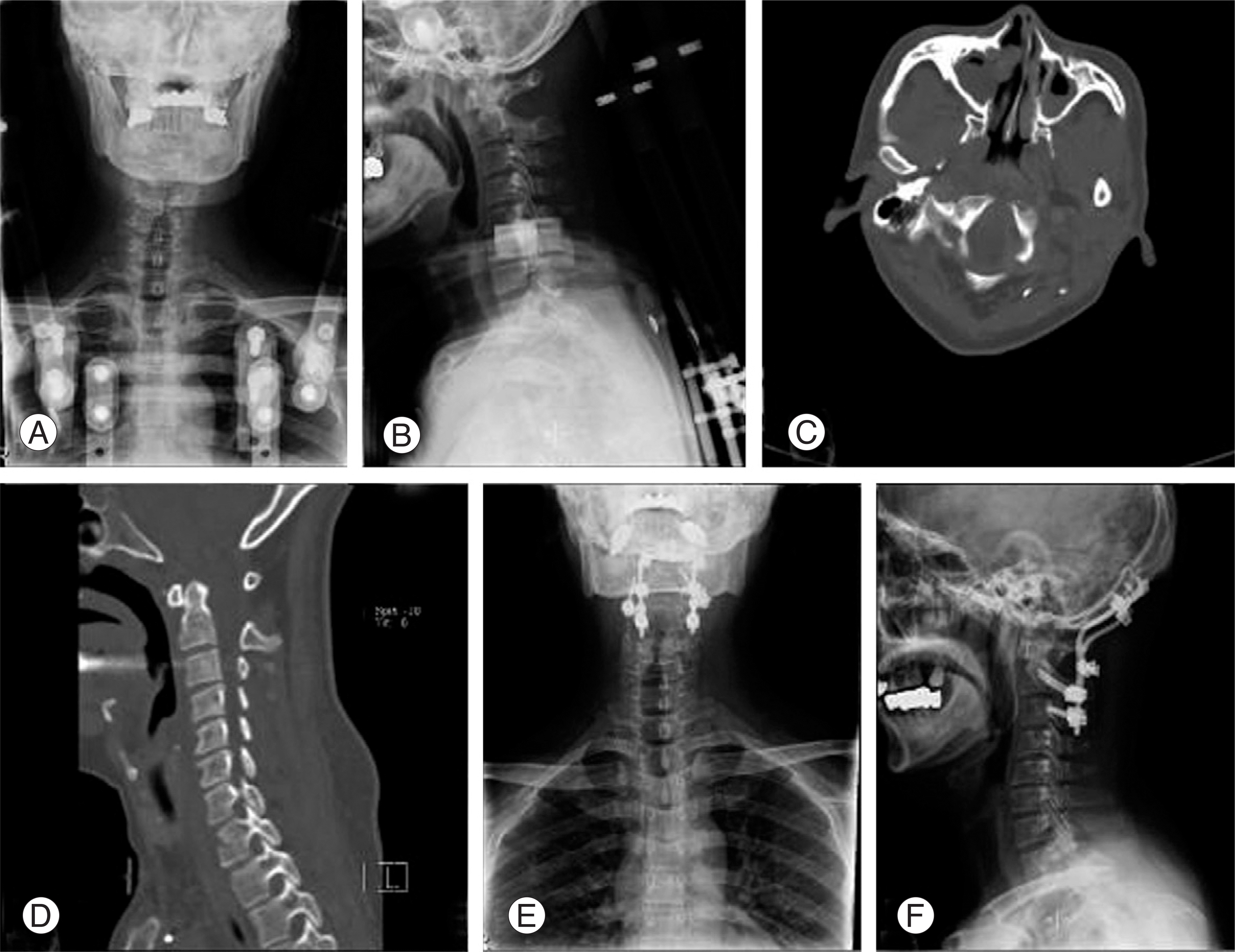
Follow-up roentgenography and CT scans. (A, B) Anterior-posterior and lateral roentgenographic views at 3 months after Halo-vest Application. (C, D) Axial and sagittal CT scans at 3 months after Halo-vest application. (E, F) Anterior-posterior and lateral roentgenographic views after occipito-cervical fusion.
 XML Download