

 PDF
PDF ePub
ePub Citation
 Print
Print


Abstract
Objectives
The aim of this study is to analyze the signals and configurations of the MRI findings of osteoporotic vertebral fractures and the clinical consequences of each type of the MRI findings.
Summary of the Literature Review
There have been some reports that have focused on the MR findings for the differentiation of osteoporotic and metastatic fractures, but there are few reports on the characteristics of the early stage of osteoporotic vertebral fractures.
Materials and Methods
From July 2002 to April 2008, the MRI findings and medical records of 97 patients who were diagnosed with acute or subacute osteoporotic vertebral fractures and who were followed-up for more than 1 year were analyzed. The patients with minor trauma within 3 months before obtaining MRIs and they had decreased bone density were included in this study. Those with fractures due to severe trauma or pathologic causes or normal bone density were excluded. Three spine surgeons evaluated, at three times per each surgeon, the T1-weighted, T2-weighted and fat suppression T1-enhanced sagittal images for the signal of the vertebral body bone marrow and the type of the intravertebral body lesion shape. The relationships between the type of MRI findings and the time from the trauma and the follow up clinical consequences were analyzed.
Results
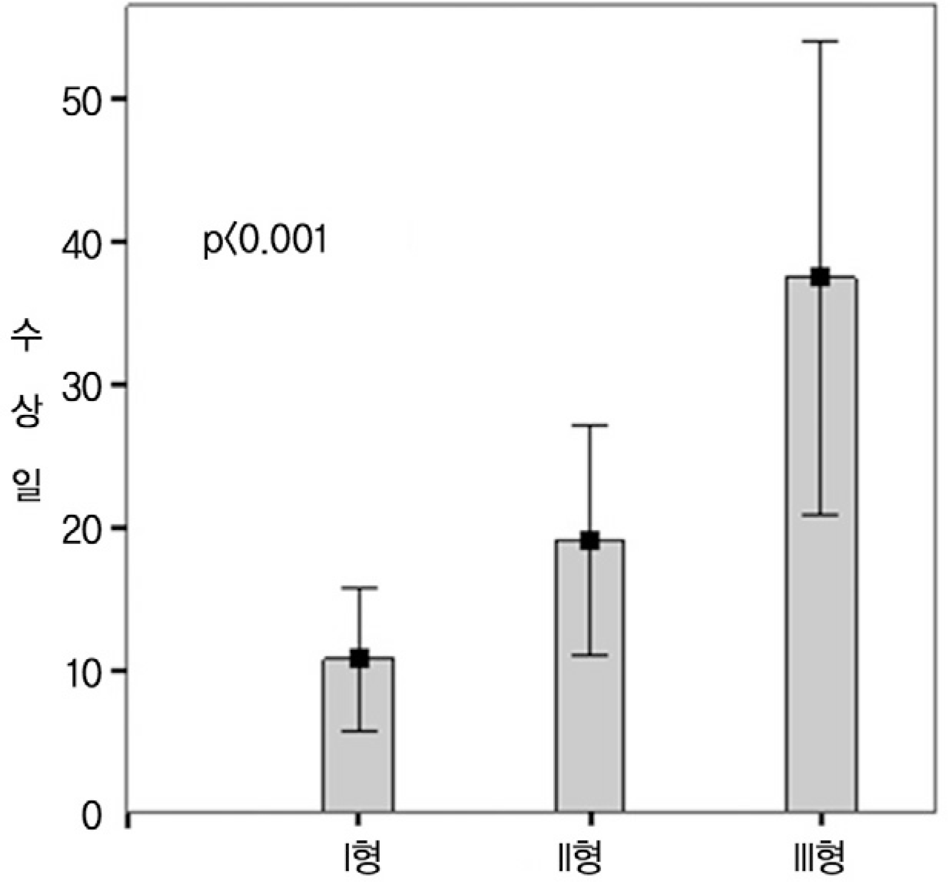
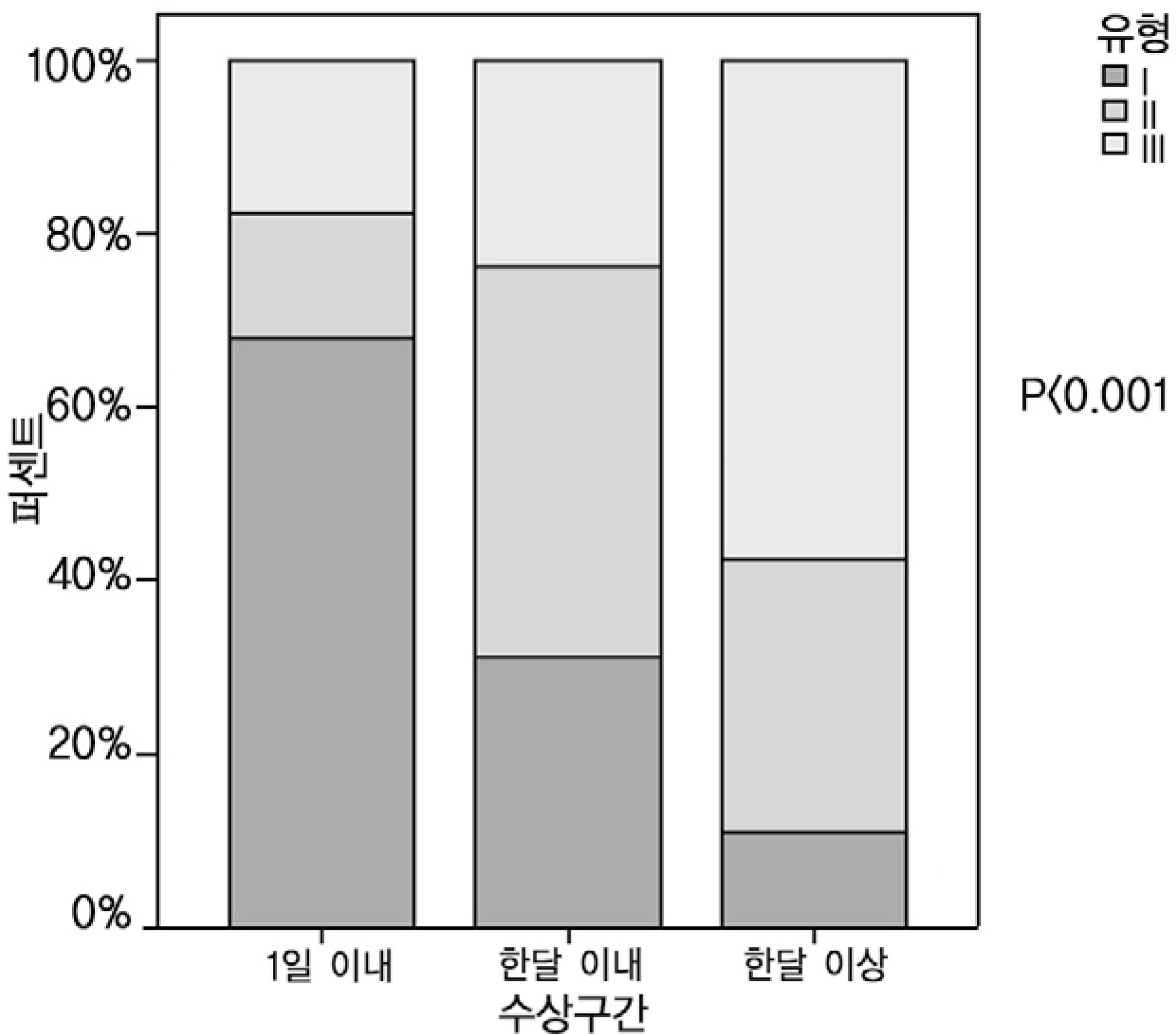
The MRI patterns of 97 patients with 111 fractures of the vertebrae were divided into three types. There were 56 cases of Type I (50.5%), which was defined as diffuse typical signal intensity in the vertebral body, 39 cases (35.1%) of Type II, which was defined as geographic low signal in the center of the vertebral body with typical signal changes, and 16 cases (14.4%). of type III, which was defined as atypical signal intensity or a shape of lesion that did not correspond to type 1 nor type 2. The average time from trauma was 10.8±19.0days (0~90) for type I, 19.1±24.9days (0~90) for type III and 37.5±31.1days (0~90) for type III, which showed differences among each types (p<0.001).
Conclusions
The analysis of the relationship between the time from trauma and the signal intensity and the type of lesion on MRI examination revealed that the low signal intensity in the typical vertebral body signal or an atypical signal or shape were poor prognostic factors of osteoporotic vertebral fracture
REFERENCES
01). Cohen LD. Fractures of the osteoporotic spine. Orthop Clin North Am. 1990. 21:143–150.

02). Kim DH., Vaccaro AR. Osteoporotic compression fractures of the spine; current options and considerations for treatment. Spine J. 2006. 6:479–487.
03). Lippuner K. Medical treatment of vertebral osteoporosis. Eur Spine J. 2003. 12:132–141.
04). Riggs BL., Melton LJ 3rd. The worldwide problem of osteoporosis: insights afforded by epidemiology. Bone. 1995. 17:505–511.
05). Gold DT. The clinical impact of vertebral fractures: quality of life in women with osteoporosis. Bone. 1996. 18:185–189.
06). Nevitt MC., Ettinger B., Black DM, et al. The association of radiologically detected vertebral fractures with back pain and function: a prospective study. Ann Intern Med. 1998. 128:793–800.
07). Lee YL., Yip KM. The osteoporotic spine. Clin Orthop Relat Res. 1996. 323:91–97.
08). Kim KT., Suk KS., Kim JM., Lee SH. Delayed vertebral collapse with neurological deficits secondary to osteoporosis. Int Orthop. 2003. 27:65–69.
09). Kado DM., Browner WS., Palermo L., Nevitt MC., Genant HK., Cummings SR. Vertebral fractures and mortality in older women: a prospective study. Study of Osteoporotic Fractures Research Group. Arch Intern Med. 1999. 159:1215–1220.
10). Schlaich C., Minne HW., Bruckner T, et al. Reduced pulmonary function in patients with spinal osteoporotic fractures. Osteoporos Int. 1998. 8:261–267.
11). Tezer M., Erturer RE., Ozturk C., Ozurk I., Kuzgun U. Conservative treatment of fractures of thoracolumbar spine. International Orthopedics. 2005. 29:78–82.
12). Diamond TH., Champion B., Clark WA. Management of acute osteoporotic vertebral fractures: a nonrandomized trial comparing percutaneous vertebroplasty with conservative therapy. Am J Med. 2003. 114:257–265.
13). Hochmuth K., Proschek D., Schwarz W., Mack M., Kurth AA., Vogl TJ. Percutaneous vertebroplasty in the therapy of osteoporotic vertebral compression fractures: a critical review. Eur Radiol. 2006. 16:998–1004.
14). Watts NB. Is percutaneous vertebral augmentation (vertebroplasty) effective treatment for painful vertebral fractures? Am J Med. 2003. 114:326–328.
15). Legroux-Ge′rot I., Lormeau C., Boutry N., Cotten A., Duquesnoy B., Cortet B. Long-term follow-up of vertebral osteoporotic fractures treated by percutaneous vertebroplasty. Clin Rheumatol. 2004. 23:310–317.
16). Garfin SR., Yuan HA., Reiley MA. New technologies in spine: kyphoplasty and vertebroplasty for the treatment of painful osteoporotic compression fractures. Spine. 2001. 26:1511–1515.
17). Baur A., Sta¨bler A., Bru¨ning R, et al. Diffusion-weighted MR imaging of bone marrow: differentiation of benign versus pathologic compression fractures. Radiology. 1998. 207:349–356.
18). An HS., Andreshak TG., Nguyen C., Williams A., Daniels D. Can we distinguish between benign versus malignant compression fractures of the spine by magnetic resonance imaging? Spine. 1995. 20:1776–1782.
19). Spuentrup E., Buecker A., Adam G., van Vaals JJ., Guen-ther RW. Diffusion-weighted MR imaging for differentiation of benign fracture edema and tumor infiltration of the vertebral body. AJR Am J Roentgenol. 2001. 176:351–358.
20). Yamato M., Nishimura G., Kuramochi E., Saiki N., Fujio-ka M. MR appearance at different ages of osteoporotic compression fractures of the vertebrae. Radiat Med. 1998. 16:329–334.
21). Sung MS., Park SH., Lee JM, et al. Sequential changes of traumatic vertebral compression fracture on MR imaging. J Korean Med Sci. 1995. 10:189–194.
22). Landis JR., Koch GG. The measurement of observer agreement for categorical data. Biometrics. 1997. 33:159–174.
23). Mathis JM., Barr JD., Belkoff SM., Barr MS., Jensen ME., Deramond H. Percutaneous vertebroplasty: a developing standard of care for vertebral compression fractures. AJNR Am J Neuroradiol. 2001. 22:373–381.
24). Cyteval C., Sarrabe �re MP., Roux JO, et al. Acute osteoporotic vertebral collapse: open study on percutaneous injection of acrylic surgical cement in 20 patients. AJR Am J Roentgenol. 1999. 173:1685–1690.
25). Lyritis GP., Mayasis B., Tsakalakos N, et al. The natural history of the osteoporotic vertebral fracture. Clin Rheumatol. 1989. 8:66–69.
26). Ismail AA., Cooper C., Felsenberg D, et al. Number and type of vertebral deformities: epidemiological characteristics and relation to back pain and height loss. European Vertebral Osteoporosis Study Group. Osteoporos Int. 1999. 9:206–213.
27). Baur A., Sta¨bler A., Arbogast S., Duerr HR., Bartl R., Reiser M. Acute osteoporotic and neoplastic vertebral compression fractures: fluid sign at MR imaging. Radiology. 2002. 225:730–735.
28). Oner FC., van Gils AP., Dhert WJ., Verbout AJ. MRI findings of thoracolumbar spine fractures: a categorisation based on MRI examinations of 100 fractures. Skeletal Radiol. 1999. 28:433–443.
29). Yu CW., Hsu CY., Shih TT., Chen BB., Fu CJ. Vertebral osteonecrosis: MR imaging findings and related changes on adjacent levels. AJNR Am J Neuroradiol. 2007. 28:42–47.
30). Kanchiku T., Taguchi T., Kawai S. Magnetic resonance imaging diagnosis and new classification of the osteoporotic vertebral fracture. J Orthop Sci. 2003. 8:463–466.

 XML Download