Abstract
Objectives
We wanted to analyze the frequency of instrument breakage and the long term reduction loss for patients who received pedicle screw fixation for thoracolumbar fractures. Summary of the Literature Review: A pedicle screw fixation system for thoracolumbar fractures has become popular since the late 1980s, but it is difficult to find articles mentioning its mid and long term results.
Materials and Methods
Twenty-seven patients those received pedicle screw fixation for thoracolumbar fractures and dislocations and who were followed up more than 5 years were included. The average follow-up period was 139.0 months. We compared the anterior column height, the kyphotic angle and the local kyphotic angle on serial radiographs, and we measured the changes of the intervertebral disc height and the changes of the angle between screws. We also investigated the breakage and loosening of instruments.
Results
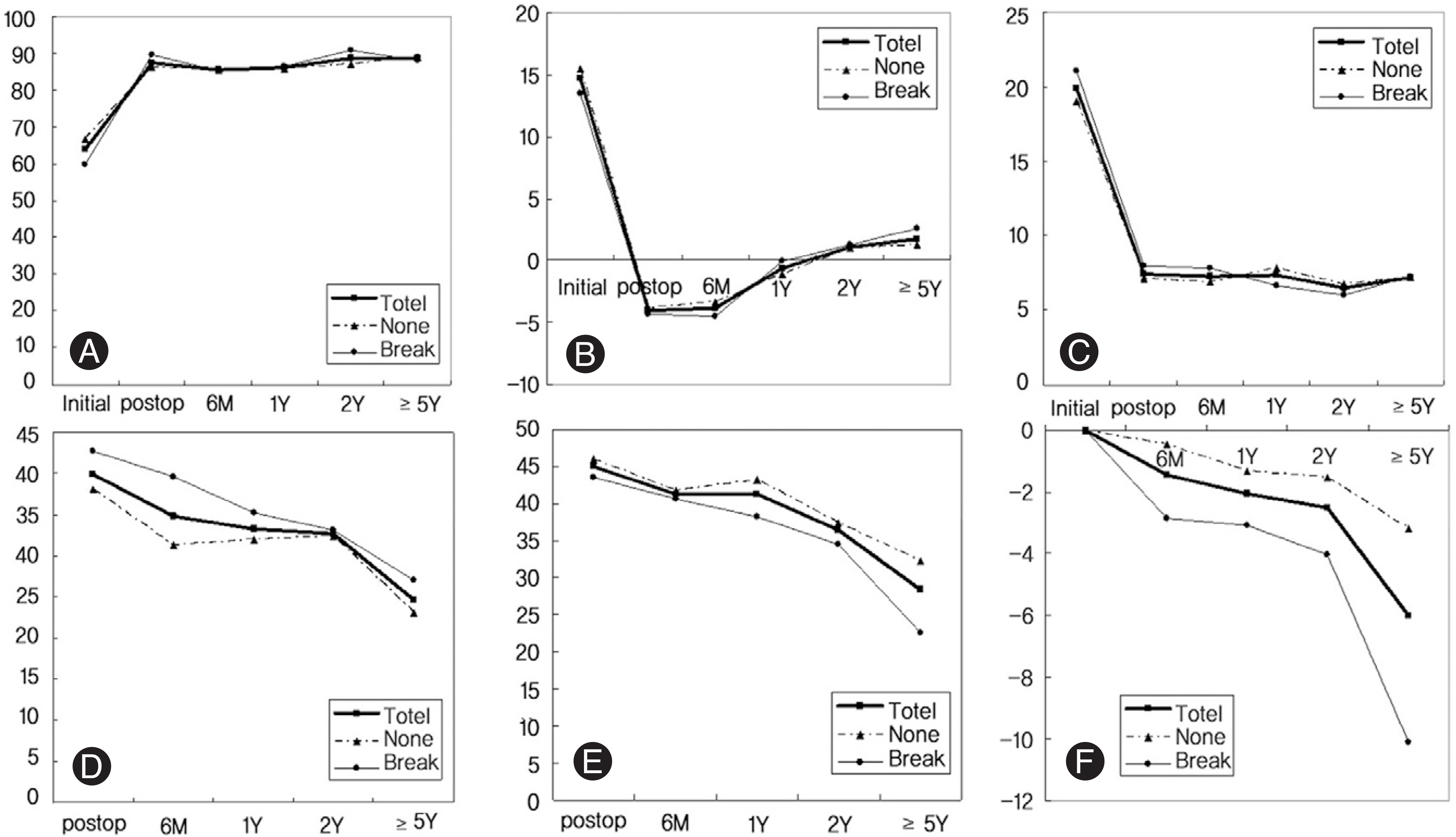
The breakage of screws was observed in 11 cases (40.7%) and it had a statistical correlation with the loss of the lower intervertebral disc height and the loss of angles between the screws. During the follow-up, the kyphotic angle, the upper and lower disc height and the interscrew angle were decreased over time, whereas the anterior column height and wedge angle of the vertebra were maintained after the operation. There was no statistical correlation between the breakage of instruments and the degree of lower back pain.
Conclusions
On the mid and long-term follow-up of the patients who were treated by pedicle screws for thoracolumbar fractures, the correction of the kyphotic angle was lost over time and breakage of screws may eventually occur. The loss of the kyphotic angle was mainly due to the continuous loss of the intervertebral disc height.
Go to : 
REFERENCES
01). Denis F. The three column spine and its significance in the classification of acute thoracolumbar spinal injuries. Spine. 1983. 8:817–831.

02). McAfee PC., Yuan HA., Fredrickson BE., Lubicky JP. The value of computed tomography in thoracolumbar fractures. An analysis of one hundred consecutive cases and a new classification. J Bone Joint Surg Am. 1983. 65:461–473.

03). Sjo¨stro¨m L., Jacobsson O., Karlstro¨m G., Pech P., Rauschning W. CT analysis of pedicles and screw tracts after implant removal in thoracolumbar fractures. J Spinal Disord. 1993. 6:225–231.
04). Yue JJ., Sossan A., Selgrath C, et al. The treatment of unstable thoracic spine fractures with transpedicular screw instrumentation: a 3-year consecutive series. Spine. 2002. 27:2782–2787.

05). Mahar A., Kim C., Wedemeyer M, et al. Short-segment fixation of lumbar burst fractures using pedicle fixation at the level of the fracture. Spine. 2007. 32:1503–1507.

06). Carl AL., Tromanhauser SG., Roger DJ. Pedicle screw instrumentation for thoracolumbar burst fractures and fracture-dislocations. Spine. 1992. 17:317–324.

07). Knop C., Fabian HF., Bastian L., Blauth M. Late results of thoracolumbar fractures after posterior instrumentation and transpedicular bone grafting. Spine. 2001. 26:88–99.

08). McLain RF., Burkus JK., Benson DR. Segmental instrumentation for thoracic and thoracolumbar fractures: prospective analysis of construct survival and five-year follow-up. Spine J. 2001. 1:310–323.

09). McLain RF., Sparling E., Benson DR. Early failure of short-segment pedicle instrumentation for thoracolumbar fractures. A preliminary report. J Bone Joint Surg Am. 1993. 75:162–167.

10). Wang XY., Dai LY., Xu HZ., Chi YL. Kyphosis recurrence after posterior short-segment fixation in thoracolumbar burst fractures. J Neurosurg Spine. 2008. 8:246–254.

11). Butler JS, Walsh A, O'Byrne J. Functional outcome of burst fractures of the first lumbar vertebra managed surgically and conservatively. Int Orthop. 2005. 29:51–54.
12). Blumenthal SL., Gill K. Can lumbar spine radiographs accurately determine fusion in postoperative patients? Correlation of routine radiographs with a second surgical look at lumbar fusions. Spine. 1993. 18:1186–1189.
13). Dai LY., Yao WF., Cui YM., Zhou Q. Thoracolumbar fractures in patients with multiple injuries: dagnosis and treatment. J Trauma. 2004. 56:348–355.
14). Cinotti G., Rocca CD., Romeo S., Vittur F., Toffanin R., Trasimeni G. Degenerative changes of porcine intervertebral disc induced by vertebral endplate injuries. Spine. 2005. 30:174–180.

15). Osti OL., Vernon-Roberts B., Fraser RD. 1990 Volvo Award in experimental studies. Anulus tears and intervertebral disc degeneration. An experimental study using an animal model. Spine. 1990. 15:762–767.
16). Daniaux H. Transpedicular repositioning and spongio-plasty in fractures of the vertebral bodies of the lower thoracic and lumbar spine. Unfallchirurg. 1986. 89:197–213.
17). Karl R., Megan W., Anna M., Zachary V. Clinical and radiographic results after implant removal in idiopathic scoliosis. Spine. 2007. 32:2184–2188.

18). Andress HJ., Braun H., Helmberger T., Schu¨rmann M., Hertlein H., Hartl WH. Long-term results after posterior fixation of thoracolumbar burst fractures. Injury. 2002. 33:357–365.

19). Louis CA., Gauthier VY., Louis RP. Posterior approach with Louis plates for fractures of the thoracolumbar and lumbar spine with and without neurologic deficits. Spine. 1998. 23:2030–2040.

Go to : 
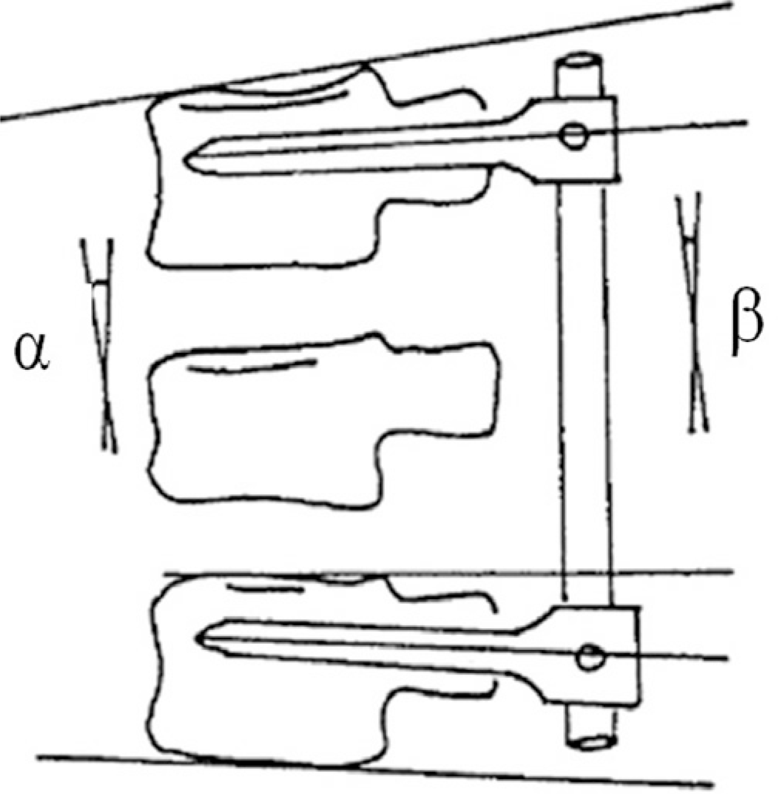
 | Fig. 2.Measuring methods of kyphotic and screw angle. α : kyphotic angle β: interscrew angle |
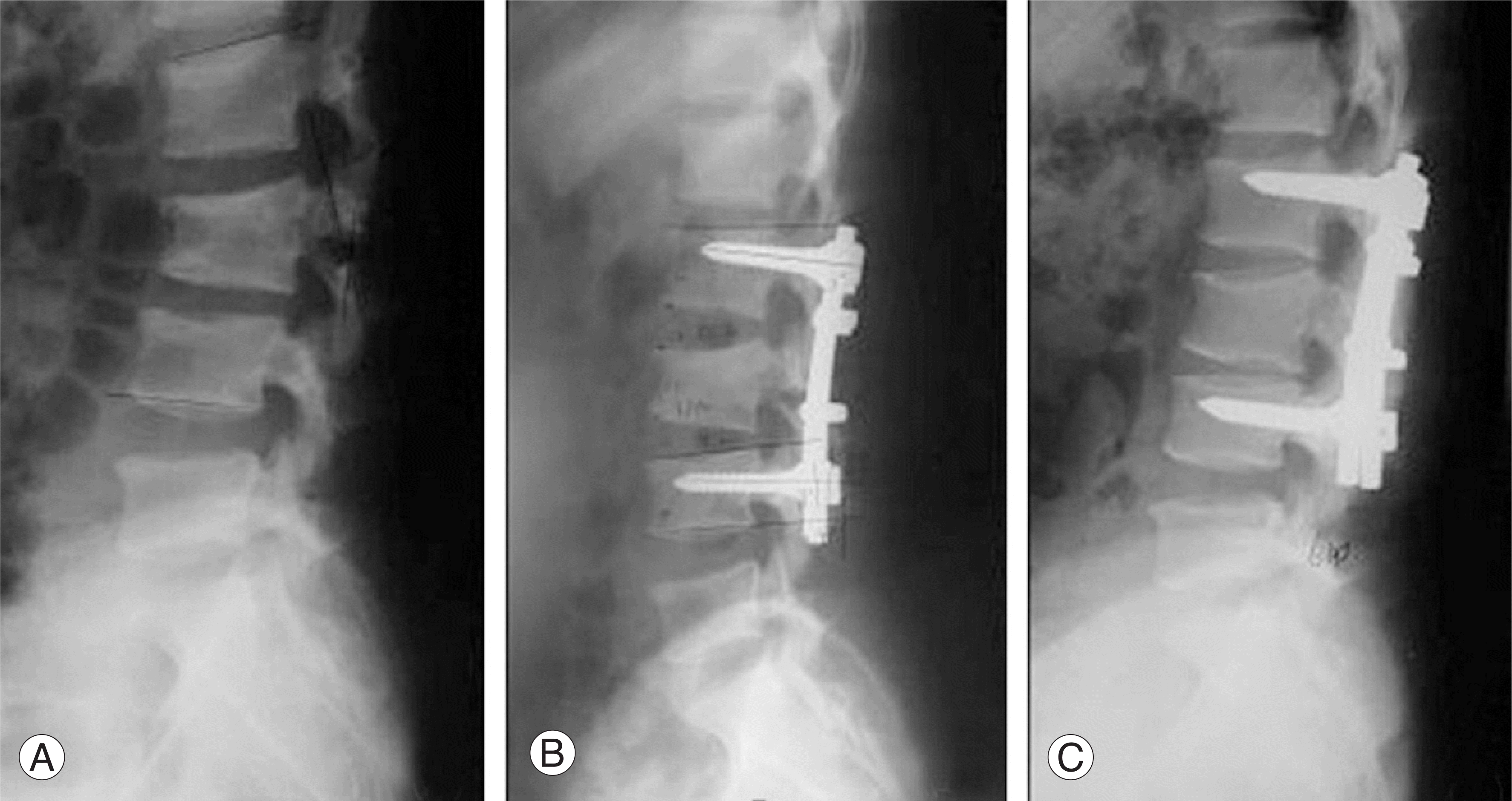
 | Fig. 3. (A)Preoperative radiograph shows burst fracture of L2 vertebra. (B) Immediate postoperative radiograph shows a good correction of kyphosis by posterior instrumented fusion. (C) On postoperative 6 year radiograph, decrease of upper and lower disc height and breakage of lower pedicle screws. |
 | Fig. 4. (A)Changes of anterior body height of fractured vertebra (B) Changes of kyphotic angle of fractured vertebra (C) Changes of wedge angle of fractured vertebra (D) Changes of upper disc height of fractured vertebra (E) Changes of lower disc height of fractured vertebra (F) Changes of interpedicular angle of fractured vertebra. Total : whole patients, None: patients without screw breakage, Failure: patients with pedicle screw breakage. |


 PDF
PDF ePub
ePub Citation
 Print
Print


 XML Download