

 PDF
PDF ePub
ePub Citation
 Print
Print


Abstract
Study Design
This is a retrospective review of 10 consecutive patients with spinal cord injury without radiographic evidence of abnormalities (SCIWORA) and 17 spinal cord injury patients without radiographic evidence of trauma (SCIWORET).
Objectives
We wanted to assess the MRI and clinical findings, the prognosis and effect of anterior decompression of the spinal cord in SCIWORET patients.
Summary of the Literature Review
SCIWORET is not uncommon among middle-age and elderly people. It is less reported in adults as compared with children. There are no studies on the method for the treatment or the effectiveness of anterior decompression of the spinal cord.
Materials and Methods
From February 1994 to December 2005, this study included 27 patients who had cervical spinal cord injury without radiographic evidence of trauma on the plain roentgenography and MRI. Ten patients had no spinal cord compression (SCIWORA patients, group 1) and 17 had their spinal cord compressed from the anterior (SCIWORET patients, group 2), We conservatively treated the group 1 patients and 10 of the group 2 patients, and anterior decompression and fusion were done for 7 of the group 2 patients. Neurological evaluation was performed initial and at last follow up using an ASIA motor score and the Frankel grade.
Results
The patients who had spinal cord edema on MRI had a better prognosis than those who had contusion (p=0.06). There is no statistical difference between the two groups for the neurologic changes at the initial period and the last follow up period (p=0.06, 0.61). Decompression of the spinal cord anteriorly was not effective for the neurologic recovery (p=0.25) and the involved segments were not related to the neurologic changes (p=0.34, 0.25).
Conclusions
It was presumed that patients with edema of the spinal cord had a better prognosis than those with contusion of the spinal cord. There was no difference between the SCIWORA and SCIWORET groups for the neurologic changes and anterior decompression was not effective for the recovery of neurologic symptoms. This study was limited by its retrospective nature and the small number of patients, so a multi-center study is needed.
REFERENCES
01). Pang D., Wilberger JE Jr. Spinal cord injury without radiographic abnormalities in children. J Neurosurg. 1982. 57:114–129.

02). Choi JU., Hoffman HJ., Hendrick EB., Humphreys RP., Keith WS. Traumatic infarction of the spinal cord in children. J Neurosurg. 1986. 65:608–610.
03). Hill SA., Miller CA., Kosnik EJ., Hunt WE. Pediatric neck injuries: A clinical study. J Neurosurg. 1984. 60:700–706.
04). Ruge JR., Sinson GP., McLone DG., Cerullo LJ. Pediatric spinal injury: the very young. J Neurosurg. 1988. 68:25–30.
05). Levitt MA., Flanders AE. Diagnostic capabilities of magnetic resonance imaging and computed tomography in acute cervical spinal column injury. Am J Emerg Med. 1991. 9:131–135.
06). Mirvis SE., Geisler FH., Jelinek JJ., Joslyn JN., Gellad F. Acute cervical spine trauma: evaluation with 1.5T MR imaging, Radiology. 1988. 166:307–316.
07). Silberstein M., Hennessy O. Implicatinons of focal spinal cord lesions following trauma: evaluation with magnetic resonance imaging. Paraplegia. 1993. 31:160–167.
08). Frankel HL., Hancock DO., Hyslop G, et al. The value of postural reduction in the initial management of closed injuries of the spine with paraplegia and tetraplegia. I. Paraplegia. 1969. 7:179–192.
09). Scher AT. Cervical spinal cord injury without evidence of fracture or dislocation. An assessment of the radiological features. S Afr Med J. 1976. 12:962–965.
10). Gupta SK., Rajeev K., Khosla VK, et al. Spinal cord injury without radiographic abnormality in adults. Spinal cord. 1999. 37:726–729.
11). Crooks F., Birkett AN. Fractures and dislocations of the cervical spine. BR J Surg. 1944. 31:252–265.
12). Chirossel JP., Vanneuville G., Passagia JG, et al. Biomechanics and classification of traumatic lesions of the spine. Adv Tech Stand Neurosurg. 1995. 22:55–135.
13). Lyness SS., Wagman AD. Neurological deficits following cervical manipulation. Surg Neurol. 1974. 2:121–124.
14). Wider BL. Hypothesis: the etiology of midcervical quadriplegia after operation with the patient in the sitting position. Neurosurg. 1982. 11:530–531.
15). Taylor AR. The mechanism of injury to the spinal cord in the neck without damage to the vertebral column. J Bone Joint Surg Br. 1951. 33:543–547.
16). Kobrine AI. The neuronal theory of experimental traumatic spinal cord dysfunction. Surg Neurol. 1975. 3:261–264.
17). Simpson RK Jr., Robertson DP., Narayan PK. Penetrating spinal cord injury. Narayan RK, Wilberger JE, Povlishock JT, editors. Neurotrauma. New York: McGraw-Hill Inc;p. 1289–1300. 1996.
18). Holmes G. Spinal injuries of warfare. Br Med J. 1915. 2:769–774.
19). Firooznia H., Ahn JH., Rafii M., Raqnarsson KT. Sudden quadriplegia after a minor trauma. The role of preexisting spinal stenosis. Surg Neurol. 1985. 23:165–168.
20). Hayashi K., Yone K., Ito H., Yanase M., Sakou T. MRI findings in patients with a cervical spinal cord injury who do not show radiographic evidence of a fracture or dislocation. Paraplegia. 1995. 33:212–215.
21). Bhatoe HS. Cervical spinal cord injury without radiological abnormality in adults. Neurol India. 2000. 48:243–248.
22). Chen TY., Lee ST., Lui TN, et al. Efficacy of surgical treatment in traumatic central cord syndrome. Surg Neurol. 1997. 48:435–440.
23). Dare AO., Dias MS., Li V. Magnetic resonance imaging correlation in pediatric spinal cord injury without radiographic abnormality. J Neurosurg. 2002. 97:33–39.
24). Bondurant FJ., Cortler HB., Kulkarni MV., McArdle CB., Harris JH Jr. Acute spinal cord injury; A study using physical examination and magnetic resonance imaging. Spine. 1990. 15:161–168.
25). Kulkarni MV., McARDLE CB., Kopanicky D, et al. Acute spinal cord injury; MR imaging at 1.5 T. Radiology. 1987. 164:837–843.
26). Marciello MA., Flanders AE., Herbison GJ., Schaefer DM., Friedman DP., Lane JI. Magnetic resonance imaging related to neurologic outcome in cervical spinal cord injury. Arch Phys Med Rehabil. 1993. 74:940–946.
27). Flanders AE., Schaefer DM., Doan HT., Mishkin MM., Gonzalez CF., Northrup BE. Acute cervical spine trauma: Correlation of MR imaging findings with degree of neurological deficit. Radiology. 1990. 177:25–33.
28). Allen AR. Surgery of experimental lesion of spinal cord equivalent to crush injury of fracture dislocation of spinal column. A preliminary report. JAMA. 1911. 146:878–880.
29). Collins WF. A review and update of experiment and clinical studies of spinal cord injury. Paraplegia. 1983. 21:204–219.
30). Tator CH., Fehlings MG. Review of the secondary injury theory of acute spinal cord trauma with emphasis on vascular mechanisms. J Neurosurg. 1991. 75:15–26.
31). Senter HJ., Venes JL. Loss of autoregulation and posttraumatic ischemia following experimental spinal cord trauma. J Neurosurg. 1979. 50:198–206.
32). Geisler FH., Coleman WP., Grieco G., Poonian D. The Sygen multicen-ter acute spinal cord injury study. Spine. 2001. 26:87–98.
33). Geisler FH., Dorsey FC., Coleman WP. Recovery of motor function after spinal cord injury; a randomized placebo-controlled trial with G-1 ganglioside. N Engl J Med. 1991. 324:1892–1838.
34). Laud NS., Ramani PS. Patterns of spinal injuries. Ramani PS, editor. Spinal Surgery. 1:Dept of Neuro and Spinal Surgery, Mumbai, LTM Medical College;p. 185–192. 1996.
Fig. 1.
58-year old man with acute spinal cord injury. MRI of the cervical spine was obtained trauma day(A, B) and 12 months (C, D) after injury. (A) Sagittal midline T1-weighetd image shows isointensity cord lesion at C3-4. (B) T2-weighted image shows diffuse hyperintensity cord lesion at C3-4, indicating the existence of edema. (C) Sagittal midline T1-weighted image shows isointensity lesion at C3-4 level. (D) T2-weighted image shows ill defined diffuse hyperintensity lesion at C3-4 level, indicating gliosis.
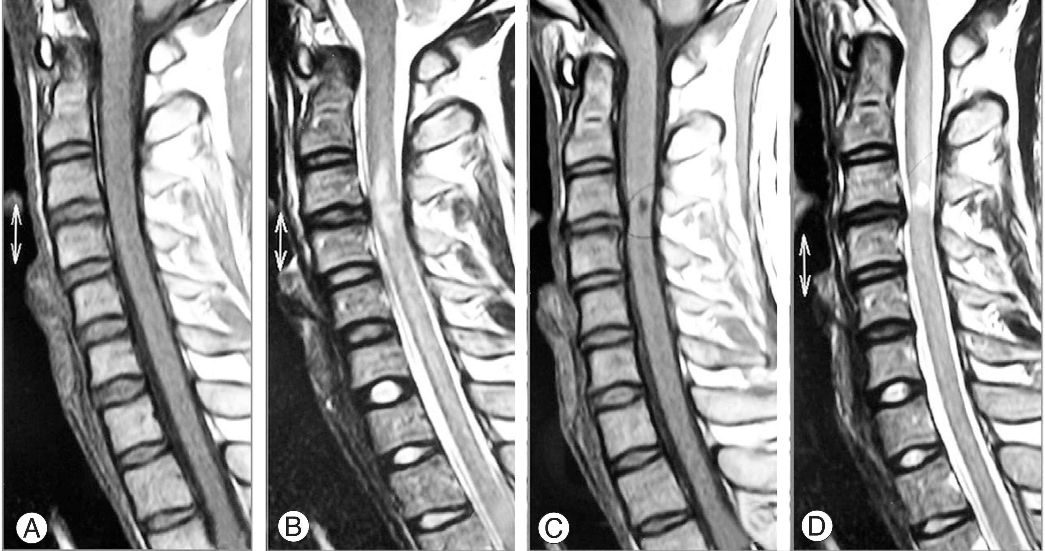
Fig. 2.
A 48-year old woman presenting Frankel C. MRI of the cervical spine was obtained 2 days (A, B) after injury and 1 year (C, D, E) after anterior decompression and fusion. (A) Sagittal T1-weighted image shows slightly hypointensity cord lesion at C4-5. (B) T2-weighted image shows relatively well defined hyperintensity cord lesion at C4-5, indicating existence of cord contusion, and slightly protruded disc. (C) Sagittal midline T1-weighted image shows isointensity lesion at C4-5 level. (D) T2-weighted image shows ill defined diffuse hyperintensity lesion at C4-5 level. (E) T1-weighted Gadullium enhanced image shows hypointensity(not enhanced) lesion at C4-5 level, indicating gliosis.
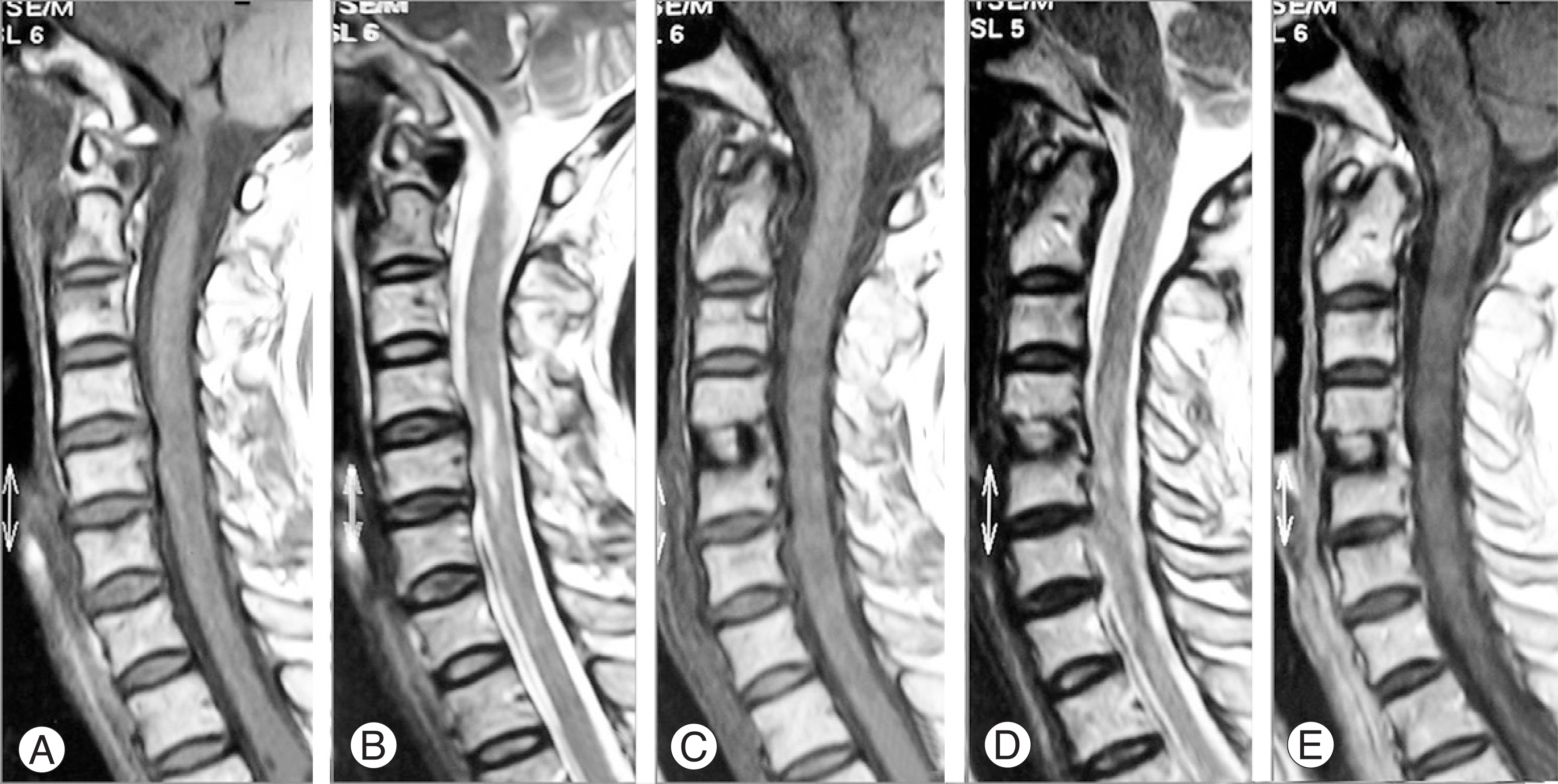
Table 1.
Neurologic changes between the groups (ASIA score)
| Initial | Follow up | Improvement | |
|---|---|---|---|
| Edema (n=12) | 63.0(±32.8) | 89.7(±14.5) | 26.7(±27.7) |
| Contusion (n=15) | 40.3(±26.7) | 73.0(±24.8) | 32.7(±17.2) |
| p value | 0.06 | 0.06 | 0.51 |
| ∗SCIWORA (n=10) | 47.3(±31.6) | 78.3(±25.3) | 31.0(±21.7) |
| ‡SCIWORET (n=17) | 57.8(±30.0) | 82.8(±19.7) | 29.1(±21.8) |
| p value | 0.60 | 0.61 | 0.83 |
| Decompression (n=8) | 66.9(±20.1) | 88.4(± 8.2) | 21.6(±15.1) |
| Non operation (n=9) | 44.6(±33.3) | 78.9(±24.5) | 34.3(±24.9) |
| p value | 0.14 | 0.27 | 0.25 |
| Single leve l(n=18) | 54.7(±27.3) | 85.2(±16.2) | 30.5(±21.8) |
| Multiple level (n=9) | 41.7(±37.9) | 69.6(±31.2) | 27.7(±21.6) |
| p value | 0.34 | 0.25 | 0.77 |
 XML Download