

 PDF
PDF ePub
ePub Citation
 Print
Print


Abstract
Objectives
To evaluate the early clinical results of percutaneous endoscopic lumbar discectomy (PELD) and microdiscectomy (MD) using a tubular retractor.
Summary of the Literature Review
There are few reports comparing the clinical results of different minimal invasive surgical procedures for disc herniation.
Materials and Methods
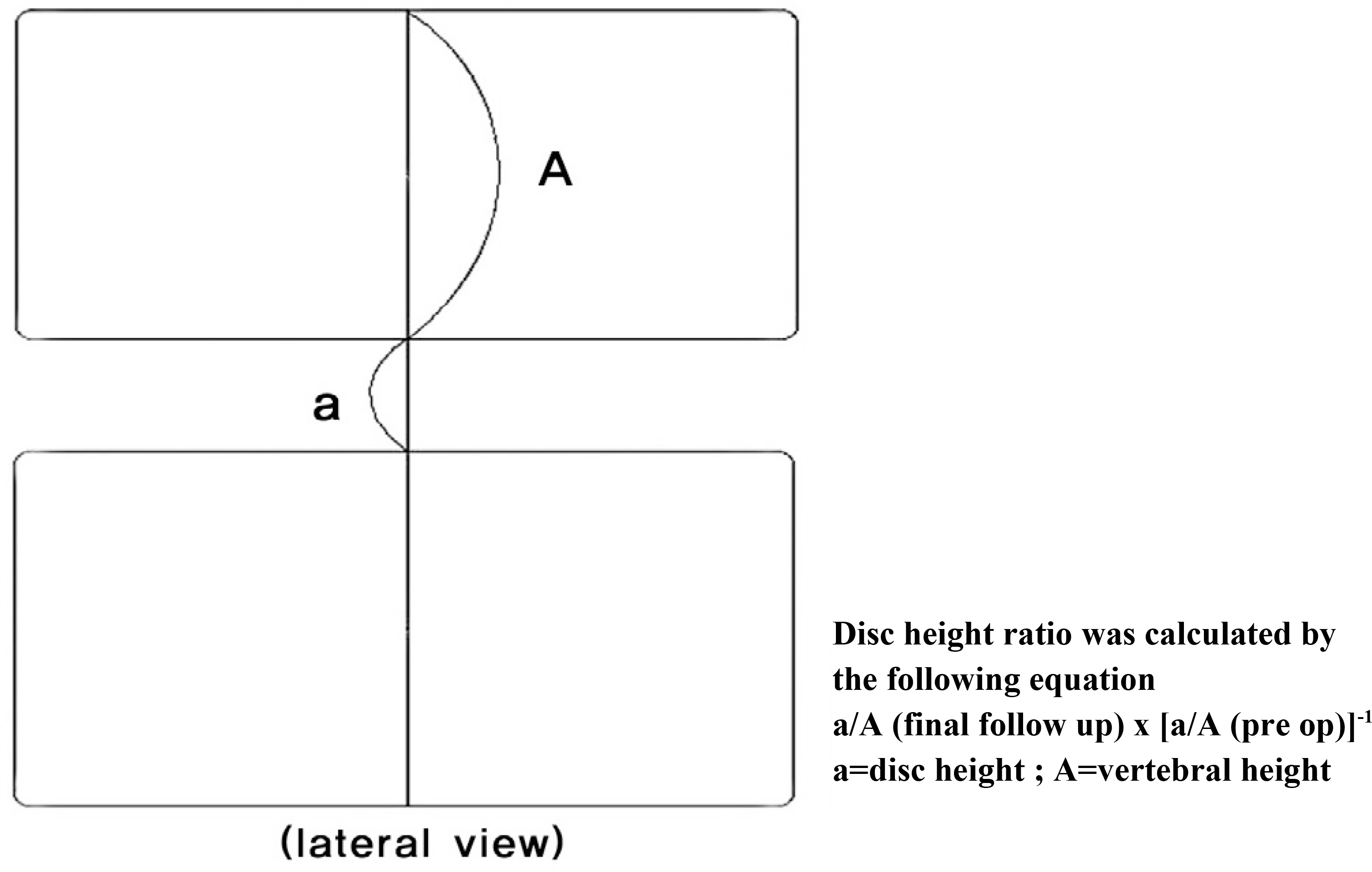
Out of 41 patients who underwent a discectomy at the L4-5 level, 16 patients (Group I) underwent PELD and 25 patients (Group II) underwent MD. The surgical techniques were based on the patient's selection. The characteristics of the operation(operation time, time for C-arm, amount of removed disc) were compared with the clinical outcomes by evaluating the SLR (straight leg raising test), leg VAS (visual analogue scale), ODI (Oswestry Disability Index), hospital day, changes in disc height.
Results
Group I showed a larger amount of disc removed and exposure time for the C-arm than group II (p<0.05). However, the hospital day was shorter in group I than in group II (p<0.05). There were no differences in the leg VAS, ODI, the change in disc height and surgery time between the two groups at the last follow up. One case in group I had a neuropraxia of the L5 root that had recovered fully at postoperative 3months. In group II, there was one case of a postoperative hematoma and 2 cases of a dural tear.
Go to : 
REFERENCES
1). Davis H. Increasing rates of cervical and lumbar spine surgery in the United States, 1979-1990. Spine. 1994; 19:1117–1123.

2). Dandy WE. Loose cartilage from intervertebral disk simulating tumor of the spinal cord. By Walter E. Dandy, 1929. Clin Orthop Relat Res. 1989; 238:4–8.
3). Kambin P, Brager MD. Percutaneous posterolateral discectomy. Anatomy and mechanism. Clin Orthop Relat Res. 1987; 223:145–154.
4). Maroon JC, Abla A, Bost J. Association between peridural scar and persistent low back pain after lumbar discectomy. Neurol Res. 1999; 21:43–46.
5). Smith L. Enzyme dissolution of the nucleus pulposus in humans. JAMA. 1964; 187:137–140.
6). Hijikata S. Percutaneous nucleotomy: A new concept technique and 12 years experience. Clin Orthop Relat Res. 1989; 238:9–23.
7). Smith MM, Foley KT. Microendoscopic discectomy system: surgical technique and initial clinical results. Tech Neurosurg. 1997; 3:301–307.
8). Xiaotao W, Suyang Z, Zubin M, Hui C. Microendoscopic discectomy for lumbar disc herniation: Surgical technique and outcome in 873 consecutive cases. Spine. 2006; 31:2689–2694.
9). Anthony T, Christopher A. Minimally invasive techniques for the management of lumbar disc herniation. Orthop Clin N Am. 2007; 38:363–372.
10). Casper GD, Hartman VL, Mullins LL. Laser assisted disc decompression: an alternative treatment modality in the Medicare population. J Okla State Med Assoc. 1996; 89:11–15.
11). Hermantin FU, Peters T, Quartararo L, Kambin P. A prospective, randomized study comparing the results of open discectomy with those of video-assisted arthroscopic microdiscectomy. J Bone Joint Surg Am. 1999; 81:958–965.
12). Mochida J, Nishimura K, Nomura T. Appropriate procedures of posterior herniotomy and percutaneous nucleto-my in lumbar disc herniation. Rinsho Seikei Geka. 1994; 29:423–430.
13). Yeung AT, Tsou PM. Posterolateral endoscopic excision for lumbar disc herniation: Surgical technique, outcome, and complications in 307 consecutive cases. Spine. 2002; 27:722–731.
14). Chung JY, Lee JJ. Percutaneous endoscopic discectomy for lumbar disc herniation. J Korean Soc Spine Surg. 2007; 14:212–219.
15). Kang YH, Lee WS, Yune SH. Comparison of the results between standard discectomy and microdiscectomy of the herniated lumbar disc. J Korean Soc Spine Surg. 2000; 7:228–233.
16). Matsui H, Kitagawa H, Kawaguchi Y, Tsuji H. Physiologic changes of nerve root during posterior lumbar discectomy. Spine. 1995; 20:654–659.
17). Hijikata S, Yamagishi M, Nakayama T. Percutaneous discectomy: A new treatment method for lumbar disc herniation. J Toden Hosp. 1975; 5:5–13.
18). Onik G, Helms CA, Ginsberg L, Hoaglund FT, Morris J. Percutaneous lumbar discectomy using a new aspiration probe. AJNR. 1985; 6:1137–1140.
19). Onik G, Shang YL, Maroon JC. Automated percutaneous biopsy in postoperative diskitis: a new method. AJNR. 1990; 11:391–393.
20). Nakagawa H, Kamimura M, Uchiyama S, Takahara K, Itsubo T, Miyasaka T. Microendoscopic discectomy (MED) for lumbar disc prolapsed. J Clin Neurosci. 2003; 10:231–235.
21). Kambin P, Sampson S. Posterolateral percutaneous suction-excision of herniated lumbar intervertebral discs. Report of interim results. Clin Orthop Relat Res. 1986; 207:37–43.
22). Kikuchi S, Hasue M, Nishiyama K, Ito T. Anatomic features of the furcal nerve and its clinical significance. Spine. 1986; 11:1002–1007.
23). Schick U, Dohnert J, Richter A, Ko¨nig A, Vitzthum HE. Microendoscopic lumbar discectomy versus open surgery: an intraoperative EMG study. Eur spine J. 2002; 11:20–26.
Go to :
Figures and Tables%
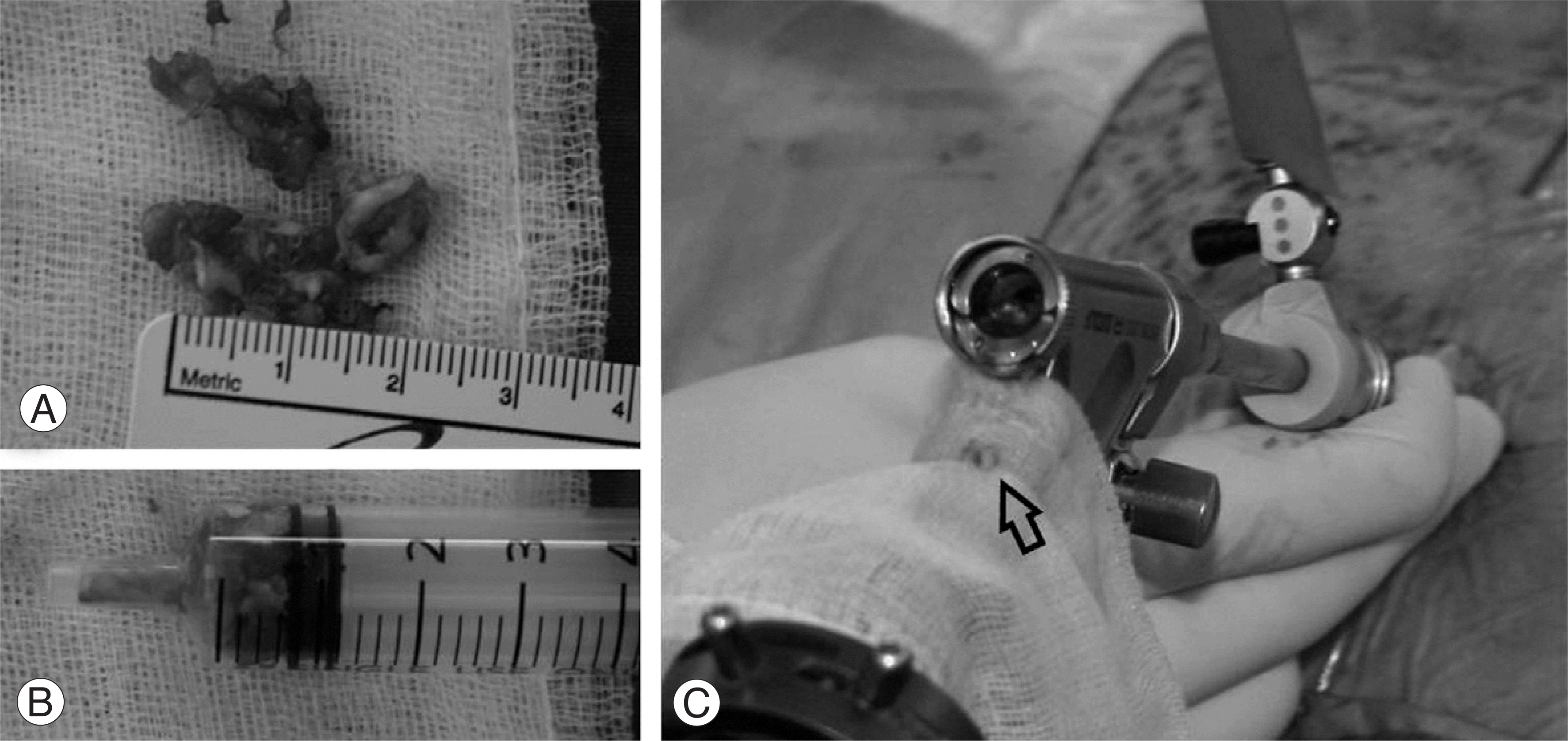 | Fig. 1.Measurement of the amount of removed disc using 5cc syringe. Disc materials extruded through working portal of arthroscopy was added to measure the amount of removed disc in PELD group. arrow: disc material flowed out through the working portal under surgery. |
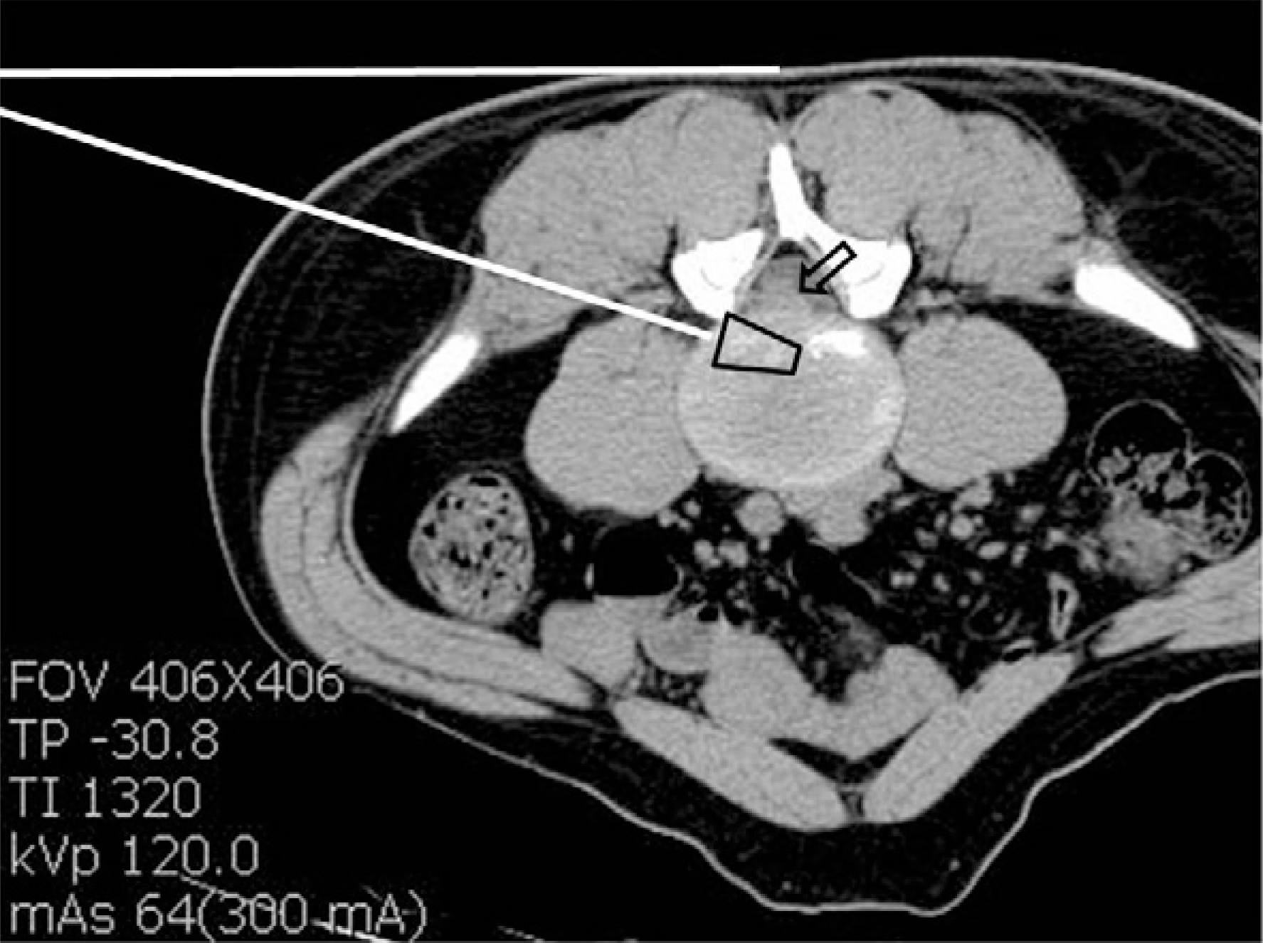 | Fig. 3.Computed tomographic scan at L4-5 level in prone position preoperatively. white lines: the preoperative trajectories for deciding entry points and angle of guide wire, arrow: the extruded disc, black square: the portion which will be removed disc in disc space to reach the true pathology (black arrow). |
Table 1.
Demographic data between two groups
| $PELD (N=16) | %MD (N=25) | P -VALUE | |
|---|---|---|---|
| *Sex (male:female) | 9:7 | 17:8 | 0.61 |
| Age (yrs) | 37(20~69) | 42(25~62) | 0.52 |
| *Disc type (protrusion:extrusion) | 10:6 | 14:11 | 0.24 |
| Sx duration (months) | 3.4(1~18) | 4.2(2~22) | 0.80 |
| †SLR (involved leg) | 43(30~70) | 45(35~65) | 0.35 |
| Pre op †VAS (leg) | 7.3(5~10) | 7.1(6~10) | 0.32 |
| Pre op #ODI | 48(40~62) | 50(38~68) | 0.20 |
| Mean followup (months) | 13(6~19) | 11(6~18) | 0.53 |
Table 2.
The comparison of perioperative factor and clinical outcomes between two groups
| $PELD (N=16) | %MD (N=25) | P-VALUE | |||||
|---|---|---|---|---|---|---|---|
| Pre op | Discharge | Last F/U | Pre op | Discharge | Last F/U | ||
| *SLR (involved leg) | 43(30~70) | 84(70~90) | 87(80~90) | 45(35~65) | 87(60~90) | 87(80~90) | |
| †VAS (leg) | 7.3(5~10) | 1.5(0~4) | 1.6(0~4) | 7.1(6~10) | 1.3(1~4) | 1.1(0~5) | |
| †ODI | 48(40~62) | - | 1 (2~14) | 50(38~68) | - | 8(2~12) | |
| Change of the disc height (%) | 93(68~100) | 94(70~100) | |||||
| Complication case | 1 | 3 | |||||
| Mean operating time(min) | 89(45~180) | 97(75~150) | 0.13 | ||||
| #Mean C-arm time(sec) | 41(17~120) | 15(6~41) | 0.03 | ||||
| #Removed disc(cc) | 1.9(0.8~3.1) | 1.1(0.5-2) | 0.02 | ||||
| #Mean hospital stay | 4(2~8) | 8(4~14) | 0.03 | ||||
 XML Download