Abstract
Objectives
To analyze and compare the cervical and thoracic sagittal curves between normal adolescents and patients with thoracic adolescent idiopathic scoliosis (AIS).
Summary of Literature Review
There are no reports on cervical sagittal curves and its correlation with thoracic sagittal curves in AIS.
Materials and Methods
The sagittal curves were analyzed in normal adolescents (N-adol group, n=23) and patients with thoracic AIS (AIS group, n=26) who had thoracic curves ≥ 45°. Lateral standing radiographs of the cervical spine with a elbow straight and the whole spine with the hands on the clavicles were taken. The sagittal curves and balance were measured in the following segments; C2-C7, T2-T5, T5-12, T2-12, T12-S1. Cervical lordosis (C2-C7) was measured in both cervical spine radiographs and whole spine radiographs.
Results
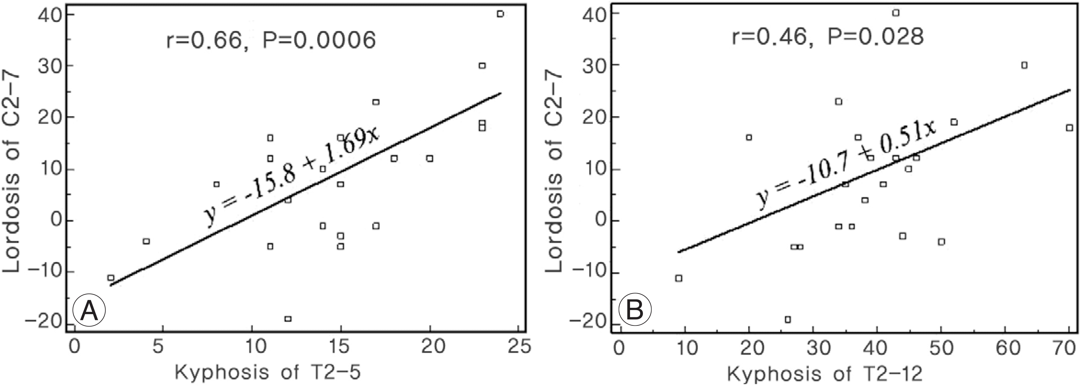
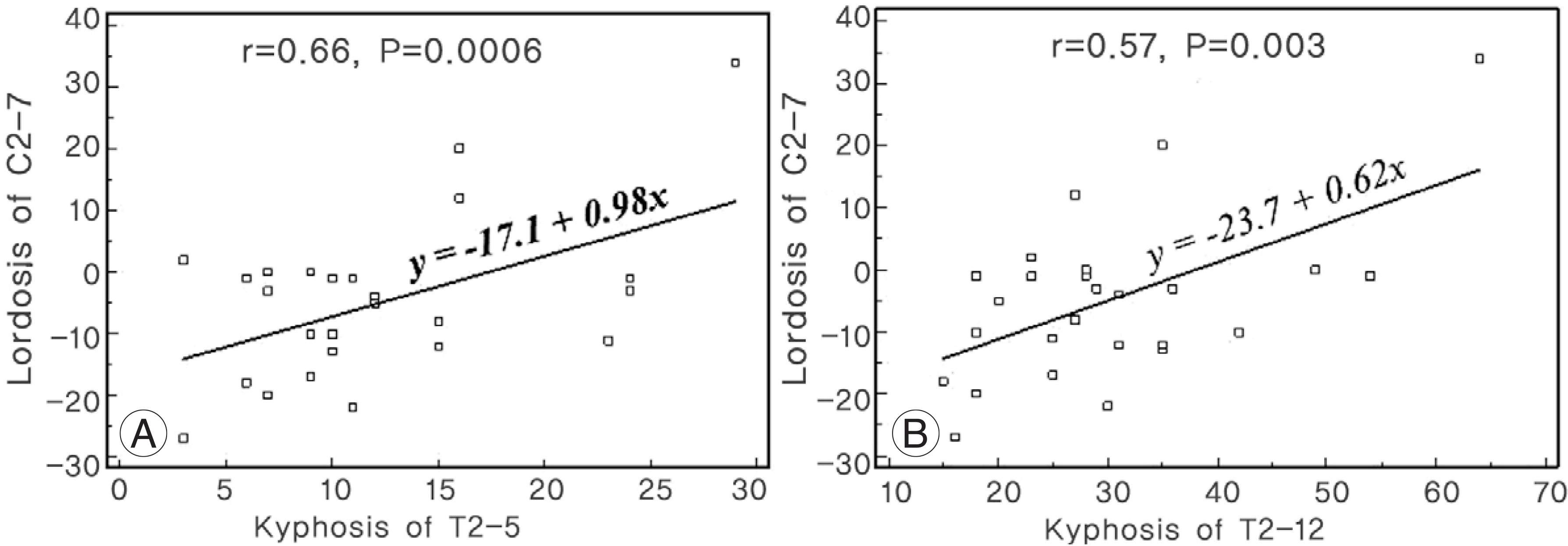
In the N-adol group, the cervical lordosis was 9.2±14.6°in the cervical spine radiographs and -0.6±12.9°(‘-’ means kyphosis) in whole spine radiographs. In the AIS group, cervical lordosis was -5.0±12.9°in the cervical radiographs and -8.1± 12.7° in the whole radiographs. The AIS group had significantly less cervical lordosis than the N-adol group. Thoracic kyphosis of T5-12 and T2-12 was 24.1±10.6°and 38.9±13.1°in the N-adol group, respectively, and 17.8±9.4°and 30.1±11.8°in the AIS group, respectively. There was a significant difference between the two groups (Ps<0.05). There was no significant difference in thoracic kyphosis of T2-T5, lumbar lordosis and sagittal balance between the two groups (Ps>0.05). In the AIS group, the cervical lordosis measured in the cervical spine radiograph showed a positive correlation with thoracic kyphosis of T2-5 (r=0.50, P=0.009) and T2-12 (r=0.57, P=0.003).
Go to : 
REFERENCES
1). Suk SS, Lee SM, Chung ER, Kim JH, Kim SS. Selective thoracic fusion with segmental pedicle screw fixation in the treatment of thoracic idiopathic scoliosis. Spine. 2005; 30:1602–1609.

2). Horton WC, Brown CW, Bridwell KH, Glassman SD, Suk SI, Cha CW. Is there an optimal patient stance for abtaining a lateral 36” radiograph? A critical comparison of three techniques. Spine. 2005; 30:427–433.
3). O'Brien MF, Kuklo TR, Blanke KM, et al. .:. Radiographic Measurement Manual. Spinal Deformity Study Group (SDSG). Medtronic Sofamor Danek;2004.
4). Ohara A, Miyamot K, Naganawa T, Matsumoto K, Shimizu K. Reliabilities of and correlations among five standard methods of assessing the sagittal alignment of the cervical spine. Spine. 2006; 31:2585–2591.

Go to : 
Figures and Tables%
 | Fig. 1.In the normal adolescent group, cervical lordosis of C2-7 measured in cervical spine radiographs had a positive correlation with kyphosis of T2-5 (A) and T2-12 (B). |
 | Fig. 2.In the AIS group, cervical lordosis of C2-7 measured in cervical spine radiographs had a positive correlation with kyphosis of T2-5 (A) and T2-12 (B). |
Table 1.
Parameters measured in radiographs


 PDF
PDF ePub
ePub Citation
 Print
Print


 XML Download