Abstract
Objectives
To examine the effect of transpedicular screw fixation on fractured vertebrae about the vertebral wedge angle (VWA) after posterior instrumentation of the thoracolumbar fracture, determine the effect of reduced VWA on the change in the Kyphotic angle (KA), and minimize loss of reduction of KA.
Summary of the literature review
Maintenance of the KA of a thoracolumbar fracture after surgery is important for the radiologic and functional outcome.
Materials and methods
Forty patients, who had undergone posterior instrumentation in a thoracolumbar fracture between February 2006 and February 2008 and followed-up for more than one year, were enrolled in this study. The patients were divided into two groups according to transpedicular screw fixation (Group A) or not (Group B) including fractured vertebrae. The evaluation was performed by measuring the changes in the KA and VWA taken after the injury, immediate after surgery and 1 year after surgery.
Results
There was correlation between groups A (transpedicular screw fixation on fractured vertebrae) and B (no transpedicular screw fixation on the fractured vertebrae) regarding the correction of the VWA and the loss of correction KA, (p<0.05).
Conclusions
Reduction of the VWA is an important factor for preventing reduction loss of the KA, and transpedicular screw fixation including fractured vertebrae would help reduce the VWA. Therefore, the operator must pay attention to the increase in VWA to maintain the KA through short segment transpedicular screw fixation including fractured vertebrae.
Go to : 
REFERENCES
1). Siebenga J, Leferink VJ, Segers MJ, et al. .:. Treatment of traumatic thoracolumbar spine fractures: a multicenter prospective randomized study of operative versus nonsurgical treatment. Spine. 2006; 31:2881–2890.

2). Reinhold M, Knop C, Lange U, Bastian L, Blauth M. Non-operative treatment of thoracolumbar spinal fractures. Longterm clinical results over 16 years. Unfallch-irurg. 2003; 106:566–576.
3). Stadhouder A, Buskens E, de Klerk LW, et al. .:. Traumatic thoracic and lumbar spinal fractures: operative or nonoperative treatment: comparison of two treatment strategies by means of surgeon equipoise. Spine. 2008; 33:1006–1017.
4). Vaccaro AR, Kim DH, Brodke DS, et al. .:. Diagnosis and management of thoracolumbar spine fractures. Instr Course Lect. 2004; 53:359–373.

5). Yue JJ, Sossan A, Selgrath C, et al. .:. The treatment of unstable thoracic spine fractures with transpedicular screw instrumentation: a 3-year consecutive series. Spine. 2002; 27:2782–2787.

6). Shen WJ, Liu TJ, Shen YS. Nonoperative treatment versus posterior fixation for thoracolumbar junction burst fractures without neurologic deficit. Spine. 2001; 26:1038–1045.

7). Mikles MR, Stchur RP, Graziano GP. Posterior instrumentation for thoracolumbar fractures. J Am Acad Orthop Surg. 2004; 12:424–435.

8). Ahn JS, Lee JK, Hwang DS, Kim YM, Kim WJ, Byun KH. The change of kyphotic angle and anterior vertebral height after posterior or posterolateral fusion with transpedicular screws for thoracolumbar bursting fractures. J Korean Fracture Soc. 2003; 2:379–387.

9). Kim YM, Kim DS, Choi ES. Results of non-fusion method in thoracolumbar and lumbar spinal fractures. J Korean Soc Spine Surg. 2005; 12:132–139.

10). Wang XY, Dai LY, Xu HZ, Chi YL. Kyphosis recurrence after posterior short-segment fixation in thoracolumbar burst fracture. J Neurosurg Spine. 2008; 8:246–254.
11). Scholl BM, Theiss SM, Kirkpatrick JS. Short segment fixation of thoracolumbar burst fractures. Orthopedics. 2006; 29:703–708.

12). Dai LY, Jiang SD, Wang XY, Jiang LS. A review of the management of thoracolumbar burst fractures. Surg Neurol. 2007; 67:221–231.

13). Inamasu J, Guiot BH, Nakatsukasa M. Posterior instrumentation surgery for thoracolumbar junction injury causing neurologic deficit. Neurol Med Chir (Tokyo). 2008; 48:15–21.

14). Lee JY, Kim GL. Posterior instrumentation of thoracolumbar fracture. J Korean Soc Spine Surg. 2001; 8:423–427.

15). Mahar A, Kim C, Wedemeyer M, et al. .:. Short segment fixation of lumbar burst fractures using pedicle fixation at the level of the fracture. Spine. 2007; 32:1503–1507.
16). Min HJ, Kim KW, Kim YH, Yoon US, Hwang JS. The comparison of loss of reduction at the thoracolumbar fracture according to insertion of screw including fractured vertebra or not in short segment posterolateral fusion. J Korean Soc Spine Surg. 2002; 9:19–26.
Go to : 
Figures and Tables%
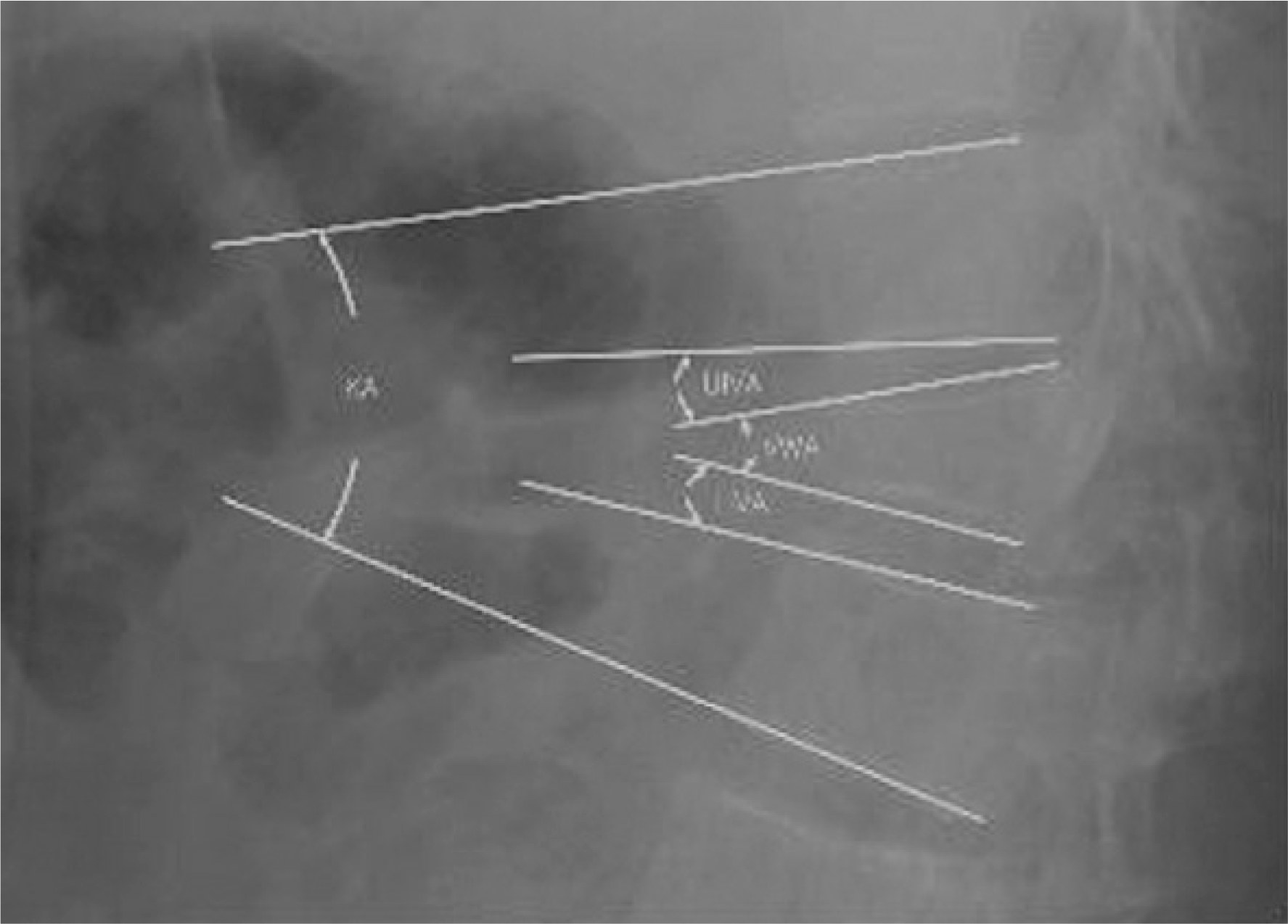
 | Fig. 1.Schematic diagram of geometric parameters measured on a lateral radiograph. KA: Kyphotic angle, VWA: Vertebral wedge angle, UIVA: Upper intervertebral angle, LIVA: Lower intervertebral angle |
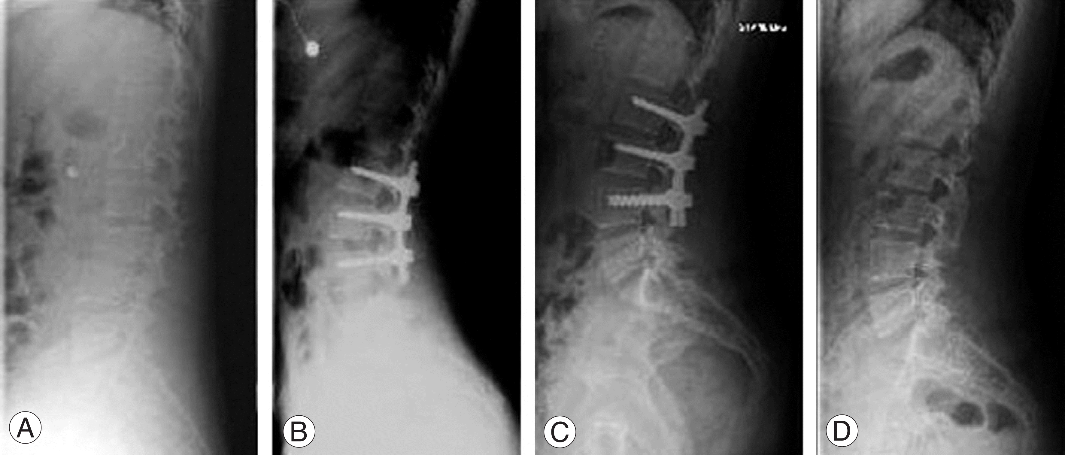
 | Fig. 2.Change of KA & VWA over time (screw fixation including a fractured vertebra) The patient was 24 years old female. Fracture type was Flexion-Distraction. Spinous process of L2 spine was bisected. Transpedicular fixation including a fractured vertebra was done. KA and VWA was initial 7°, 16° and immediate postoperation -22°, 2°, last followup -20°, 2°, After implant removal -19°, 2°. Initial reduction of VWA was 14°. So, reduction loss of KA and VWA (2°, 0°) was minimal. Reduction loss KA and VWA (3°, 0°) after implant removal was minimal, too. (A) Preoperative lateral radiography (B) immediate postoperative lateral radiography (C) Followup lateral radiography after 15 months (D) Followup lateral radiography after implant removal. |
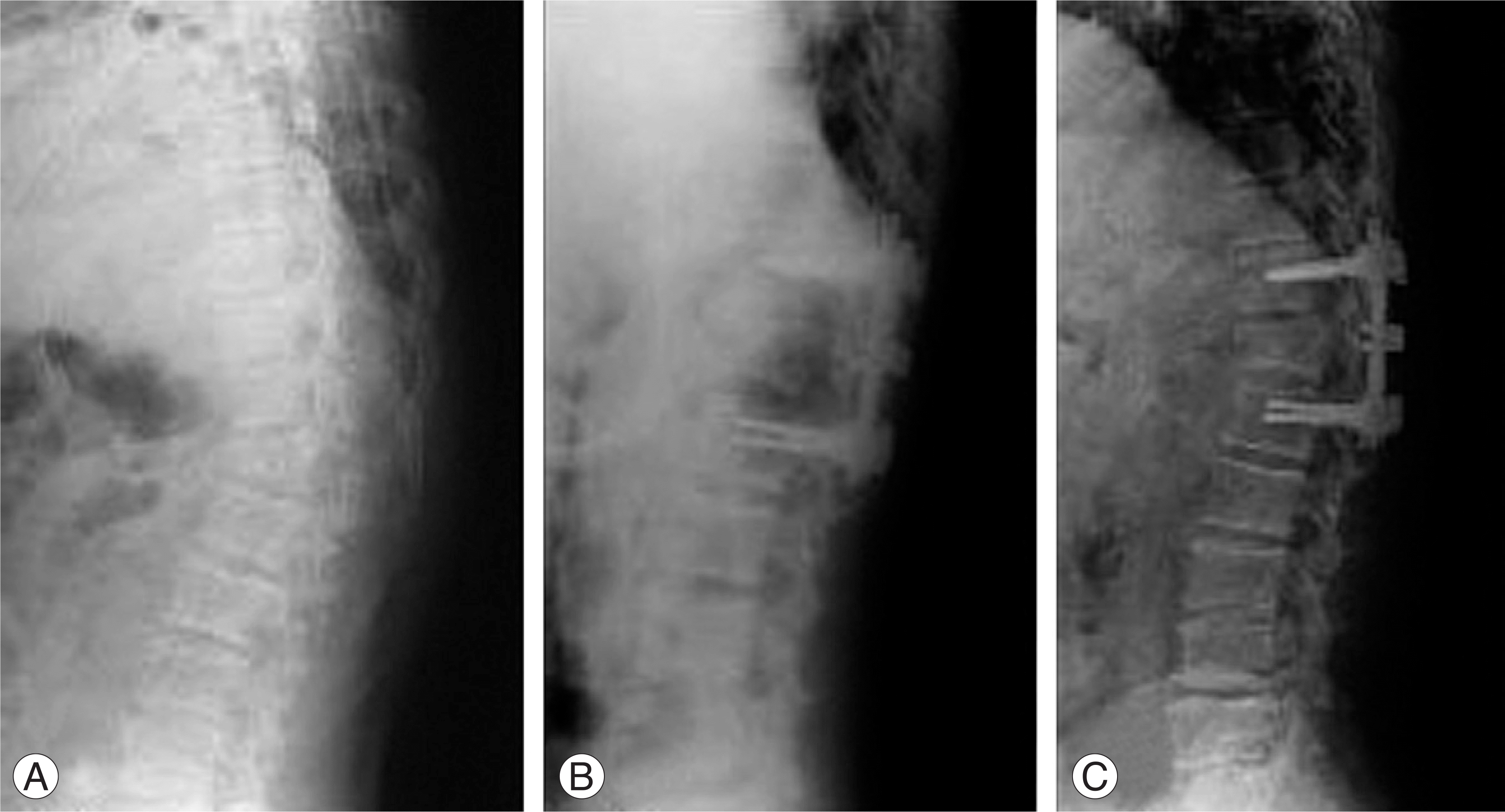
 | Fig. 3.Change of KA & VWA over time (screw fixation not including a fractured vertebra) The patient was 54 years old male. Fracture type was Flexion-Distraction. Transpedicular fixation not including a fractured vertebra was done. KA and VWA was initial 34°, 27°and immediate postoperation 15°, 7°, last followup 20°, 17°. Initial reduction of VWA was 20°. Reduction loss of KA and VWA (5°, 10°) was progressed. (A) Preoperative lateral radiography (B) immediate postoperative lateral radiography (C) Followup lateral radiography after 27 months. |
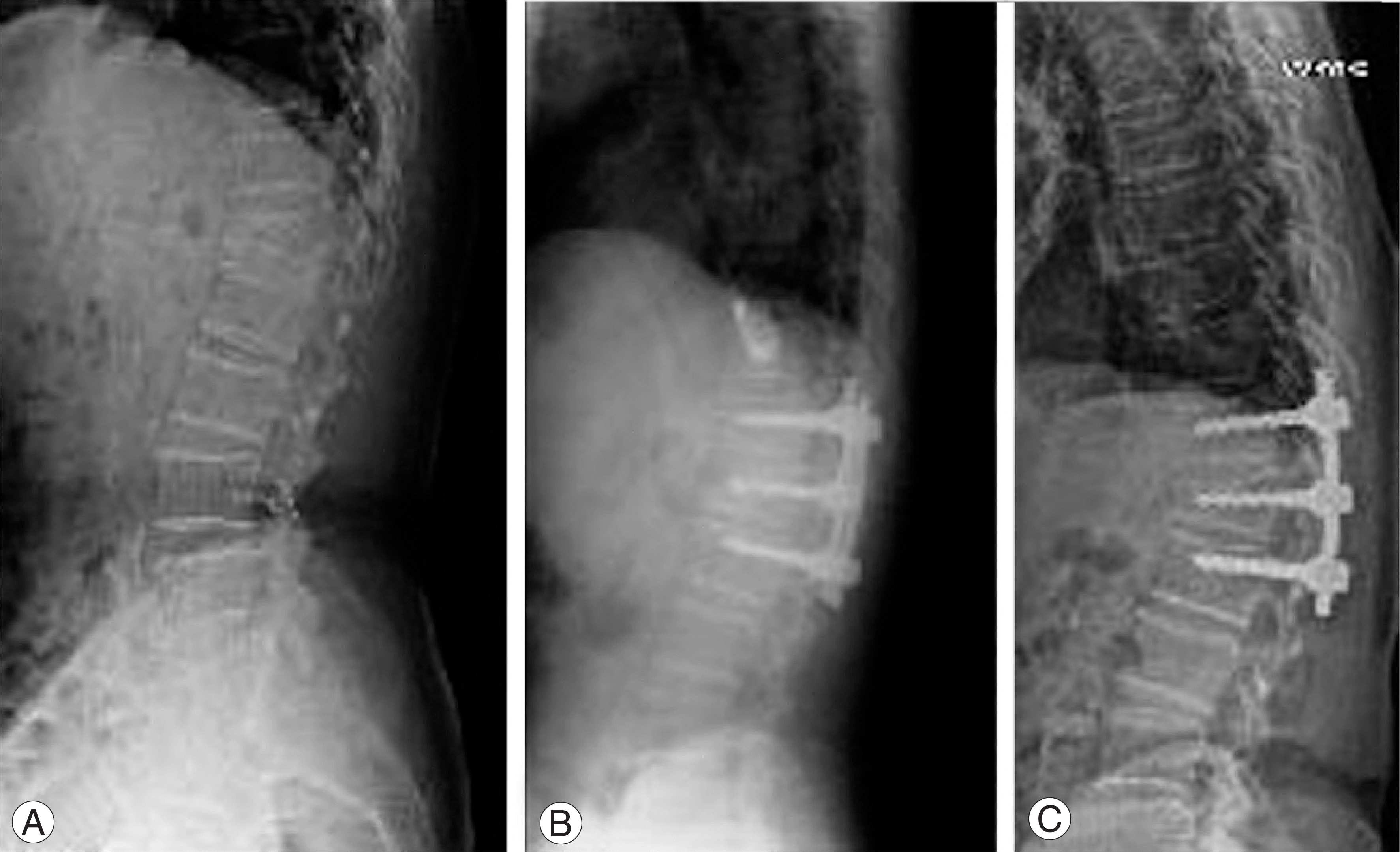
 | Fig. 4.Change of KA & VWA over time (screw fixation including a fractured vertebra) The patient was 69 years old male. Fracture type was Flexion-Distraction. Transpedicular fixation including a fractured vertebra was done. KA and VWA was initial 32°, 26°and immediate postoperation 21°, 20°, last followup 30°, 25°. Initial reduction of VWA was 6°. So, reduction loss of KA and VWA (9°, 5°) was progressed. (A) Preoperative lateral radiography (B) immediate postoperative lateral radiography (C) Followup lateral radiography after 23 months. |
Table 1.
The changes in radiologic parameters over time
preop.; preoperative, Immpo.; parameters of immdiate postoperative, (reduction); average of amount of reduction, 1yr; parameters after 1 year at least, (loss); average of amount of reduction loss, KA; Kyphotic angle, VWA; Vertebral wedge angle, UIVA; Upper Intervertebral angle, LIVA; Lower Intervertebral angle


 PDF
PDF ePub
ePub Citation
 Print
Print


 XML Download