Abstract
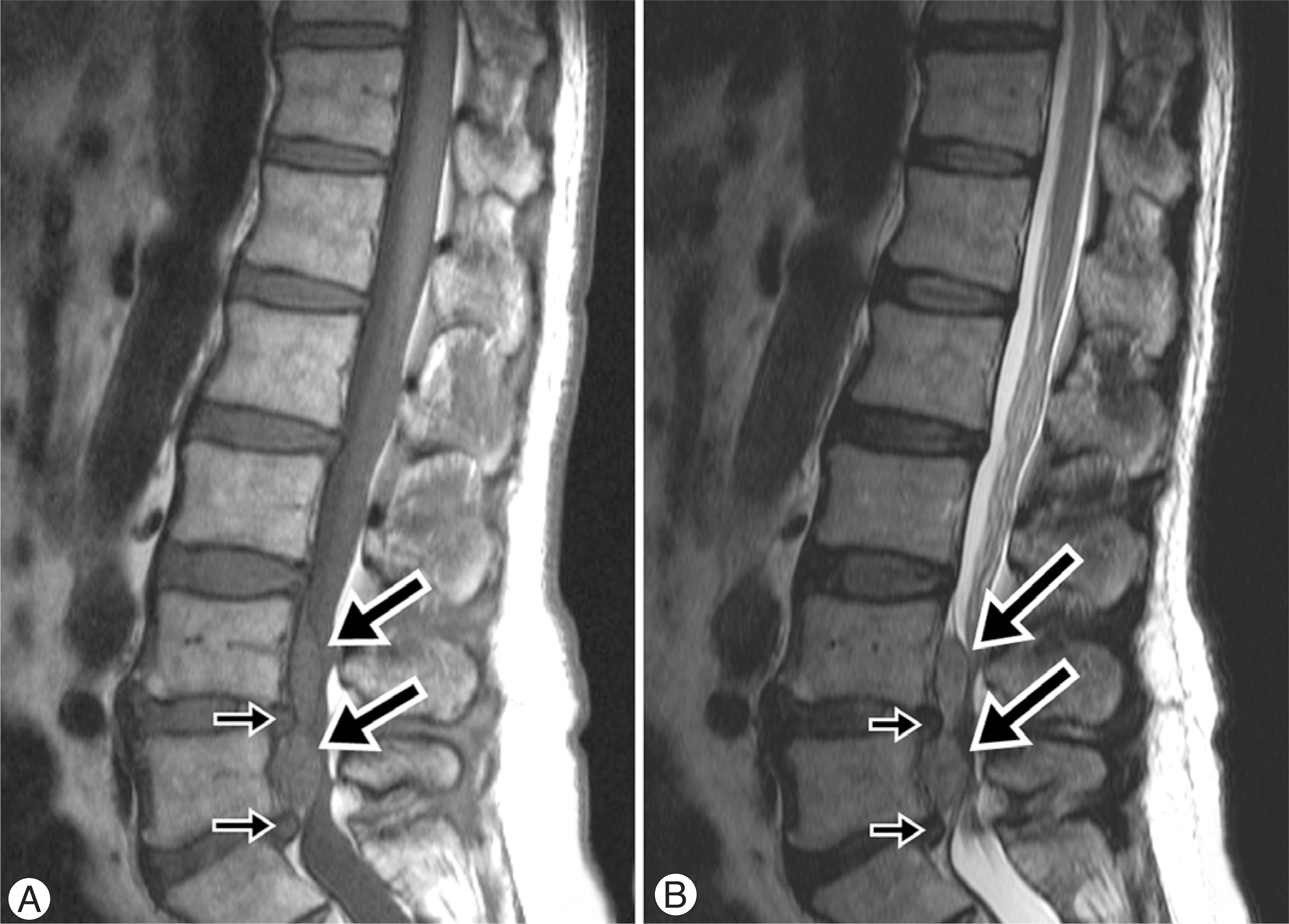
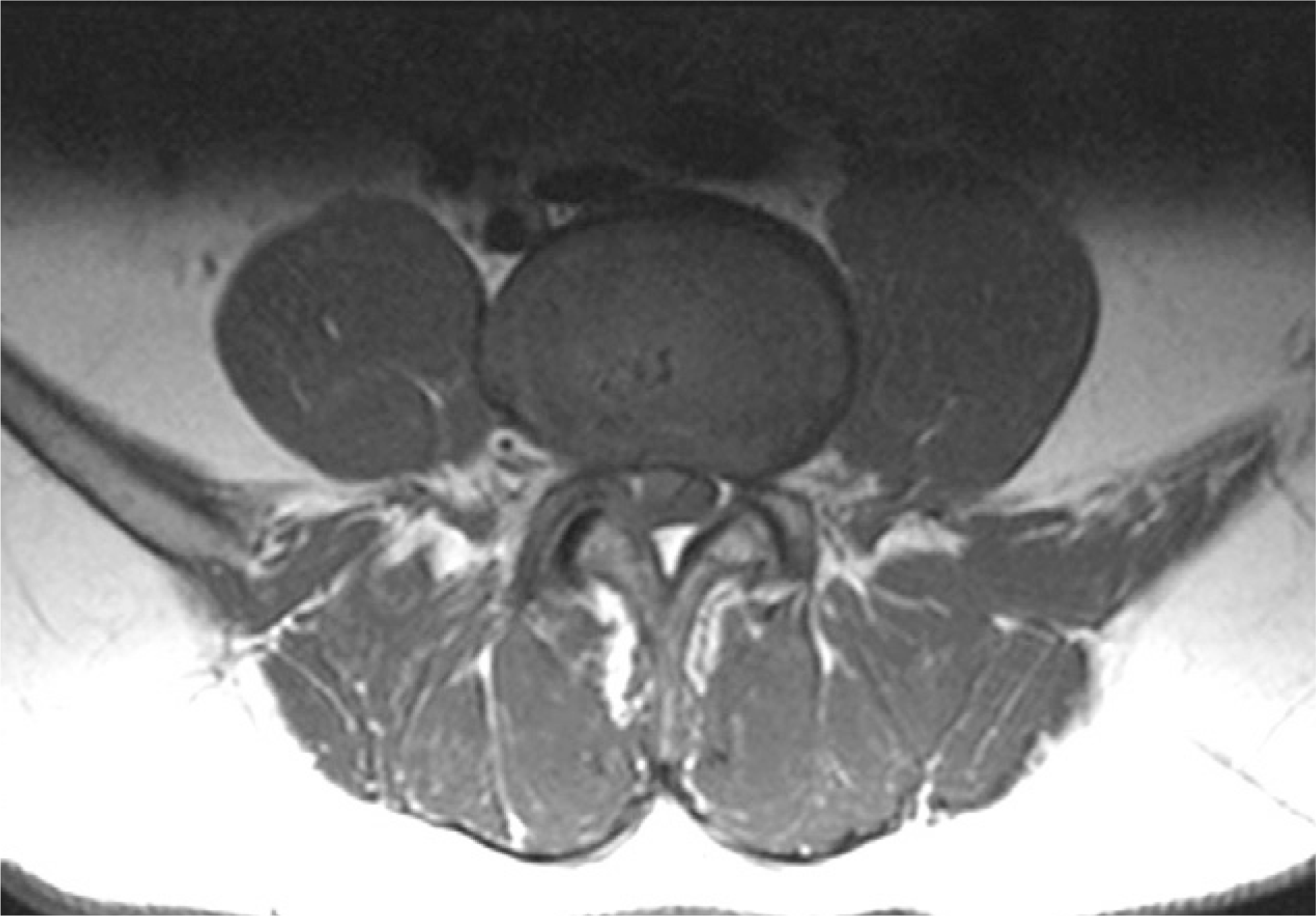
Cauda equina syndrome after epidural block is a rare complication, but it requires emergency surgery when it is diagnosed. A 65-year-old man who underwent epidural block at a local clinic was admitted with right lower leg weakness and decreased leg sensation, severe lower radiating pain, dysuria and decreasing sensation in the perianal region. Magnetic resonance image showed protruded disc material between L4-L5 and a hematoma that occupied most of the spinal canal and this was compressing the spinal cord. These findings were diagnostic for cauda equina syndrome after epidural block and so laminectomy, excision of the herniated disc and removal of the hematoma were done. At 6 months followup, the neurologic symptoms were resolved except for the dorsiflextion of the ankle and the big toe. We report here on a case of cauda equina syndrome as a rare complication after epidural anesthesia.
Go to : 
REFERENCES
1). Waldman SD. Cervial epidural nerve block. Interventional Pain Management. 2nd ed.Waldman SD, editor. Philadelphia: WB Saunders company;pp. p. 373–381. 2001.
2). Bent U, Gniffke S, Reinbold WD. An epidural hematoma following single shot epidural anesthesia, Anaesthesist. 1994; 43:245–248.
3). Dickman CA, Shedd SA, Spetzler RF, Shetter AG, Sonntag VK. Spinal epidural hematoma associated with epidural anesthesia: complication of systemic heparinization in patient receiving peripheral thrombolytic therapy. Anesthesiology. 1990; 72:947–950.
4). Skilton RW, Justice W. Epidural hematoma following anticoagulant treatment in a patient with an indwelling epidural catheter, Anaesthesia. 1998; 53:691–695.
5). Park BM, Won YY. Clinical observation on 8 cases of cauda equina syndrome. J of Korean orthop Assoc. 1988; 23:184–192.

6). Ayten B, Sacit G. Cauda equina syndrome after epidural steroid injection: A case report. J. of Manipulative and Physiological Therapeutics. 2006; 29:492. .e1-492.e3.
7). Kostuik JP, Harrington L, Alexander D, Rand W, Evans D. Cauda equina syndrome and lumbar disc herniation. J. Bone joint Surgery Am. 1986; 68:386–391.
Go to : 
 | Fig. 1.(A) Sagital T1-weighted magnetic resonance image shows protruded disc material at L4-L5 and L5-S1 (short arrow) and large epidural lesion (hematoma) showing intermediate high signal, compressing thecal sac at L4-5 anterior epidural region (long arrow) (B) Sagital T2-weighted magnetic resonance image shows large epidural lesion showing mixed high signal. |


 PDF
PDF ePub
ePub Citation
 Print
Print


 XML Download