Abstract
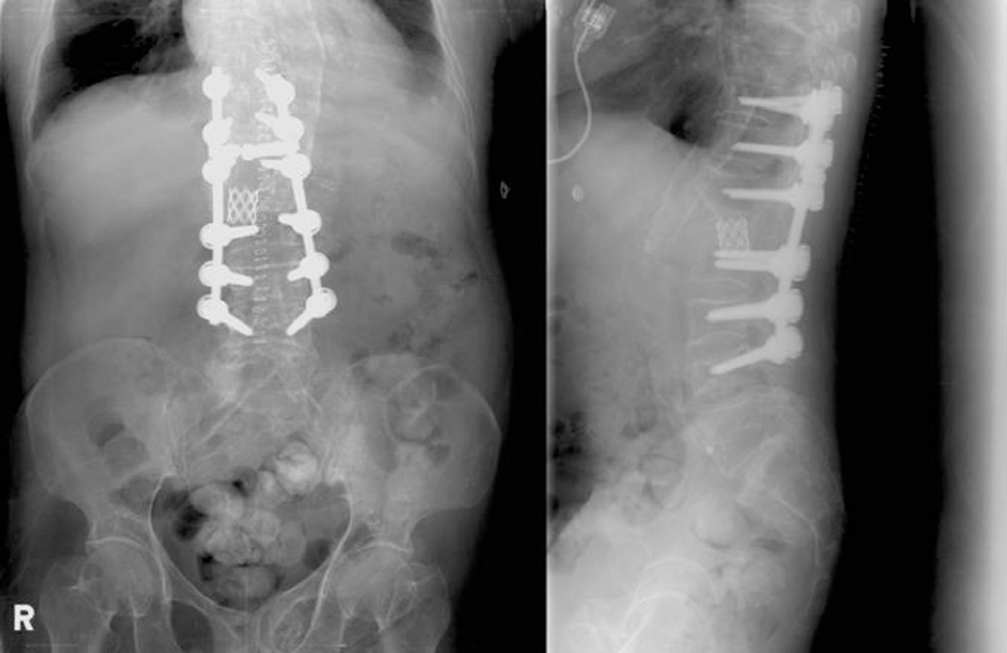
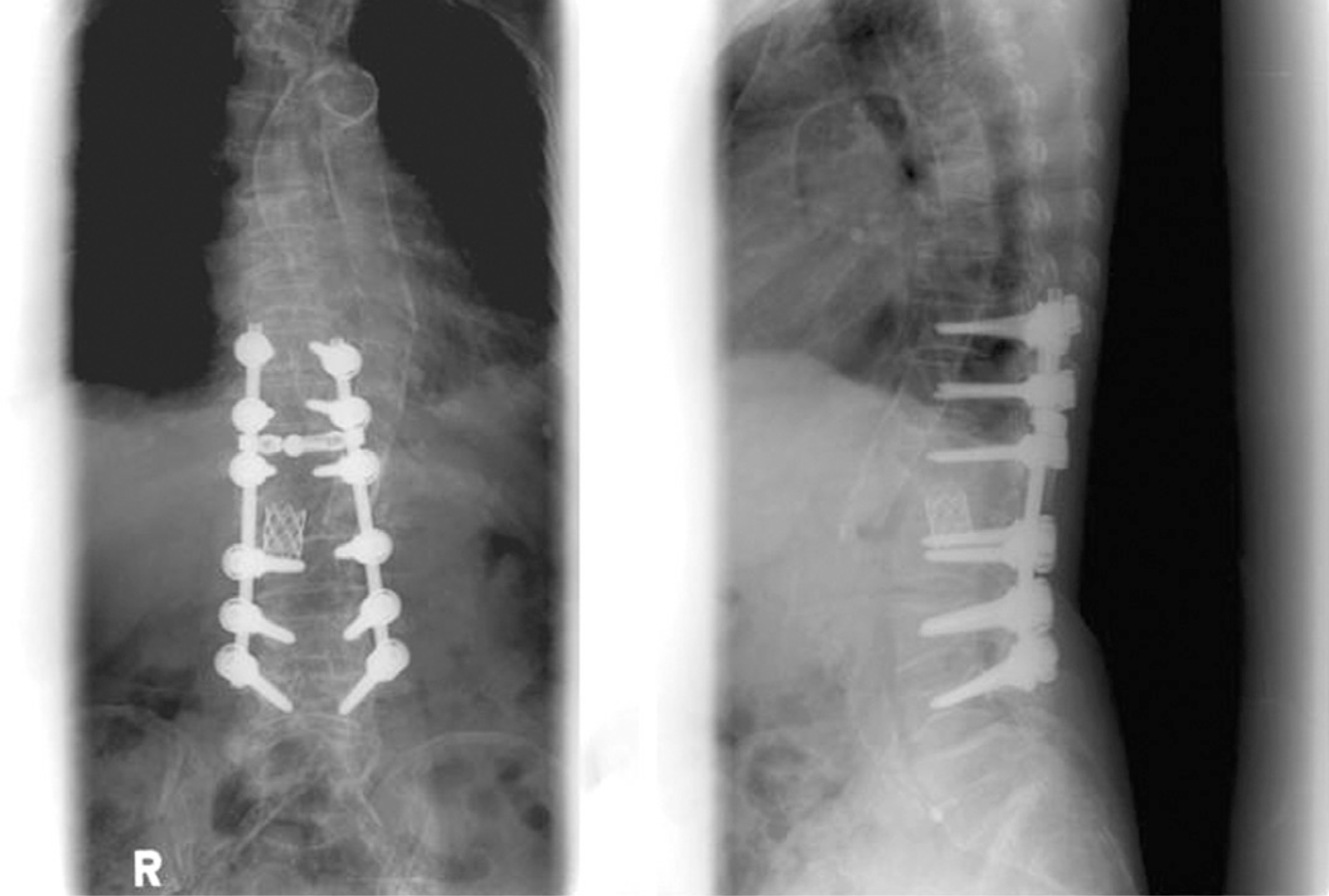
The spinal kyphosis caused by bony ankylosis is ankylosing spondylitis and tuberculous spondylitis. There are some reports on spinal fractures through the fused vertebral body in ankylosing spondylitis, but there is no report of spinal fractures occurring in a fused vertebral body after tuberculous spondylitis. The authors report a case of spinal fracture at the apex of acute angular kyphosis after tuberculous spondylitis, which resulted in a spontaneous correction of kyphosis without neurological deficits. The fracture was stabilized by posterior interbody fusion using a mesh cage after a posterior vertebral column resection and posterolateral fusion.
REFERENCES
1). Suk SI. Spinal surgery 2nd ed. Seoul. Newest Medical Publishing Co;p. 423. 2004.
2). Lonstein JE. Cord compression. (in. Bradford DS, editor. Moe’ textbook of scoliosis and other spinal deformities. 3rd. Philadelphia: WB saunders Co;p. 534–540. 1995.
3). Leatherman KD, Dickson RA. Two-stage corrective surgery for congenital deformities of the spine. J Bone Joint Surg Br. 1979; 61:324–328.

4). Bradford DS, Boachie-Adjei O. One-stage anterior and posterior hemivertebral resection and arthrodesis for congenital scoliosis. J Bone Joint Surg Am. 1990; 72:536–540.

5). Bradford DS, Trbus CB. Vertebral column resection for the treatment of rigid conronal decompensation. Spine. 1997; 22:1590–1599.
Fig. 1.
Radiography (A) and MRI (B) of the thoracolumbar spine taken before injury showed acute angular kyphosis following tuberculous spondylitis.

Fig. 2.
Preoperative radiography after fall down showed spinal fracture through the fused vertebral body following tuberculous spondylitis between T12 and L2, which resulted in spontaneous correction of kyphosis.

Fig. 3.
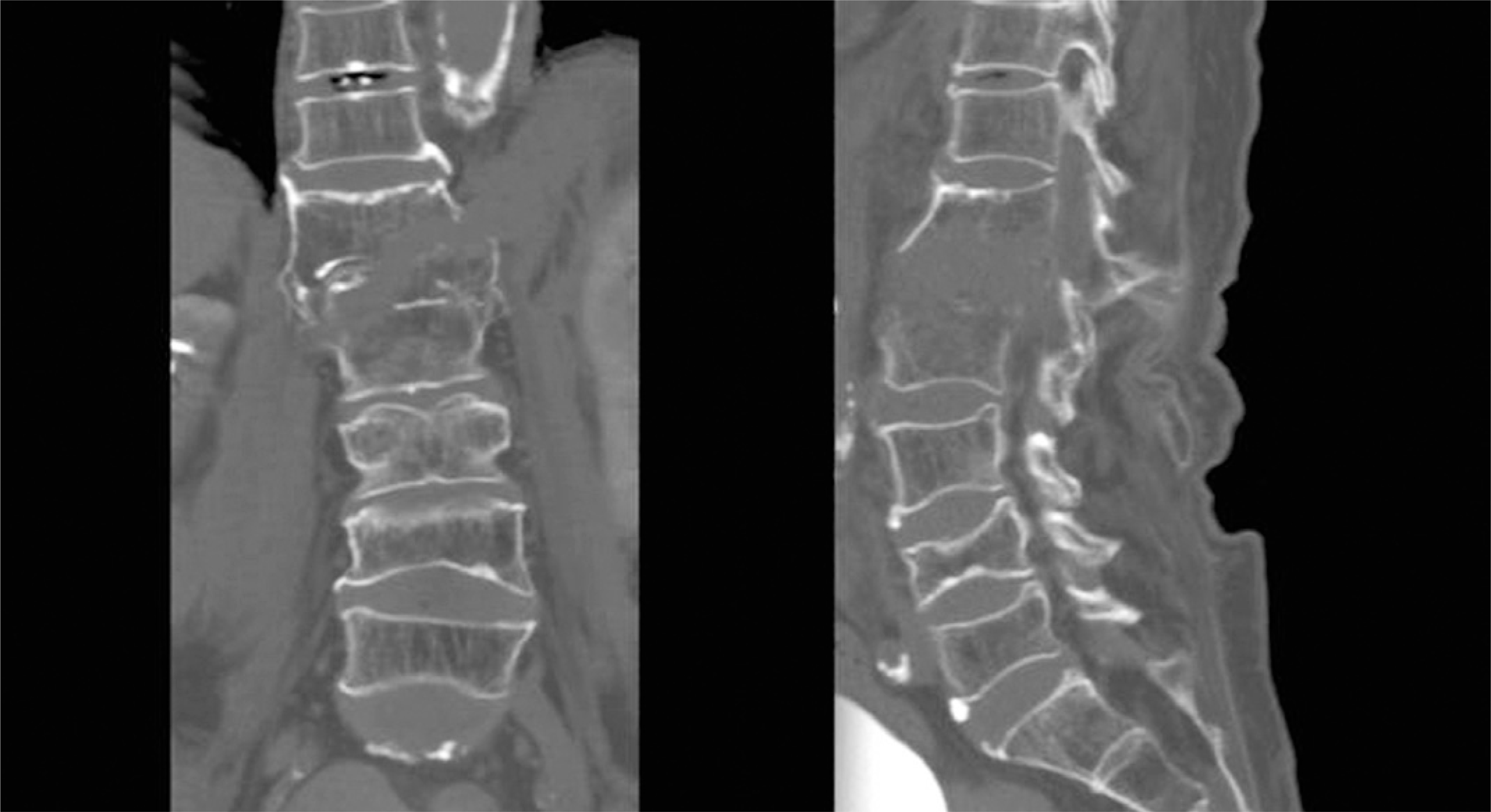
Preoperative CT scan showed bony gap and oblique fracture from right side to left side and lateral displacement.

Fig. 4.
T2-weighted sagittal MR image (A) showed hematoma with mixed signal intensity but no definitive cord discontinuity. Gadolinium enhanced sagittal MR image (B) showed well defined irregular shape hematoma with enhancing lesion.


 PDF
PDF ePub
ePub Citation
 Print
Print


 XML Download