

 PDF
PDF ePub
ePub Citation
 Print
Print


Abstract
Objectives
The aim of this study was to determine if unilateral TLIF is comparable to conventional PLIF with regard to radiologic and clinical outcomes, and to examine the viability of local bone for bone grafting in lumbar interbody fusion.
Summary of Literature Review
TLIF, a modified form of PLIF, is a new spinal fusion technique that avoids the typical complications of PLIF.
Materials and Methods
We analyzed 32 cases of single-level TLIF or PLIF in patients with degenerative or isthmic spondylolisthesis, who were followed for more than 1 year. The patients in group 1 underwent TLIF, and the patients in group 2 underwent PLIF. The fusion rate, changes in disc height, and degree of anterolisthesis in the fused segment were analyzed radiologically. The clinical results were evaluated using the Oswestry Disability Index and visual analog scale. We also analyzed operative time, blood loss, and complications in both groups.
Results
Radiologically and clinically, there were no significant differences between the two groups in terms of fusion rate, changes in disc height, or degree of anterolisthesis in the fused segment. The mean operative time was 200 minutes in group 1 and 240 minutes in group 2. The mean blood loss was 854 ml in group 1 and 1102 ml in group 2(p®0.05).
Go to : 
REFERENCES
1). Cloward RB. Spondylolisthesis: treatment by laminectomy and posterior interbody fusion. Clin Orthop Relat Res. 1981; 154:74–82.
2). Lin PM. Posterior lumbar interbody fusion technique: complications and pitfalls. Clin Orthop Relat Res. 1985; 193:90–102.
3). Suk SI, Lee CK, Kim WJ, Lee JH, Cho KJ, Kim HG. Adding posterior lumbar interbody fusion to pedicle screw fixation and posterolateral fusion after decompression in spondylolytic spondylolisthesis. Spine. 1997; 22:210–219.

4). Evans JH. Biomechanics of lumbar fusion. Clin Orthop Relat Res. 1985; 193:38–46.
5). Humphreys SC, Hodges SD, Patwardhan AG, Eck JC, Murphy RB, Covington LA. Comparison of posterior and transforaminal approaches to lumbar interbody fusion. Spine. 2001; 26:567–571.
6). Okuyama K, Abe E, Suzuki T, Tamura Y, Chiba M, Sato K. Posterior lumbar interbody fusion: a retrospective study of complications after facet joint excision and pedicle screw fixation in 148 cases. Acta Orthop Scand. 1999; 70:329–334.
7). Harms JG, Jeszenszky D. The unilateral, transforaminal approach for posterior lumbar interbody fusion. Orthop Traumatol. 1998; 6:88–99.
8). Hackenberg L, Halm H, Bullmann V, Vieth V, Schneider M, Liljenqvist U. Transforaminal lumbar interbody fusion: a safe technique with satisfactory three to five year results. Eur Spine J. 2005; 14:551–558.
9). Harris BM, Hilibrand AS, Savas PE, et al. Transforaminal lumbar interbody fusion: the effect of various instrumentation techniques on the flexibility of the lumbar spine. Spine. 2004; 29:65–70.
10). Lowe TG, Tahernia AD, O'Brien MF, Smith DA. Unilateral transforaminal posterior lumbar interbody fusion (TLIF): indications, technique, and 2-year results. J Spinal Disord Tech. 2002; 15:31–38.
11). Salehi SA, Tawk R, Ganju A, LaMarca F, Liu JC, Ondra SL. Transforaminal lumbar interbody fusion: surgical technique and results in 24 patients. Neurosurgery. 2004; 54:368–374.
12). Arai Y, Takahashi M, Kurosawa H, Shitoto K. Comparative study of iliac bone graft and carbon cage with local bone graft in posterior lumbar interbody fusion. J Orthop Surg (Hong Kong). 2002; 10:1–7.
13). Miura Y, Imagama S, Yoda M, Mitsuguchi H, Kachi H. Is local bone viable as a source of bone graft in posterior lumbar interbody fusion? Spine. 2003; 28:2386–2389.
14). Trouillier H, Birkenmaier C, Rauch A, Weiler C, Kauschke T, Refior HJ. Posterior lumbar interbody fusion (PLIF) with cages and local bone graft in the treatment of spinal stenosis. Acta Orthop Belg. 2006; 72:460–466.
15). Cse´csei GI, Klekner AP, Dobai J, Lajgut A, Sikula J. Posterior interbody fusion using laminectomy bone and transpedicular screw fixation in the treatment of lumbar spondylolisthesis. Surg Neurol. 2000; 53:2–6.
16). Boden SD, Sumner DR. Biologic factors affecting spinal fusion and bone regeneration. Spine. 1995; 20:102–112.
17). Onesti ST, Ashkenazi E. The Ray Threaded Fusion Cage for posterior lumbar interbody fusion. Neurosurgery. 1998; 42:200–204.
18). La Rosa G, Conti A, Cacciola F, et al. Pedicle screw fixation for isthmic spondylolisthesis: does posterior lumbar interbody fusion improve outcome over posterolateral fusion? J Neurosurg. 2003; 99:143–150.
19). Brantigan JW, Steffee AD, Lewis ML, Quinn LM, Persenaire JM. Lumbar interbody fusion using the Brantigan I/F cage for posterior lumbar interbody fusion and the variable pedicle screw placement system. Spine. 2000; 25:1437–1446.
20). Chastain CA, Eck JC, Hodges SD, Humphreys SC, Levi P. Transforaminal lumbar interbody fusion: a retrospective study of long-term pain relief and fusion outcomes. Orthopedics. 2007; 30:389–392.
21). Houten JK, Post NH, Dryer JW, Errico TJ. Clinical and radiographically/neuroimaging documented outcome in transforaminal lumbar interbody fusion. Neurosurg Focus. 2006; 20:8.
22). Kwon BK, Berta S, Daffner SD, et al. Radiographic analysis of transforaminal lumbar interbody fusion for the treatment of adult isthmic spondylolisthesis. J Spinal Disord Tech. 2003; 16:469–476.
23). Lauber S, Schulte TL, Liljenqvist U, Halm H, Hackenberg L. Clinical and radiologic 2-4-year results of transforaminal lumbar interbody fusion in degenerative and isthmic spondylolisthesis grades 1 and 2. Spine. 2006; 31:1693–1698.
24). Chung SS, Lee CS, Chung KH. Comparison of unilateral and bilateral approaches for posterior lumbar interbody fusion. J of Korean Orthop Assoc. 2004; 39:642–647.
25). Rosenberg WS, Mummaneni PV. Transforaminal lumbar interbody fusion: technique, complications, and early results. Neurosurgery. 2001; 48:569–574.
Go to :
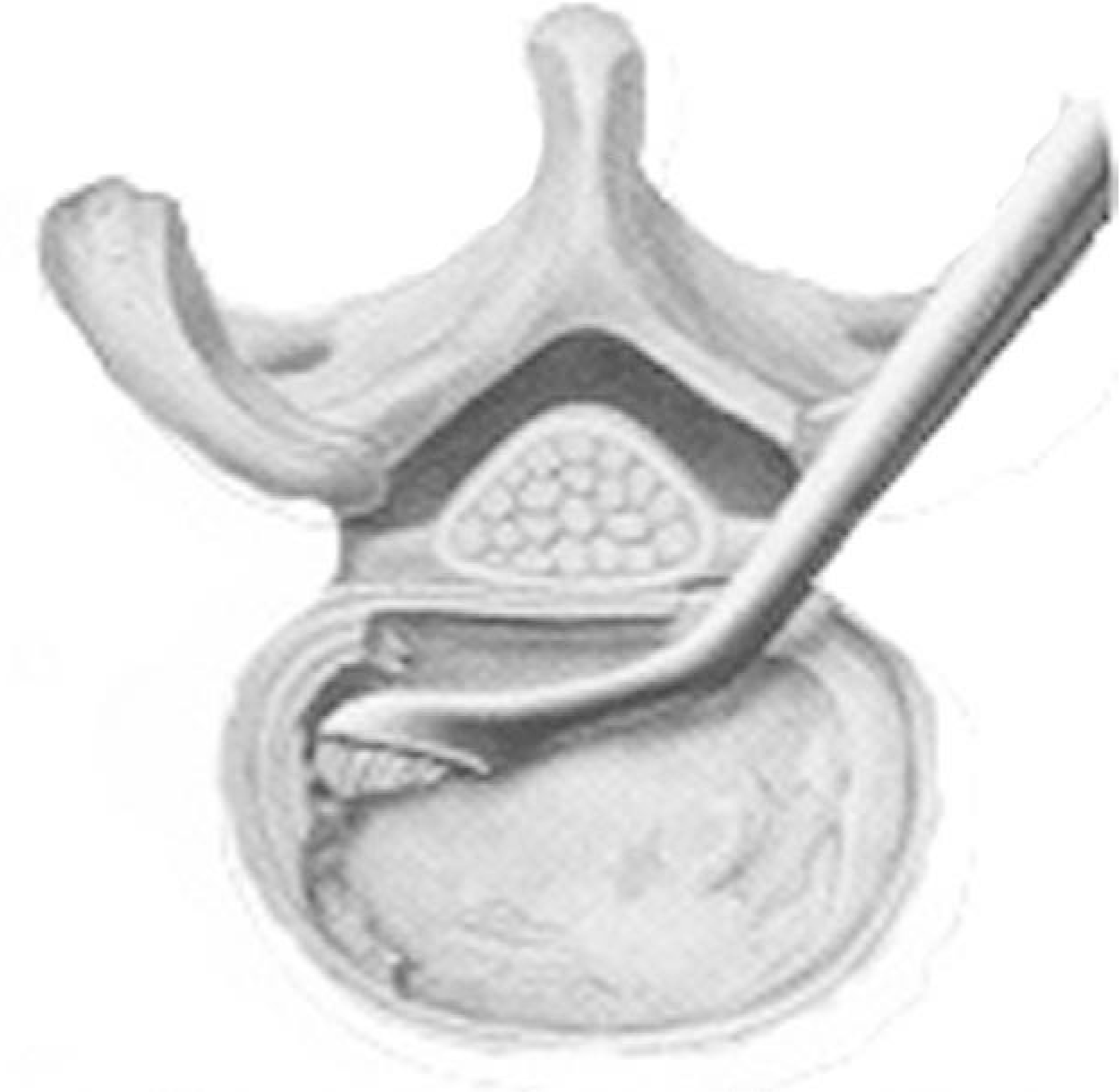 | Fig. 1.Various instruments are utilized to thoroughly clean the disc and cartilaginous endplate from the disc space. Special angled curets are helpful in reaching the contralateral protions of the disc. |
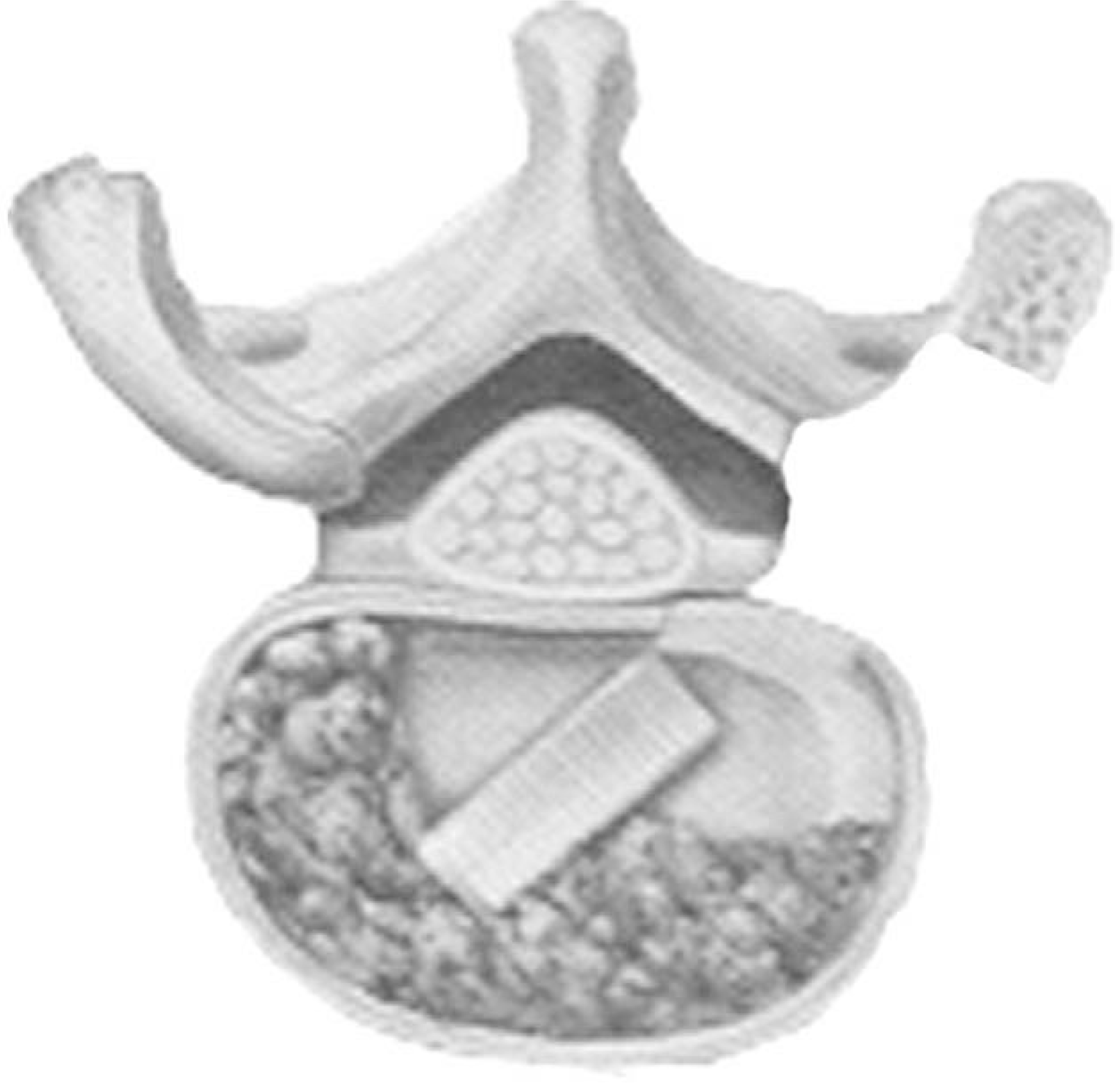 | Fig. 2.A Structural interbody implant is placed into the disc space to support the loads of the spine and promote fusion. In this figure, an oblique implant is shown. |
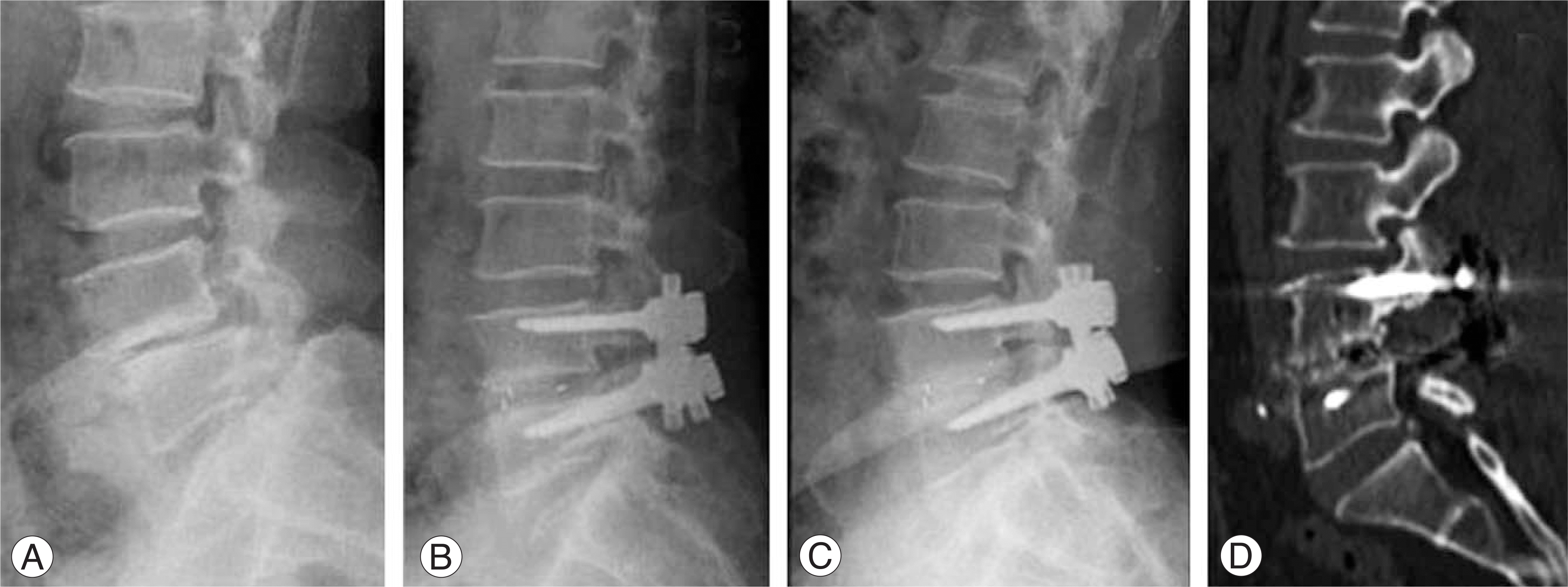 | Fig. 3.Postoperative radiological studies obtained in a 59 year old woman after one level fusion for spondylolithesis. (A) Preoperative lateral radiograph show narrowing of L4-5 disc space and anterior translation of L4 body. (B) Postoperative lateral radiograph shows restoration of the intervertebral disc height and reduction of anterolisthesis. (C) Lateral radiography obtained 12 months postoperatively. Note that the intervertebral height and reduction of anterolisthesis is well preserved. Note also that the bone density the entire disc space is well preserved, indicating probable progression toward solid arthrodesis. (D) Sagittal reconstructed CT scan obtained at 7 months postoperatively, demonstrating excellent interbody fusion with bone columns. |
Table 1.
Patient Data
Table 2.
Radiological Results
 XML Download