Abstract
Objective
To compare one and two-caged posterior lumbar interbody fusion (PLIF) with local bone grafting for spondylolisthesis.
Summary of Literature Review
Even though there are many reports on PLIF using cages and local bone grafting, Studies comparing one and two-caged PLIFs are rare.
Materials and Methods
Sixty-three patients who underwent pedicle screw fixated PLIF using cages and local bone grafts were followed for more than 1 year. Twenty-five patients had one cage (group I), and 38 patients had two cages (group II). Sampling error, disc height, sagittal Cobb angle, coronal Cobb angle, fusion rate, Oswestry disability index (ODI), operation time, blood loss, and neurologic complications were assessed.
Results
There was no sampling error between the two groups, except with regard to diagnosis: degenerative spondylolisthesis, 15 cases in group I and 9 cases in group II; spondylolytic spondylolisthesis, 10 cases in group I and 29 cases in group II (p=0.004). Fusion rates were 87.5% and 88.2% for groups I and II, respectively (p=1.000). More disc height loss occurred in group I (0.6 mm) than in group II (0.0 mm) (p=0.041). Over-3mm-disc height-losses were noted more frequently in group I (20%) than in group II (2.6%) (p=0.022). ODI improved from 28.1 to 12.3 (72.1% improvement) in group I and from 29.2 to 12.7 (79.3% improvement) in group II. There were no significant differences in operation time, amount of blood loss, or neurologic complications between the two groups.
Conclusion
Unilateral one-caged PLIF with local bone grafting and posterior instrumentation was no different from bilateral two-caged PLIF with regard to fusion rates or radiologic or clinical results. The statistically significant differences in disc height seemed to be clinically insignificant. Disc height loss of greater than 3 mm was much more common in group I, with one-caged PLIF.
Go to : 
REFERENCES
1). Okuyama K, Kido T, Unoki E, Chiba M. PLIF with a titanium cage and excised facet joint bone for degenerative spondylolisthesis in augmentation with a pedicle screw. J Spinal Disord Tech. 2007; 20:53–59.
2). Miura Y, Imagama S, Yoda M, Mitsuguchi H, Kachi H. Is local bone viable as a source of bone graft in posterior lumbar interbody fusion? Spine. 2003; 28:2386–2389.

3). Nah KH, Shin JH, Choi NY, Lee YS, Ha KY. The effect of posterior lumbar interbody fusion after posterolateral fusion in degenerative spondylolisthesis. J Korean Orthop Assoc. 2005; 40:852–860.

4). Brantigan JW, Neidre A, Toohey JS. The lumbar I/F cage for posterior lumbar interbody fusion with the variable screw placement system: 10-year results of a Food and Drug Administration clinical trial. Spine. 2004; 4:681–688.

5). Enker P, Steffee AD. Interbody fusion and instrumentation. Clin Orthop Relat Res. 1994; 300:90–101.

6). Mummaneni PV, Haid RW, Rodts GE. Lumbar interbody fusion: state-of-the-art technical advances. J Neurosurg Spine. 2004; 1:24–30.

7). Wang ST, Goel VK, Fu CY, et al. Comparison of two interbody fusion cages for posterior lumbar interbody fusion in a cadaveric model. Int Orthop. 2006; 30:299–304.

8). Brantigan JW. Pseudarthrosis rate after allograft posterior lumbar interbody fusion with pedicle screw and plate fixation. Spine. 1994; 19:1271–1279.

9). Csecsei GI, Klekner AP, Dobai J, Lajgut A, Sikula J. Posterior interbody fusion using laminectomy bone and transpedicular screw fixation in the treatment of lumbar spondylolisthesis. Surg Neurol. 2000; 53:2–6.
10). Ahn DK, Jeong KW, Lee Song, Choi DJ, Cha SK. Posterior lumbar interbody fusion with chip bone and pedicle screw fixation-comparative study between local chip bone graft and autoiliac chip bone graft. J Korean Orthop Assoc. 2004; 39:614–620.
11). Kai Y, Oyama M, Morooka M. Posterior lumbar interbody fusion using local facet joint autograft and pedicle screw fixation. Spine. 2004; 29:41–46.

12). Vadapalli S, Robon M, Biyani A, Sairyo K, Khandha A, Goel VK. Effect of lumbar interbody cage geometry on construct stability: a cadaveric study. Spine. 2006; 31:2189–2194.

13). Asazuma T, Masuoka K, Motosuneya T, Tsuji T, Yasuoka H, Fujikawa K. Posterior lumbar interbody fusion using dense hydroxyapatite blocks and autogenous iliac bone: clinical and radiographic examinations. J Spinal Disord Tech(Suppl). 2005; 18:41–47.
14). Closkey RF, Parsons JR, Lee CK, Blacksin MF, Zimmerman MC. Mechanics of interbody spinal fusion. Analysis of critical bone graft area. Spine. 1993; 18:1011–1015.
15). Prolo DJ, Oklund SA, Butcher M. Toward uniformity in evaluating results of lumbar spine operations. A paradigm applied to posterior lumbar interbody fusions. Spine. 1986; 11:601–606.
16). Fogel GR, Toohey JS, Neidre A, Brantigan JW. Is one cage enough in posterior lumbar interbody fusion: a comparison of unilateral single cage interbody fusion to bilateral cages. J Spinal Disord Tech. 2007; 20:60–65.

18). Harris BM, Hilibrand AS, Savas PE, Pellegrino A, Siegler S, Albert TJ. Transforaminal lumbar interbody fusion: the effect of various instrumentation techniques on the flexibility of the lumbar spine. Spine. 2004; 29:65–70.
19). Heth JA, Hitchon PW, Goel VK, Rogge TN, Drake JS, Torner JC. A biomechanical comparison between anterior and transverse interbody fusion cages. Spine. 2001; 26:261–267.

20). Godde S, Fritsch E, Dienst M, Kohn D. Influence of cage geometry on sagittal alignment in instrumented posterior lumbar interbody fusion. Spine. 2003; 28:1693–1699.

21). Okuyama K, Abe E, Suzuki T, Tamura Y, Chiba M, Sato K. Influence of bone mineral density on pedicle screw fixation: a study of pedicle screw fixation augmenting posterior lumbar interbody fusion in elderly patients. Spine. 2001; 1:402–407.
Go to : 
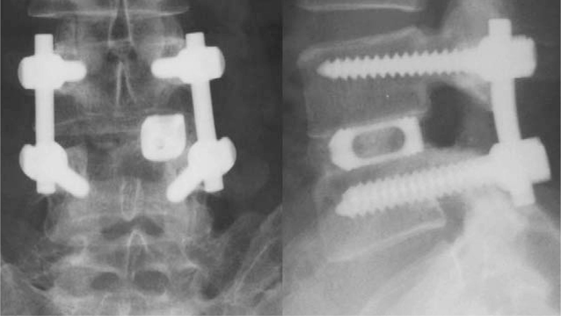
 | Fig. 1.Complete fusin, with solid bony mass around a cage at anteroposterior and lateral view |
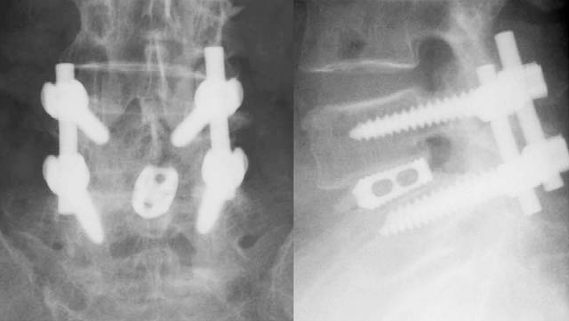
 | Fig. 2.Probable fusion, with solid bony mass in a cage with small amount of resorption at lateral view. |
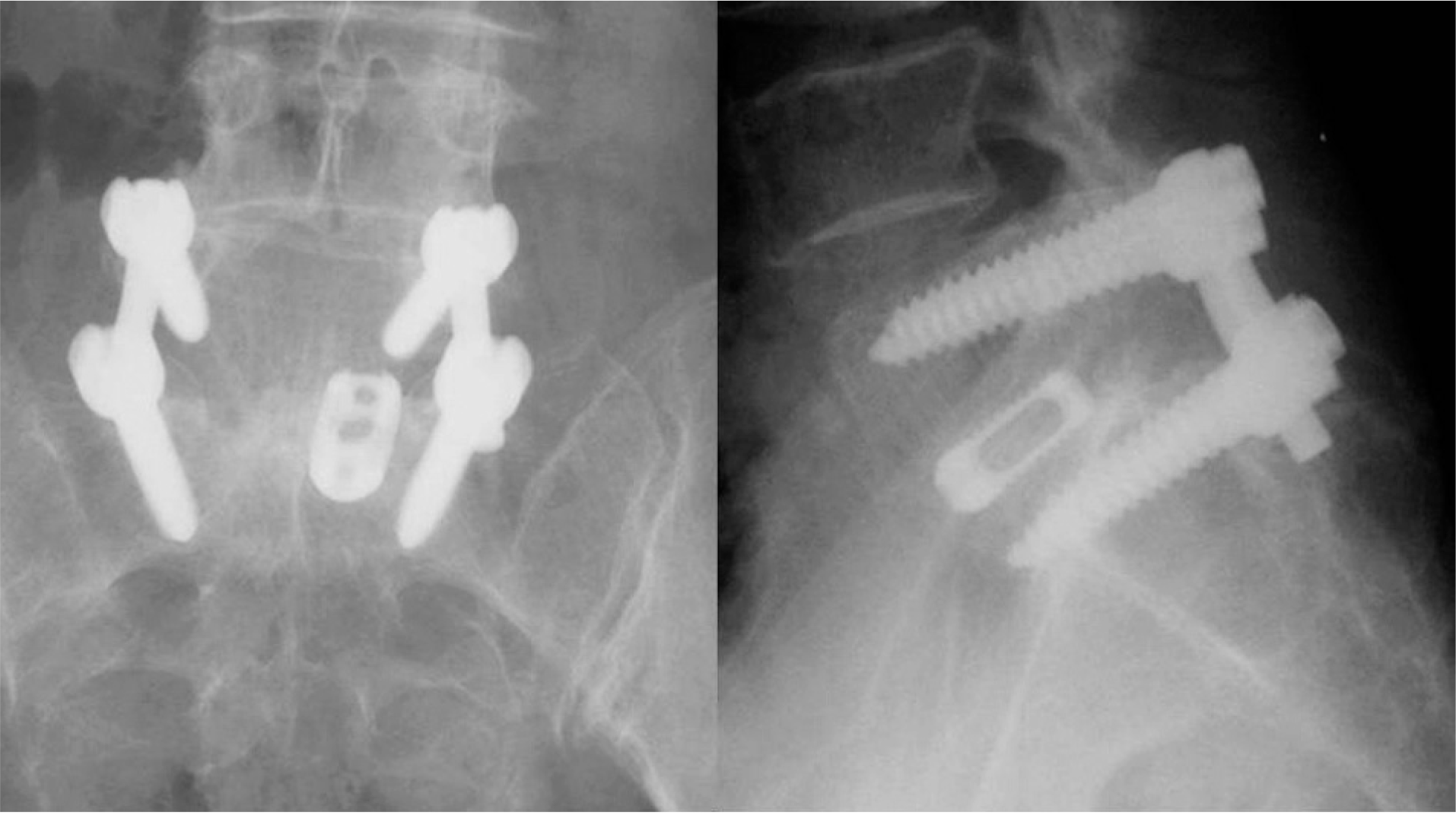
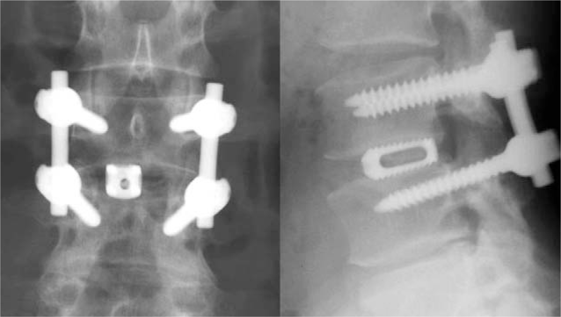
 | Fig. 4.Undecipherable, with insufficient radiologic information, masked by iliac or sacral bone. |
Table 1.
Radiologic classification
Table 2.
Demography of sampling errors
| Group I | Group II | Significance (p value) | |
|---|---|---|---|
| No. of Patients | 25 | 38 | |
| Age (years) | 56.6±7.92 | 57.3±9.62 | 0.593 |
| Sex (M/F) | 3/22 | 11/43 | 0.495∗ |
| Medical disease | |||
| Diabetes | 2 | 14 | 0.738∗ |
| Thyroid disease | 0 | 12 | 0.244∗ |
| Smoking | 1 | 18 | 0.058∗ |
| Follow-up (months) | 23.2±11.5 | 21.3±10.2 | 0.438∗ |
| Diagnosis (Spondylolisthesis) | 0.004∗ | ||
| Spondylolytic | 10 | 29 | |
| Degenerative | 15 | 19 | |
| Fusion level | 0.710∗ | ||
| L3-4 | 11 | 12 | |
| L4-5 | 15 | 26 | |
| L5-S1 | 19 | 10 | |
| Operation times (minutes) | 197±282 | 211±362 | 0.082∗ |
| Blood loss (ml) | 575±215 | 615±228 | 0.485∗ |
| Neurologic complication | 10 | 10 | - |
Table 3.
Radiological and clinical results
| Group I | Group II | Significance (p value) | |
|---|---|---|---|
| Decipherable | 24 | 34 | 0.933∗ |
| Fusion, complete | 16(66.7%) | 23(67.6%) | |
| Fusion, probable | 15(20.8%) | 7(20.6%) | |
| Resorption | 13(12.5%) | 4(11.8%) | |
| Undecipherable Oswestry disability index | 1 | 4 | |
| Preoperative | 28.1±4.91 | 129.2±4.01 | 0.364∗ |
| Postoperative | 12.3±3.61 | 112.7±3.81 | 0.768∗ |
| Recovery rate (%) Disc height (mm) | 72.1 | 79.3 | |
| Preoperative | 10.0±2.51 | 19.1±3.1 | 0.287∗ |
| Postoperative | 12.6±1.81 | 12.0±1.4 | 0.083∗ |
| Follow-up | 11.9±1.61 | 112.±1.4 | 0.947∗ |
| Reduction loss | 0.7±1.3 | 10.0±1.4 | 0.038∗ |
| Frequency of disk height loss more than 3 mm Coronal Cobb angle (degree) | 5(20.0%) | 1(2.6%) | 0.022∗ |
| Preoperative | 0.2±2.9 | 10.8±3.7 | 0.417∗ |
| Postoperative | 0.4±2.5 | 10.0±2.6 | 0.357∗ |
| Follow-up | 0.3±2.8 | 10.2±2.9 | 0.734∗ |
| Reduction loss Frequency of Coronal Cobb angle | 0.1±2.3 | 10.1±1.9 | 0.280∗ |
| loss more than 5 degrees Sagittal Cobb angle (degree) | 2(8.0%) | 1(2.6%) | 0.328∗ |
| Preoperative | 10.2±5.01 | 19.5±7.2 | 0.746∗ |
| Postoperative | 12.3±3.91 | 12.9±4.5 | 0.843∗ |
| Follow-up | 11.9±3.6 | 12.6±4.6 | 0.860∗ |
| Reduction loss | 0.4±1.4 | 10.2±1.4 | 0.746∗ |
| Frequency of Sagittal Cobb angle loss more than 5 degrees | 5(20.0%) | 7(18.4%) | 0.876∗ |
Table 4.
Results of variable graft-assemblies
| Graft-assembly | Authors | Fusion rate | Clinical result | Uniqueness |
|---|---|---|---|---|
| Allograft | Brantigan JW8) | 56% | 60% | Graft failure 28% |
| Local block bone | Csecsei GI9) | 95.7% | 87% | Segmental lordosis loss 2 degrees |
| Iliac chip bone | Ahn DK10) | 90% | 88.1% | Donor site pain 12% |
| Local chip bone & local block bone | Kai Y11) | 92.9% | 76% | Collapsed union 16.7% |
| Local chip bone | Shin BJ12) | 57% | 79% | Difficult radiologic decision 17% |
| 1 cage with local bone | Okuyama K1) | 100% | 86% (Pain) | Union 93.5% |
| 62% (Work) | Probable union 6.3% | |||
| 2 cages with local bone | Miura Y2) | 100% | 76.8% | ASD∗ 2 cases |
| Hydroxyapatite block | Asazuma T13) | 96.2% | - | Sinking 23.5% |
| & Iliac bone block | Cracking of HA block 17.6% |


 PDF
PDF ePub
ePub Citation
 Print
Print


 XML Download