Abstract
Patients with Crowned dens syndrome typically present with severe neck pain and have calcification around the axial odontoid process on radiographs. To our knowledge, Crowned dens syndrome is unreported in the Korean literature and the clinical features remain unclear. We present Crowned dens syndrome as a cause of acute cervical pain and review the literature.
REFERENCES
01). Bouvet JP., Ie Parc JM., Michalski B., Benlahrache C., Anquier L. Acute neck pain due to calcifications surrounding the odontoid process: the crowned dens syndrome. Arthritis Rheum. 1985. 28:1417–1420.

02). Malca SA., Roche PH., Pellet W., Combalbert A. Crowned dens syndrome: a manifestation of hydroxyapatite rheumatism. Acta Neurochir (Wien). 1995. 135:126–130.

03). Aouba A., Vuillemin-Bodaghi V., Mutschler C., De Bandt M. Crowned dens syndrome misdiagnosed as polymyalgia rheumatica, giant cell artertitis, meningitis or spondylitis: an analysis of eight cases. Rheumatology. 2004. 43:1508–1512.
04). Wu DW., Reginato AJ., Torriani M., Robinson DR., Reginato AM. The crowned dens syndromes as a cause of neck pain: report of two new cases and review of the literature, Arthritis Rheum. 2005. 53:133–137.
05). Goto S., Umehara J., Aizawa T., Kokubun S. Crowned Dens syndrome. J Bone Joint Surg Am. 2007. 12:2732–2736.

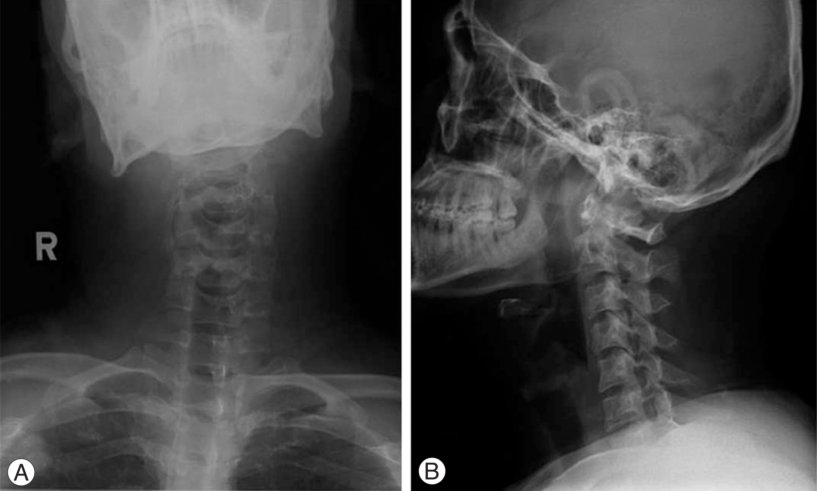
Fig. 1.
(A) Anteroposterior radiographs of the cervical spine showed scoliosis of cervical spine. (B) Lateral radiographs of the cervical spine.

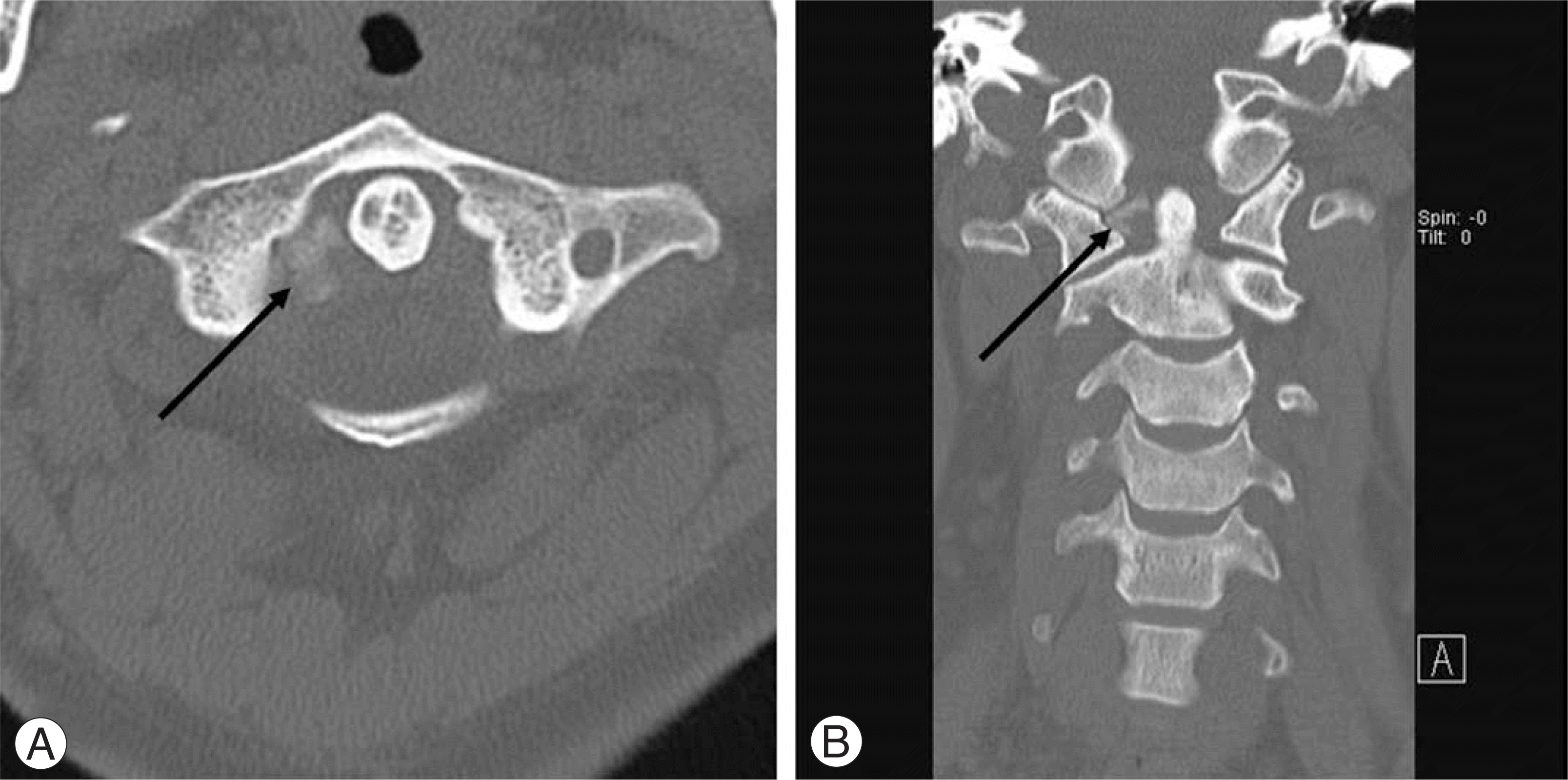
Fig. 2.
Patterns of calcifications according to localization and extent in relation to the odontoid process of the axis on computed tomography scans. (A) The axial view of computed tomography scans showed multiple calcifications (0.8×1.2×1.0 cm) in right lateral aspect of odontoid process of C2. (B) The coronal view of computed tomography scans showed calcifications (0.8 ×1.2×1.0 cm) in right lateral aspect of odontoid process of C2.


 PDF
PDF ePub
ePub Citation
 Print
Print


 XML Download