

 PDF
PDF ePub
ePub Citation
 Print
Print


Abstract
Objectives
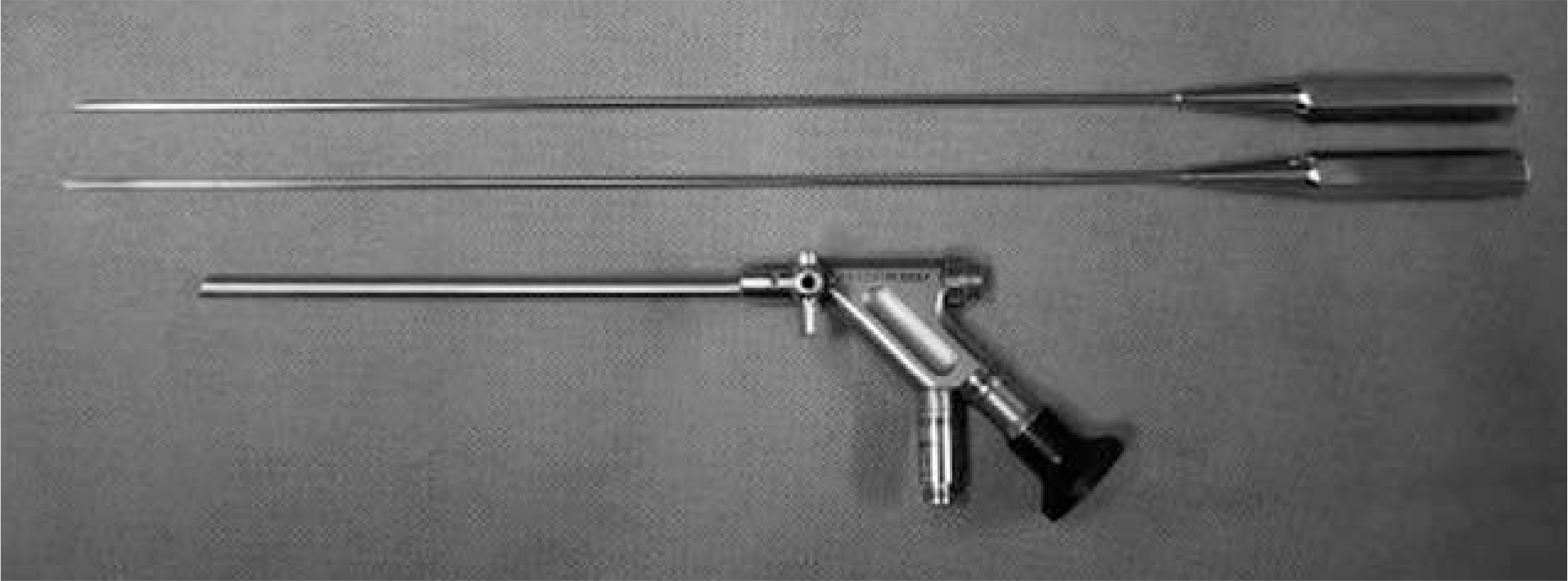
This study examined the post-operative results of interlaminar percutaneous endoscopic lumbar discectomy (PELD) with or without endoscopic laminotomy in lumbar disc herniation.
Summary of Literature Review
In addition to the technical feasibility, the indications of PELD surgery are usually the same as those for open discectomy.
Materials and Methods
From January 2005 to August 2006, 62 cases treated with PELD using an interlaminar approach due to lumbar disc herniation were examined. The mean age of the subjects was 40.1 years (18-70) and the mean follow up period was 32.6 months (24-44). Thirty-four and 28 herniated discs were extracted from L4-L5 and L5-S1, respectively. The clinical results were evaluated using MacNab's criteria.
Results
The herniated discs were accessible in all cases. Excellent and good results were obtained in 85% (53 cases) of patients but 15% of patients (9 cases) showed unsatisfactory results or needed revision. There were 4 cases of incomplete removal, 2 cases of recurrence and 4 cases of persistent low back pain due to associated degenerative pathologies. Additional surgery was required in 7 cases which were open discectomy in 5 cases (3 cases of 4 incomplete removal and 2 of recurrence). There was one case of PLIF and 1 additional decompression. Cauda equina syndrome occurred in one case who underwent subsequent wide decompression and open discectomy.
Go to : 
REFERENCES
01). Isaacs RE., Podichetty V., Fessler RG. Microendoscopic discectomy for recurrent disc herniations. Neurosurg Focus. 2003. 15:11.

02). Lee DY., Ahn Y., Lee SH. Percutaneous endoscopic lumbar discectomy for adolescent lumbar disc herniation: surgical outcomes in 46 consecutive patients. Mt Sinai J Med. 2006. 73:864–870.
03). Wu X., Zhuang S., Mao Z., Chen H. Microendoscopic discectomy for lumbar disc herniation: surgical technique and outcome in 873 consecutive cases. Spine. 2006. 31:2689–2694.
04). Nakagawa H., Kamimura M., Uchiyama S., Takahara K., Itsubo T., Miyasaka T. Microendoscopic discectomy (MED) for lumbar disc prolapse. J Clin Neurosci. 2003. 10:231–235.
05). Ozturk C., Tezer M., Aydogan M., Sarier M., Hamzaoglu A. Posterior endoscopic discectomy for the treatment of lumbar disc herniation. Acta Orthop Belg. 2006. 72:347–352.
06). Kim JM., Lee SH., Ahn Y., Yoon DH., Lee CD., Lim ST. Recurrence after successful percutaneous endoscopic lumbar discectomy. Minim Invasive Neurosurg. 2007. 50:82–85.
07). Mayer HM., Brock M. Percutaneous endoscopic lumbar discectomy (PELD). Neurosurg Rev. 1993. 16:115–120.
08). Mayer HM., Brock M. Percutaneous endoscopic discectomy: surgical technique and preliminary results compared to microsurgical discectomy. J Neurosurg. 1993. 78:216–225.
09). Yeung AT., Tsou PM. Posterolateral endoscopic excision for lumbar disc herniation: Surgical technique, outcome, and complications in 307 consecutive cases. Spine. 2002. 27:722–731.
10). Ruetten S., Komp M., Merk H., Godolias G. Full-endoscopic interlaminar and transforaminal lumbar discectomy versus conventional microsurgical technique: a prospective, randomized, controlled study. Spine. 2008. 33:931–939.
11). Ruetten S., Komp M., Godolias G. A new full-endoscopic technique for the interlaminar operation of lumbar disc herniations using 6 mm endoscopes: prospective 2-year results of 331 patients. Minim Invas Neurosurg. 2006. 49:80–87.
12). Kim EH., Shin DH., Cha JS., Jae YB. Factors affecting learning curve in endoscopic lumbar discectomy using interlaminar approach. J Kor Soc Spine Surg. 2006. 13:311–318.
13). Kambin P., Zhou L. History and current status of percutaneous arthroscopic disc surgery. Spine. 1996. 21:57–61.
14). Schreiber A., Suezawa Y., Leu H. Does percutaneous nucleotomy with discoscopy replace conventional discectomy Eight years of experience and results in treatment of herniated lumbar disc. Clin Orthop Relat Res. 1989. 238:35–42.
15). Yeung AT., Tsou PM. Posterolateral endoscopic excision for lumbar disc herniation: Surgical technique, outcome, and complications in 307 consecutive cases. Spine. 2002. 27:722–731.
16). Ruetten S., Komp M., Godolias G. An extreme lateral access for the surgery of lumbar disc herniations inside the spinal canal using the full-endoscopic uniportal transforaminal approach. Technique and prospective results of 463 patients. Spine. 2005. 30:2570–2578.
17). De Antoni DJ., Claro ML., Poehling GG., Hughes SS. Translaminar lumbar epidural endoscopy: Anatomy, technique, and indications. Arthroscopy. 1996. 12:330–334.
18). Chiu JC., Clifford TJ., Betterjee KA., Princenthal RA. Extradural transpinal percutaneous L5-S1 endoscopic dis-ectomy, the practice of minimally invasive spinal technique. American Academy of Minimally Invasive Spinal Medicine and Surgery, Lima, CSS,. 2000. 227–230.
19). Ebraheim NA., Miller RM., Xu R., Yeasting RA. The location of the intervertebral lumbar disc on the posterior aspect of the spine. Surg Neurol. 1997. 48:232–236.
Go to :
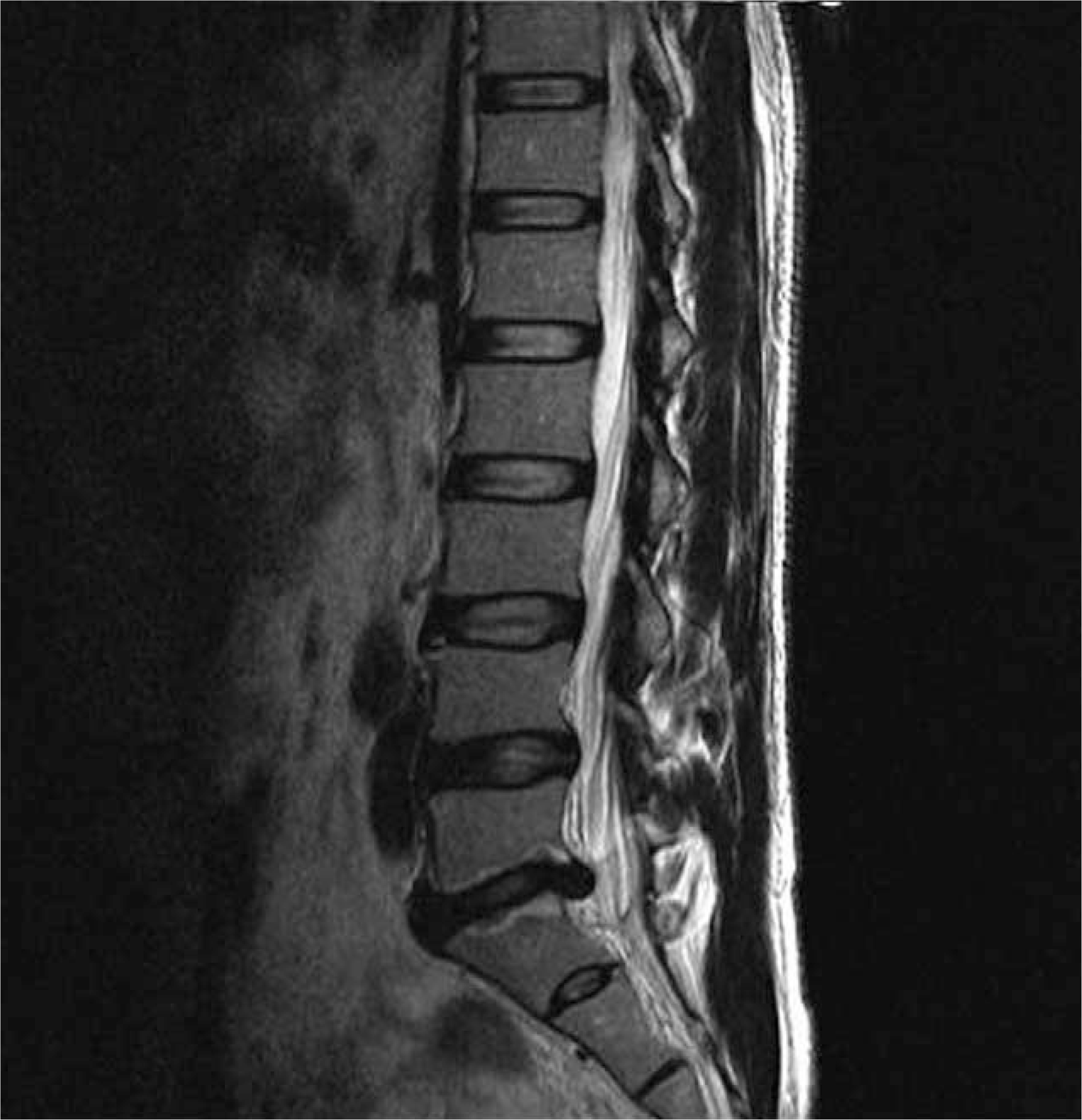 | Fig. 2.L5-S1 disc herniation with advanced disc degeneration and modic stage 1 change. This patient complaint of persistant back pain after PELD. |
 | Fig. 3.(A) Central disc herniation at L5-S1. (B) cauda equine syndrome developed after PELD due to excessive traction and protrusion of incompletely removed disc. |
Table 1.
Macnab classification
Table 2.
Poor result cases after percutaneous endoscopic lumbar discectomy (PELD) using interlaminar approach
 XML Download