

 PDF
PDF ePub
ePub Citation
 Print
Print


Abstract
Objective
To analyze the treatment results of vertebroplasty in patients who suffered osteoporotic compression fractures during conservative treatments for pre-existing degenerative lumbar disease.
Summary and Literature Review
Whilst spinal fusion has shown satisfactory clinical results, solid fusion has been reported to accelerate the degenerative changes at the unfused adjacent levels. Therefore, the level of spinal fusion in patients with compression fractures and pre-existing degenerative lumbar disease is controversial. Few studies have evaluated the outcomes of spinal fusion and adjacent segment vertebroplasty.
Materials and Methods
A retrospective review was carried out on 28 patients who suffered the osteoporotic compression fractures during conservative treatment for pre-existing degenerative lumbar disease. Posterolateral fusion and vertebroplasty were performed for degenerative disease and compression fractures. The average fusion level was 1.82. The mean compressed vertebral bodies were 1.68. The radiology results were evaluated to determine the progression of the compression rate and fractures in the adjacent segment. The clinical results were evaluated using the Denis pain scale for compression fractures and Katz satisfaction scale for degenerative lumbar disease.
Results
The average compression rate was 30.2% preoperatively, 21.4% postoperatively, and 24.6% at the final follow-up. There was no fracture in the adjacent segment. Clinically, the preoperative Denis score was P3 and P4 in 8 and 20 patients, respectively. On the other hand, the postoperative Denis score was P1, P2 and P3 in 8, 19 and 1 patients, respectively. In regard to degenerative diseases, the overall satisfaction was 82.1%.
Conclusion
The stability of fracture sites in vertebroplasty of patients with pre-existing lumbar disease was confirmed. However, further compression of the fractured vertebral body was observed after vertebroplasty in long fusion. Therefore, a followup study of more cases will be necessary to confirm the changes in the vertebroplasty site.
Go to : 
REFERENCES
01). Acosta FL Jr., Aryan HE., Taylor WR., Ames CP. Kyphoplasty-augmented short-segment pedicle screw fixation of traumatic lumbar burst fractures: initial clinical experience and literature review. Neurosurg Focus,. 2005. 18:9–14.

02). Galibert P., Deramond H., Rosat P., Le Gars D. Preliminary note on the treatment of vertebral angioma by percutaneous acrylic vertebroplasty. Neurochirurgie,. 1987. 33:166–168.
03). Jensen ME., Evans AJ., Mathis JM., Kallmes DF., Cloft HJ., Dion JE. Percutaneous polymethylmethacrylate vertebroplasty in the treatment of osteoporotic vertebral body compression fractures: technical aspects. Am J Neuroradiol,. 1997. 18:1897–1904.
04). Kang JD., Kim KY., Park SI. The effect of percutaneous vertebroplasty with bone cement in the treatment of osteoporotic thoracolumbar compression fracture. J Korean Fracture Soc,. 2001. 14:265–271.
05). Kim CW., Choi YJ., Baek SK, et al. Vertebroplasty on osteoporotic compression fracture. J Korean Fracture Soc,. 2002. 15:123–128.
06). Keller TS., Harrison DE., Colloca CJ., Harrison DD., Janik TJ. Prediction of osteoporotic spinal deformity. Spine,. 2003. 28:455–462.
07). Cortet B., Roches E., Logier R, et al. Evaluation of spinal curvatures after a recent osteoporotic vertebral fracture. Joint Bone Spine,. 2002. 69:201–208.
08). Chen CS., Cheng CK., Liu CL., Lo WH. Stress analysis of the disc adjacent to interbody fusion in lumbar spine. Med Eng Phys,. 2001. 23:483–491.
09). Schlegel JD., Smith JA., Schleusener RL. Lumbar motion segment pathology adjacent to thoracolumbar, lumbar, and lumbosacral fusions. Spine,. 1996. 21:970–981.
10). Cotten A., Dewatre F., Cortet B, et al. Percutaneous vertebroplasty for osteolytic metastases and myeloma: effects of the percentage of lesion filling and the leakage of methyl methacrylate at clinical follow-up, Radiology. 1996. 200:525–530.
11). Jensen ME., Dion JE. Percutaneous vertebroplasty in the treatment of osteoporotic compression fractures, Neuroimaging Clin N Am. 2000. 10:547–568.
12). Belkoff SM., Mathis JM., Jasper LE., Deramond H. The biomechanics of vertebroplasty. The effect of cement volume on mechanical behavior, Spine. 2001. 26:1537–1541.
13). Uppin AA., Hirsch JA., Centenera LV., Pfiefer BA., Pazianos AG., Choi IS. Occurrence of new vertebral body fracture after percutaneous vertebroplasty in patients with osteoporosis, Radiology. 2003. 226:119–124.
Go to :
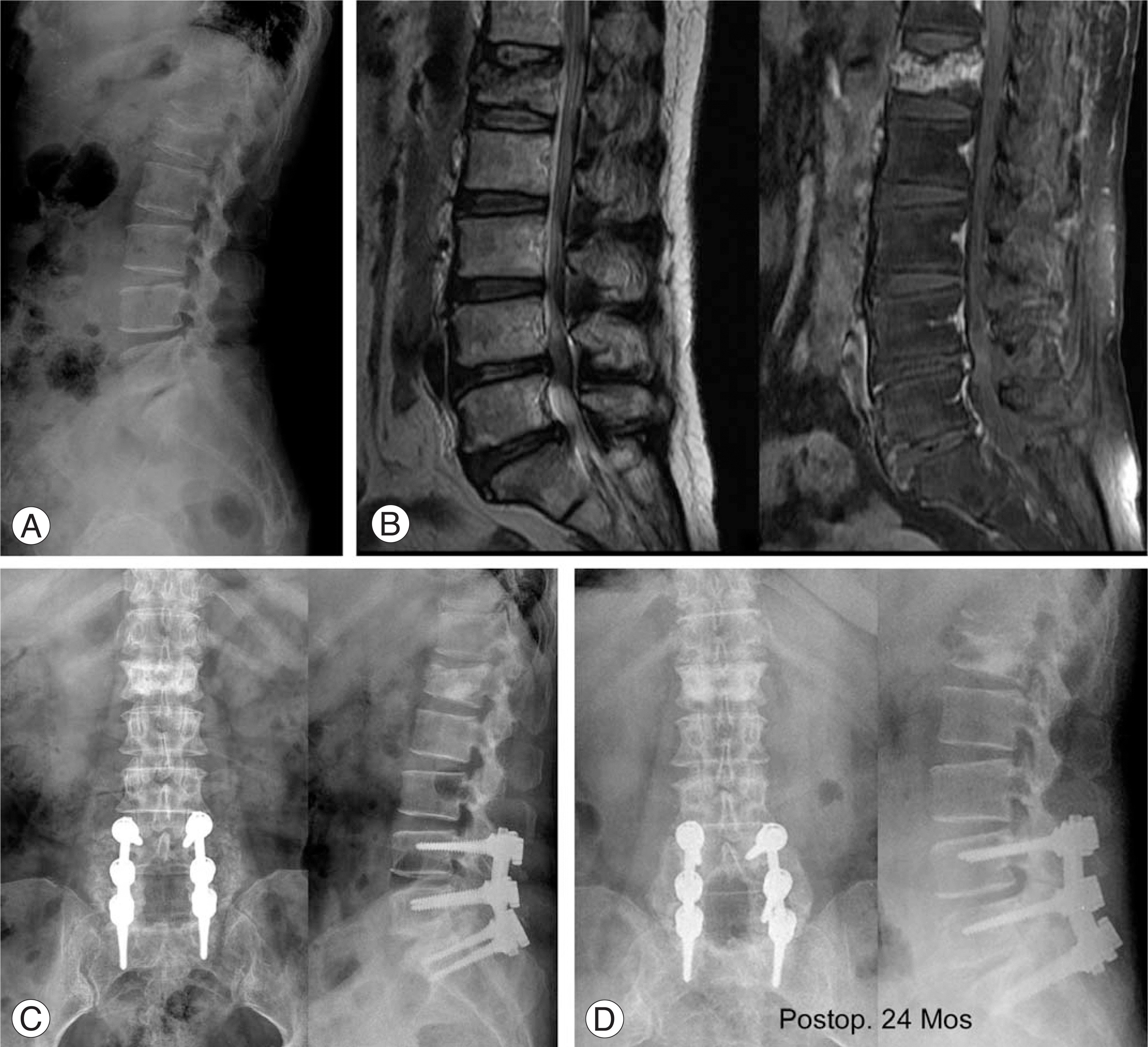 | Fig. 1.59-year old female (A) She is admitted with low back pain radiating to low extremities. Initial x-ray shows severe degenerative changes and osteoporotic compression fracture on L1. (B) MRI show degenerative spinal stenosis on lower lumbar region and osteoporotic compression fracture on L1. (C) We perform multiple decompression, transpedicular fixation, poterolateral fusion from L4-S1 and vertebroplasty on L1. (D) In postoperative 24months, follow-up x-rays show bony union, no changes of adjacent segment on L4-S1 and good augmentation of the fractured vertebral body on L1. |
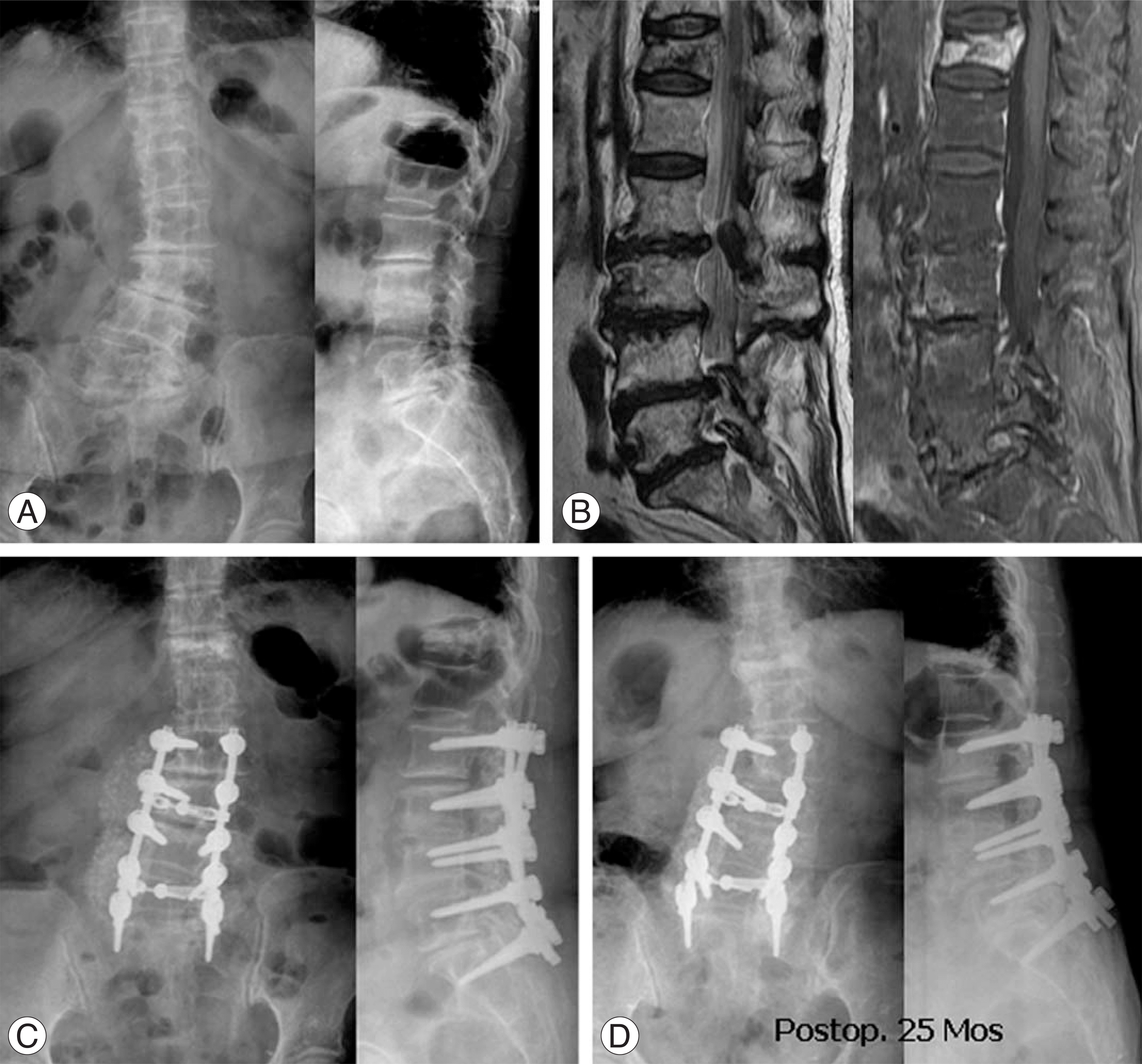 | Fig. 2.76-year old female (A) She is admitted with low back pain radiating to low extremities. Initial x-ray shows severe degenerative changes, lumbar degenerative scoliosis and osteoporotic compression fracture on T12 (B) MRI show degenerative spinal stenosis on lower lumbar region and osteoporotic compression fracture on T12. (C) We perform multiple decompression, transpedicular fixation, poterolateral fusion from L2-S1 and vertebroplasty on T12. (D) In postoperative 25 months, follow-up x-rays show bony union, no changes of adjacent segment on L2-S1 and good augmentation of the fractured vertebral body on T12 |
Table 1.
Denis’ pain scale
Table 2.
Katz's satisfaction scale
Table 3.
Summary of cases.
| No. | Age/ Sex | Diseases | Fusion level | Fracture site | Vertebroplasty approach | T-Score | Injection volume (ml) | Complication |
|---|---|---|---|---|---|---|---|---|
| 1 | F/76 | Stenosis | L4-L5 | L2 | Uni.† | -5.0 | 1.5 | none |
| 2 | F/62 | Stenosis | L3-L5 | T12 | Uni. | -4.9 | 2 | none |
| 3 | F/62 | Stenosis | L2-L5 | T11 | Bi‡ | -4.8 | 2.2 | none |
| 4 | F/76 | Stenosis | L3-L5 | L1 | Bi | -4.4 | 1.8 | none |
| 5 | F/57 | Spondylo∗ | L4-L5 | L2 | Bi | -4.0 | 1.7 | leakage |
| 6 | F/72 | Stenosis | L5-S1 | L2, 3 | Bi | -5.0 | 2 | none |
| 7 | F/62 | Stenosis | L4-L5 | T12, L1, 2, 3 | Bi | -3.7 | 1.5:2.5:2:3 | none |
| 8 | F/60 | Stenosis | L4-S1 | L2 | Bi | -4.5 | 2 | none |
| 9 | F/70 | Spondylo&Stenosis | L3-L4 | L2 | Bi | -4.6 | 2.3 | leakage |
| 10 | F/77 | Stenosis | L2-S1 | T12 | Bi | -2.7 | 1.8 | none |
| 11 | M/57 | Spondylo∗ | L4-S1 | T11, 12, L1, 2 | Bi | -5.8 | 1.5 | leakage |
| 12 | F/62 | Stenosis | L3-S1 | L1 | Bi | -3.8 | 2 | leakage |
| 13 | F/69 | Spondylo&Stenosis | L4-S1 | T12 | Bi | -4.6 | 2 | none |
| 14 | F/59 | Stenosis | L4-S1 | L1 | Bi | -3.2 | 1.7 | leakage |
| 15 | F/65 | Stenosis | L3-L5 | T12 | Bi | -4.9 | 2.1 | none |
| 16 | F/69 | Stenosis | L3-L5 | L2 | Bi | -3.9 | 2.5 | leakage |
| 17 | M/65 | Stenosis | T10-T12 | L2 | Uni | -2.2 | 3.0 | none |
| 18 | F/66 | Stenosis | T5-T9 | L1 | Bi | -2.3 | 2.5 | none |
| 19 | F/61 | Stenosis | T11-L1 | T10 | Bi | -3.3 | 1.5 | none |
| 20 | F/68 | Stenosis | T11-L1 | T10 | Bi | -1.9 | 1.5 | none |
| 21 | F/75 | Stenosis | L4-S1 | T12 | Bi | -1.6 | 2.5 | none |
| 22 | F/75 | Stenosis | L4-L5 | T10, 11, 12, | Bi | -4.1 | 2.0 | none |
| L1, 2, 3 | ||||||||
| 23 | F/62 | Stenosis | L3-L5 | L1, 2 | Bi | -2.6 | 2.0 | none |
| 24 | F/74 | Stenosis | L4-L5 | L1, 2, 3 | Bi | -1.8 | 2.0 | none |
| 25 | F/77 | Stenosis | L4-L5 | L1, 2, 3 | Bi | -4.1 | 2.5 | none |
| 26 | M/81 | Stenosis | L3-L4 | T11, 12 | Bi | -2.2 | 2.0 | none |
| 27 | F/62 | Spondylo∗ | L4-L5 | T12, L1 | Bi | -1.8 | 2.0 | none |
| 28 | F/69 | Stenosis | L5-S1 | T11 | Bi | -6.5 | 3.0 | leakege |
 XML Download