Abstract
Objectives
To evaluate the factors influencing the radiographic degenerative changes in the adjacent segments in one-level ACDF (ed note: define ACDF).
Summary of Literature Review
There is a 25% incidence of adjacent segment degeneration after 5 years.
Materials and Method
From 2002 to 2005, 34 patients (male 23, female 11) underwent anterior cervical spine fusion using a cage or bone block for degenerative cervical spine. The mean age of the patients was 51 years and the mean follow-up period was 24 months. The degenerative findings of the upper and lower adjacent segment were measured from the preoperative MRI. The fused segment curvature, disc heights of the adjacent segments, displacement of the vertebral bodies and angular mobility in the adjacent segments were measured from the pre-operative and final follow-up lateral views in the neutral position, in both flexion and extension.
Results
Degenerative changes in the adjacent segments were observed in 19 of the 34 patients. The group with degenerative changes showed significantly more lordotic angular loss of the fusion segments (11.9±3.1°) at the follow-up observation than the group with no degenerative changes (9.0±1.1°) (p=0.04). The group with degenerative change showed a significantly larger increase in disc height of the fusion segments (2.8±0.2 mm) at the follow-up observation than the group with no degenerative changes (2.2±0.3 mm) (p=0.02). The group with a Grade IV or higher level of pre-operative disc degeneration showed more degenerative changes in the adjacent segments than those with Grade III or lower.
Go to : 
REFERENCES
01). Dvorak J., Froehlich D., Penning L., Baumgartner H., Panjabi MM. Funtional radiographic dianosis of the cervical spine: Flection/Extention. Spine. 1988. 13:748–755.
02). Connolly PJ., Esses SI., Kostuik JP. Anterior cervical fusion: outcome analysis of patients fused with and without anterior cervical plates. J Spinal Disord. 1996. 9:202–206.
03). Suh Pb., Kostuik JP., Esses SI. Anterior cervical plate fixation with the titanium hollow screw plate system. A preliminary report. Spine. 1990. 10:1079–1081.

04). Bailey RW., Badgley CE. Stabilization of the cervical spine by anterior fusion. J bone Joint Surg. 1960. 42-A:565–594.

05). Robison RA., Smith GW. Anterolateral cervical disk removing and interbody fusion for cervical disk syndrome. Bull John Hopkins Hospital. 1955. 96:223–224.
06). Cloward RB. Treatment of acute fractures and fracture-dislocations of the cervical spine by vertebral body fusion. A report of eleven cases. J Neurosurg. 1958. 18:201–210.
07). Bohlman HH., Emery SE., Goodfellow DB., Jones PK. Robinson anterior cervical discectomy and arthrodesis for cervical radiculopathy: Long-term follow-up of one hundred and twenty-two patients. J Bone Joint Surg Am. 1993. 75:1298–1307.

08). Gore DR., Sepic SB. Anterior cervical fusion for degenerated or protruded discs: A review of one hundred forty-six patients. Spine. 1984. 9:667–671.
09). Goto S., Mochizuki M., Kita T, et al. Anterior surgery in four consecuitive tachnical phases for cervical spondylotic myelopathy. Spine. 1993. 18:1968–1973.
10). Braunstein EM., Hunter LY., Bailey RW. Long term radiographic changes following anterior cervical fusion. Clin Radiol. 1980. 31:201–203.

11). Hiroto N., Michael JS., Ensor ET., Jack LL. The effect of immobilization of long segments of the spine on the adjacent and distal facet force and lumbosacral motion. Spine. 1993. 18(16):2471–2479.
12). Dohler JR., Kahn MR., Hughes SP. Instability of the cervical spine after anterior interbody fusion. A study on its incidence and clinical significance in 21patients. Acta Orthop truma Surg. 1985. 104:247–250.
13). Fuller DA., Kirkpatrick JS., Emery SE., Wilber RG., Davy DT. A kinematic study after cervical spine before and after segmental arthrodesis. Spine. 1998. 23:1649–1659.
14). Jiayong Liu., Ebraheim Nabil., Haman Steven, et al. How the increase of the cervical disc space height affects the facet joint. Spine. 2006. 31:350–354.

15). Hilibrand AS., Carlson GD., Palumbo MA, et al. Radiculopathy and myelopathy at segments adjacent to the site of a previous anterior cervical arthrodesis. J bone joint Surg Am. 1999. 81:519–528.

16). Jackson RP., McMaus AC. Radiographic analysis of sagittal plane alignment and balance in standing volunteers and patients with low back pain matched for age, sex and size: a prospective controled clinical study. Spine. 1994. 19:1611–1618.
17). Oda I., Cunningham BW., Buckley RA, et al. Does spinal kyphotic deformity influence the biomechanical characteristics of the adjacent motion segments An in vivo animal model. Spine. 1999. 24:2139–2146.
18). Katsuura A., Hukuda S., Saruhashi Y., Mori K. Kyphotic malaligment after anterior cervical fusion in one of the factors promoting the degenerative process in adjacent intervetebral levels. Eur Spine. 2001. 10:320–324.
19). An HS., Evanich C., Nowicki B., Haughton V. Ideal thickness of Smith-Robinson graft for anterior cervical fusion. Spine. 1993. 18:2043–2047.

20). Gore DR., Sepic SB. Anterior discectomy and fusion for painful cervical disc disease. A report of 50 patients with an average follow-up of 21 years. Spine. 1998. 23:2047–2051.
21). Shinomiya K., Okamoto A., Kamikozuru M., Furuya K., Yamaur I. An analysis of failures in primary cervical anterior spinal cord decompression and fusion. J Spinal Disord. 1993. 6:277–288.

22). Wu W., Thuomas KA., Hedlund R., Leszniewski W., Vavruch L. Degenerative changes following anterior cervical discectomy and fusion evaluated by fast spin-echo MR imaging. Acta Radiol. 1996. 37:614–617.

23). Yonenobu K., Okada K., Fuji T., Fujiwara K., Yamashita K., Ono K. Cause df neurologic deterioration following surgical myelopathy. Spine. 1986. 11:818–823.
24). Gore DR., Gardner GM., Sepic SB., Murray MP. Roentgenographic findings following anterior cervical fusion. Skeletal Radiol. 1986. 15:556–559.

25). Hunter LY., Braunstein EM., Bailey RW. Radiographic changes following anterior cervical fusion. Spine. 1980. 5:399–401.
Go to : 
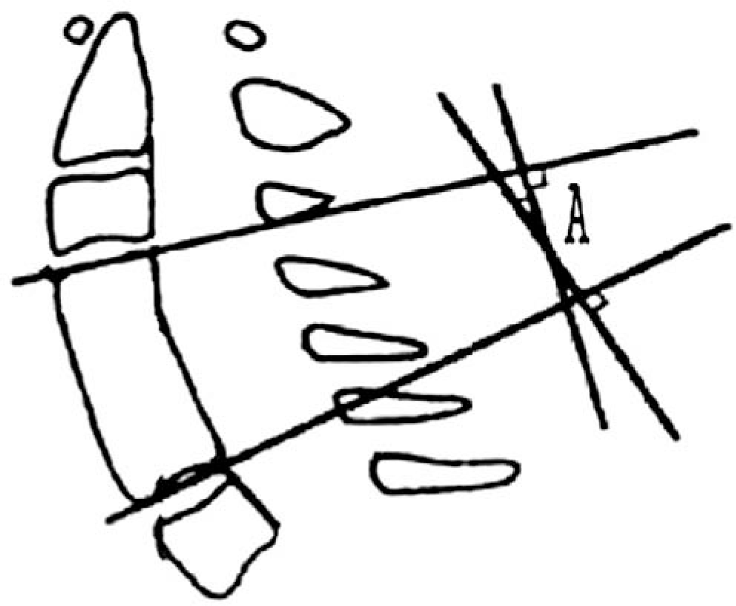
 | Fig. 1.Measurement of the alignment of the fused segment (angle A). Angle A was formed by the upper plane and lower plane of the fused segment. |
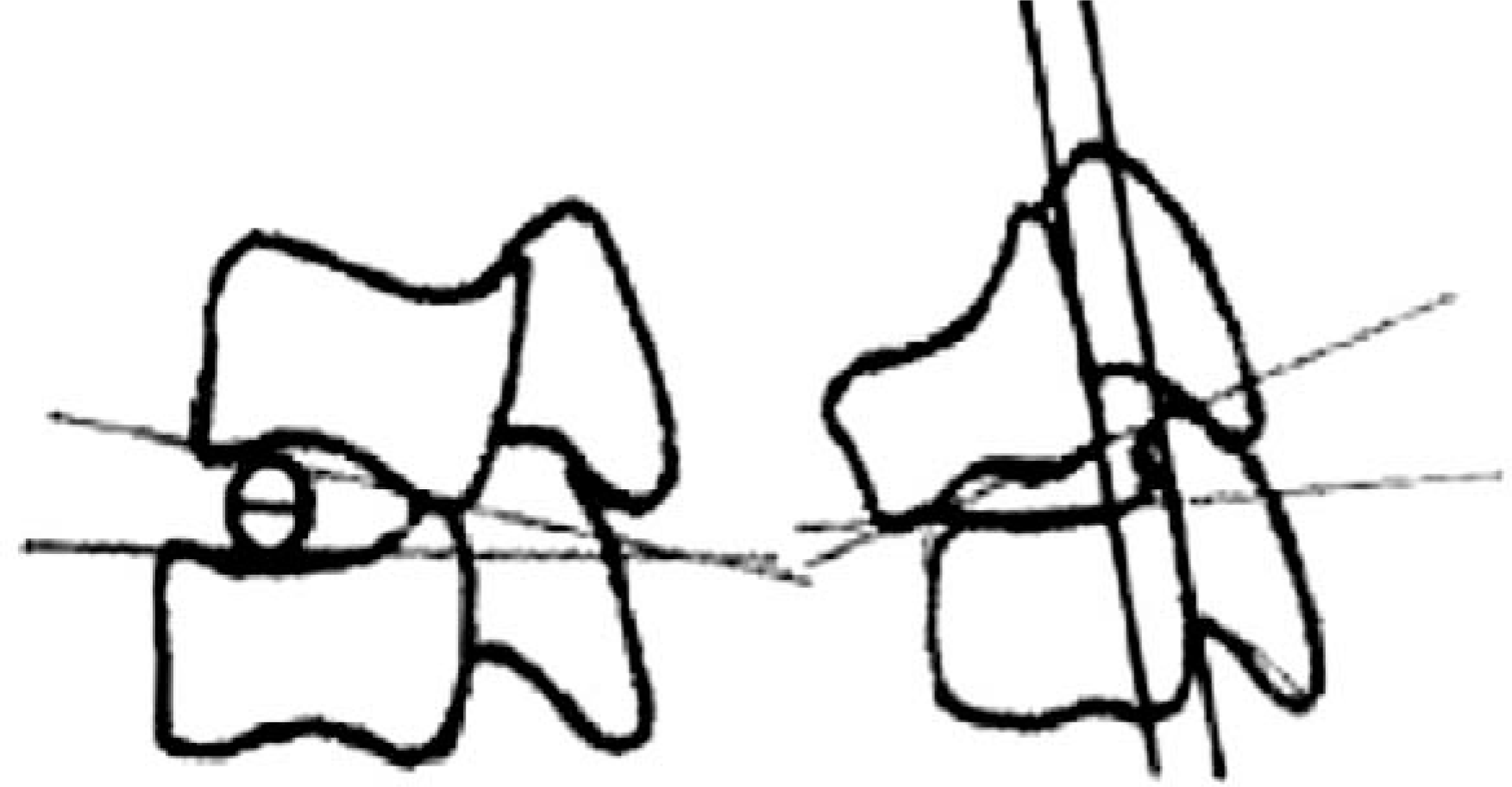
 | Fig. 2.Changes in the motion segment between vertebrae. Calculation of the angular displacement of the flexion/ extension views. |
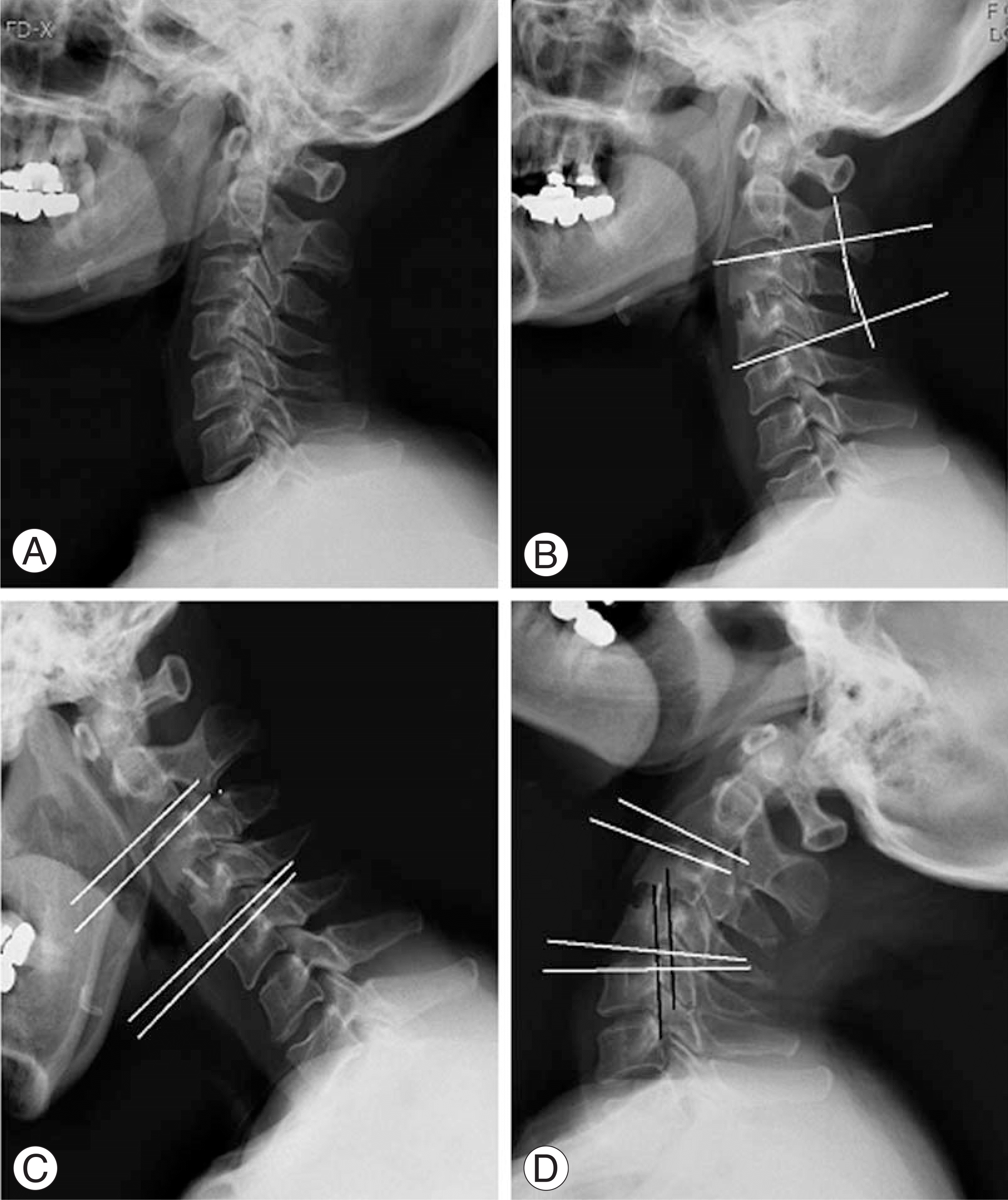
 | Fig. 3.A 42-year-old woman, disc herniation at C3-4 and treated with anterior cervical disectomy and fusion. (A) Preoperative lateral radiograph, (B) Radiograph at postoperative 2-years follow-up shows solid union and maintained lordosis at fusion segment (10°), (C, D) At last follow-up, adjacent intervertebral segment shows degenerative change, angular motion is within normal limit but sagittal translation is 3 mm. |
Table 1.
Baseline characteristics of the study population
| Total | Degenerative change (+) | Degenerative change (-) | |
|---|---|---|---|
| Mean age | 51 | 50 | 52 |
| Gender (M:F) | 23:11 | 13:6 | 10:5 |
| Mean follow up | 24 | 23 | 26 |
Table 2.
Summary of the normal values estabilished by different authors (°)
| Bakke | Buetti-Bauml | De Seze | Penning | Dvorak | |
|---|---|---|---|---|---|
| C1-C2 | 11.7 | 12 | |||
| C2-C3 | 12.6 | 11 | 0.13 | 12.5 | 10 |
| C3-C4 | 15.4 | 17 | 15.5 | 0.18 | 15 |
| C4-C5 | 15.1 | 21 | 0.19 | 0.20 | 19 |
| C5-C6 | 20.4 | 23 | 27.5 | 21.5 | 20 |
| C6-C7 | 17.0 | 19 | 17.5 | 15.5 | 19 |
Table 3.
Inflencing factors in degenerative change of adjacent segment


 PDF
PDF ePub
ePub Citation
 Print
Print


 XML Download