Abstract
Objectives
We wanted to investigate whether osteogenesis can be enhanced when a small amount of demineralized bone matrix (1 cc/segment) is mixed with local bone chips.
Summary of the Literature Review
Demineralized bone matrix (DBM) has been used for spinal arthrodesis. However, there are only a few reports about its use as a composite graft with local bone chips for posterior lumbar interbody fusion
Materials and Methods
Degenerative spine patients, who would normally be treated by decompression and posterior lumbar interbody fusion with using a pedicle screw system and one cage, were randomly, prospectively selected for whether they would be treated with using local bone chips mixed with 1cc of DBM (Group I: 15 patients and 19 segments) or local bone chips (Group II: 12 patients and 13 segments) for graft material. The sampling bias was investigated for gender, age, endocrine diseases, previous operation, habits (alcohol drinking, smoking), steroid medication, bone mineral density and the amount of local bone. The amount of bone formation was measured at 6 months after operation. On the sagittal and coronal reconstruction CT images, the bone formation outside of the cage was measured, and this was interpreted in a “blinded”fashion by 2 independent doctors who did not take part in the operations.
Results
There was no sampling bias between the 2 groups except for age (Group I= 65.3±7.1, Group II=58.9±6.0, p=0.010). The ratio of local bone chips and DBM was 5.98:1 in Group I. There was moderate concurrence between the 2 interpreters (kappa co-efficiency=0.494, p<0.001 for the sagittal plain images and kappa co-efficiency=0.467, p<0.001 for the coronal plain images) and Group I showed significantly more bone formation (p=0.003).
Conclusion
DBM that is mixed with local bone chips, even with small amount, enhanced bone formation in the posterior lumbar interbody fusion. This is regarded to act as a graft enhancer to increase the fusion rate, even when using local bone chips for graft material, for the cases that show unfavorable conditions for fusion or for the cases that are prone to loosening of hardware.
Go to : 
REFERENCES
1). Lee KJ, Roper JG, Wang GC. Demineralized bone matrix and spinal arthrodesis. Spine. 2005; 5:217–223.

2). Vaccaro AR, Chiba K, Heller JG, et al. Bone grafting alternatives in spinal surgery. Spine. 2002; 2:206–215.

3). Aspenberg P, Albrektsson T, Thorngren K. Local application of IGR-1 to healing bone Acta Orthop Scan. 1989; 60:607–610.
4). Cook DB, Dalton JE, Tan EH, Whitecloud TS, Rueger DC. In vivo evaluation of recombinant human osteogenic protein (rhOP-1) implants as a bone graft substitute for spine fusions. Spine. 1994; 19:1655–1664.
5). Cook SD, Dalton JE, Prewett AB, Whitecloud TS. In vivo evaluation of demineralized bone matrix as a bone graft substitute for posterior spine fusion. Spine. 1995; 20:877–886.
6). Louis-Ugbo J, Murakami H, Kim HS, Minamide A, Boden SD. Evidence of osteoinduction by Grafton demineralized bone matrix in nonhuman primate spinal fusion. Spine. 2004; 29:360–366.

7). Martin GJ, boden SD, Titus L, Scarborough NL. New formulations of demineralized bone matrix as a more effective in experimental posterolateral lumbar spine arthrodesis. Spine. 1999; 24:637–645.
8). Turner JA, Ersek M, Herron L, et al. Patient outcomes after lumbar spinal fusions. JAMA. 1992; 268:907–911.

9). Yahiro MA. Comprehensive literature review: pedicle screw fixation devices. Spine. 1994; 19:2274–2278.
10). Yuan HA, Garfin SR, Dickman CA, Mardjetko SM. A historical cohort study of pedicle screw fixation in thoracic, lumbar, and sacral spinal fusions. Spine. 1994; 19:2279–2296.
11). Miura Y, Imagama S, Yoda M, Mitsuguchi H, Kachi H. Is local bone viable as a source of bone graft in posterior lumbar interbody fusion? Spine. 2003; 15(28):2386–2389.

12). Okuyama K, Kido T, Unoki E, Chiba M. PLIF with a titanium cage and excised facet joint bone for degenerative spondylolisthesis in augmentation with a pedicle screw. J Spinal Disord Tech. 2007; 2:53–59.
13). Femyhough JC, Schimandle JJ, Weigel MC, Edwards CC, Levine AM. Chronic donor site pain complicating bone graft harvesting from the posterior iliac crest for spinal fusion. Spine. 1992; 17:1474–1480.

14). Russell JL, Block JE. Surgical harvesting of bone graft from the ilium: point of view. Med Hypotheses. 2000; 55:474–479.

16). Muschler GF, Lane JM. Orthopaedic surgery. Habal MB, Reddi Ah, editors. Bone grafts and bone substitutes. Philadelphia: Saunders;p. 375. 1992.
18). Canalis E, McCarthy T, Centrella M. Growth factors in the regulation of bone remodeling. J Clin Invest. 1988; 81:277–281.
19). Jungushi S, Heydemann A, Kana SK, Macey LR, Bolander ME. aFGF injection stimulates cartilage enlargement and inhibits collagen gene expression in a rat fracture healing model. J Orthop Res. 1990; 8:364–371.
20). Linkhart TA, Mohan S, Baylink DJ. Growth factors for bone growth and repair: IGF, TGF-β, BMP. Bone. 1996; 9:1–2.
22). Muller R, Bravo R, Burckhardt J, Curran T. Induction of c-fos gene and protein by growth factors precedes activation of c-myc. Nature. 1984; 312:716–720.

23). Mundy GR. Regulation of bone formation by bone morphogenetic proteins and other growth factors. Clin Orthop Rel Res. 1996; 324:24–28.

24). Wang EA, Rosen V, Cordes P, et al. Purification and characterization of other distinct bone-inducing factors. Proc Natl Acad Sci USA. 1988; 85:9484–9488.

25). Ijiri S, Yamamuro T, Nakamura T, Kotani S, Notoya K. Effect of sterilization on the osteoinductive capacity of demineralized bone matrix. Clin Orthop. 2001; 388:233–239.
26). Russell JL, Block JE. Clinical utility of demineralized bone matrix for osseous defects, arthrodesis, and reconstruction: impact of processing techniques and study methodology. Orthopedics. 2004; 22:524–531.
27). Sassard WR, Eidman DK, Gray PM, et al. Augmenting local bone with Grafton demineralized bone matrix for posterolateral lumbar spine fusion: avoiding second site autologous bone harvest. Orthopedics. 2000; 23:1059–1064.

28). Shin BJ, Kin GJ, Ha SS, Chung SH, Kwon H, Kin YI. Posterior lumbar interbody fusion using laminar bone block. J Korean Soc Spine Surg. 1999; 6:110–116.
29). Shin BJ, Kim GJ, Kwon H, Suh YS, Kim YI, Rah SK. Results of PLIF using laminar chips in spinal lesions. J Korean Soc Spine Surg. 1998; 5:284–292.
30). Ahn DK, Jeong KW, Lee S, Choi DJ, Cha SK. Posterior Lumbar Interbody Fusion with chip bone and pedicle screw fixation. J Korea Orthop Assoc. 2004; 39:614–620.
31). Harris BM, Hilibrand AS, Savas PE, et al. Transforaminal lumbar interbody fusion: the effect of various instrumentation techniques on the flexibility of the lumbar spine. Spine. 2004; 15(29):65–70.
32). Wang J, Glimcher M. Characterization of matrixinduced osteogenesis in rat calvarial bone defects: I. Differences in the cellular response to demineralized bone matrix implanted in calvarial defects and in subcutaneous sites. Calcif Tissu Int. 1999; 65:156–165.

33). Wang J, Glimcher MJ. Charaterization of matrix induced osteogenesis in rat calvarial bone defects II Origins of bone-forming cells. Clcif Tissue Int. 1999; 65:486–493.
34). Wang J, Tang R, Gerstenfeld LC, Glimcher MJ. Characterization of demineralized bone matrix-induced osteogenesis in rat calvarial bone defects. Gene and protein expression. Calcif Tissue Int. 2000; 67:314–320.
Go to : 
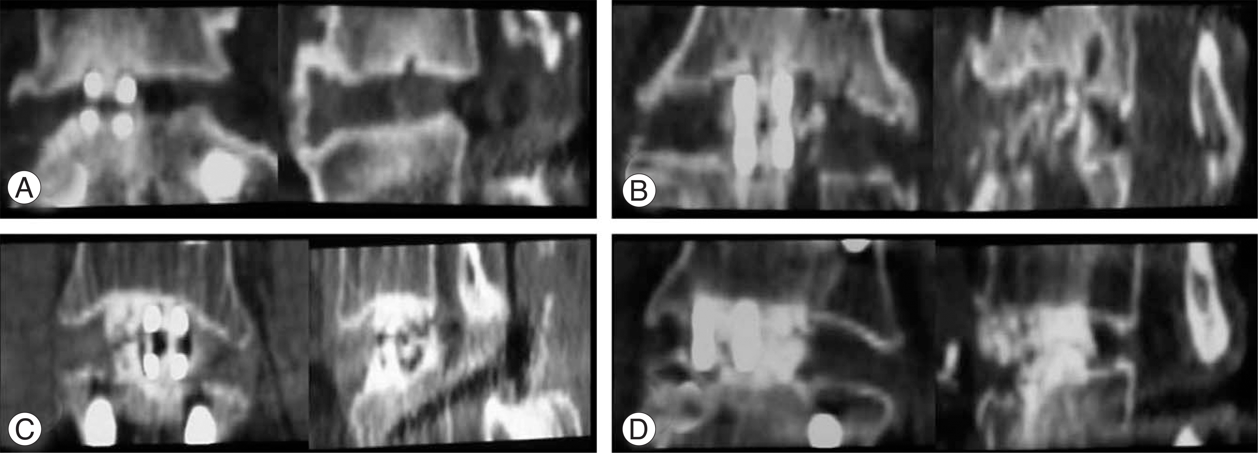
 | Fig. 3.Bone formation grading system which is measured at out of cage, mid-sagittal and anterior one third coronal plane CT reconstruction view. (A) Grade I: no or little bone formation at graft site. (B) Grade II: bone formation less than 50% at graft site. (C) Grade III: bone formation more than 50% at graft site. (D) Grade IV: full bone formation at graft site and continuing to adjacent end plates. |
Table 1.
Sampling bias
| Characteristics | Group I | Group II | p-value |
|---|---|---|---|
| Age (years) | 65.32±7.06 | 58.92±5.98 | 0.010∗ |
| Sex | 5:14 | 4:9 | 0.545 |
| Alcohol | 3 | 4 | 0.364 |
| Smoking | 3 | 3 | 0.470 |
| Previou op. | 2 | 0 | 0.195 |
| DM® | 5 | 4 | 0.545 |
| Endocrine disorder | 0 | 1 | 0.406 |
| BMD® | -2.24±0.80 | -1.84±0.80 | 0.178 |
| Amount of local bone | -5.98±0.51 | -6.39±0.58 | 0.054 |
Table 2.
Bone formation score


 PDF
PDF ePub
ePub Citation
 Print
Print


 XML Download