

 PDF
PDF ePub
ePub Citation
 Print
Print


Abstract
Objectives
We wanted to analyze the risk factors related to deep infection and removing an implant after thoracic and lumbar spinal arthrodesis.
Summary of literature reviews
The relationship between deep infection and implant removal is controversial.
Materials and Methods
We retrospectively compared the infection group with the non-infection group for the rates of deep infection, the preoperative diagnosis, the number of fused segments, the operative methods, the graft materials, the operating time and the blood loss. Moreover, we classified the deep infection patients into two groups: those who underwent implant removal and those who did not, and we compared the microorganisms that were cultured out of the patients. We also compared the relationship of deep infection with the risk factors, the mean hospital stay and the mean number of operations.
Results
There were 18 cases (2.46%) of deep infection. The factors that did not show a significant difference were the preoperative diagnosis, the graft material, the increased number of fused segments, age, gender and BMI. The factors that were significant were the operating time (p=0.001), the amount of blood loss (p<0.000), DM (p=0.021), and PLF (p=0.054). The incidence of implant removal was higher for the cases with deep infection caused by MRSA. We were able to see a significant difference of between the group that had undergone implant removal and the group that had not undergone implant removal.
Conclusions
The incidence of deep infection after thoracic and lumbar spinal athrodesis increased as the operating time and blood loss increased, and it was also higher when either PLF or DM were present. Implant removal causes bad clinical results, so physicians should be very cautious when operating on a case of implant removal.
Go to : 
REFERENCES
1). Kim EH, Song IS. Deep Wound Infection after Lumbar Spine Fusion with Pedicular Screw Fixation. J Korean Soc Spine Surg. 2000; 7:535–543.
2). Kim JH, Kim BJ, Choo SK, Cho JH, Kim YJ. Deep Infection following Instrumented Posterior Fusion. J Korean Orthop Assoc. 2006; 41:617–622.

3). Kim KS, Ko SH, Im CI, Kim DY. Postoperative Wound Infections in Patients with Posterior Spinal Instrumentations. J Korean Soc Spine Surg. 1996; 3:77–82.
4). Kim YM, Won CH, Choi ES, Seo JB, Lee HS, Cho BK. Management of Deep Infection after Posterior Spinal Instrumentation with Prolonged Suction Drainage. J Korean Soc Spine Surg. 2001; 8:504–512.
5). McCarthy RE, Peek RD, Morrissy RT, Hough AJ Jr. Allograft bone in spinal fusion for paralytic scoliosis. J Bone Joint Surg Am. 1986; 68:370–375.
6). Lin PM, Cautilli RA, Joyce MF. Posterior lumbar interbody fusion. Clin Orthop Relat Res. 1983; 180:154–168.
7). Lee HM, Park MS, Moon SH. Infection after Cervical Spine Surgery. J Korean Soc Spine Surg. 2000; 7:456–461.
8). Viola RW, King HA, Adler SM, Wilson CB. Delayed infection after elective spinal instrumentation and fusion. A retrospective analysis of eight cases. Spine. 1997; 15(22):2444–2450.
9). Rah JH, Park HJ, Ahn JI. A Clinical Study of Postoperative Infection in Posterior Spinal Surgery with Pedicle Screw System. J Korean Orthop Assoc. 1994; 29:994–1003.
10). Shim JI, Kim TS, Lee SJ, et al. Late Infection of Spinal Instrumentation. J Korean Soc Spine Surg. 2000; 7:29–36.
11). Lonstein J, Winter R, Moe J, Gaines D. Wound infection with Harrington instrumentation and spine fusion for scoliosis. Clin Orthop Relat Res. 1973; 96:222–233.
12). Massie JB, Heller JG, Abitbol JJ, McPherson D, Garfin SR. Postoperative posterior spinal wound infections. Clin Orthop Relat Res. 1992; 284:99–108.
13). Park WW, Park YS, Cheon SJ, Jung JY. Posterior lumbar interbody fusion in the pyogenic discitis. J Korean Soc Spine Surg. 2001; 8:39–45.
14). Richards BS. Delayed infections following posterior spinal instrumentation for the treatment of idiopathic scoliosis. J Bone Joint Surg Am. 1995; 77:524–529.
15). Sepica FL, Montgomerie JZ. Vertebral osteomyelitis. Infect Dis Clin North Am. 1990; 4:539–550.
16). Weinstein MA, McCabe JP, Cammisa FP Jr. Postoperative spinal wound infection. A review of 2391 consecutive index procedures. J Spinal Disord. 2000; 13:422–426.
17). Boachie-Adjei O, Bradford DS, Ogilvie JW, Transfeldt EJ. Treatment of adult spinal deformity with fusion to the sacrum using CD instrumentation. J Spinal Disord. 1991; 4:1–14.
18). McAfee PC, Bohlman HH. Complications following Harrington instrumentation for fractures of the thoracolumbar spine. J Bone Joint Surg Am. 1985; 67:672–685.
Go to :
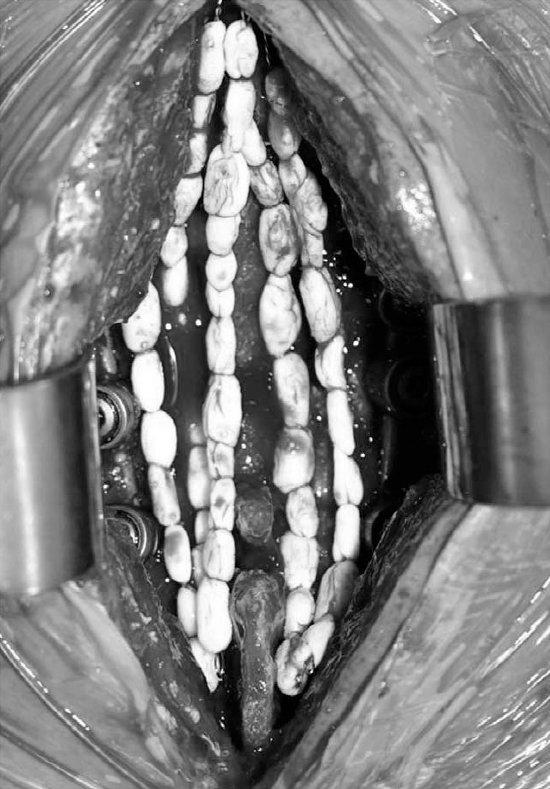 | Fig. 1.Antibiotic bead insertion was made up of cement beads impregrnated susceptible antibiotics on twisted wires. |
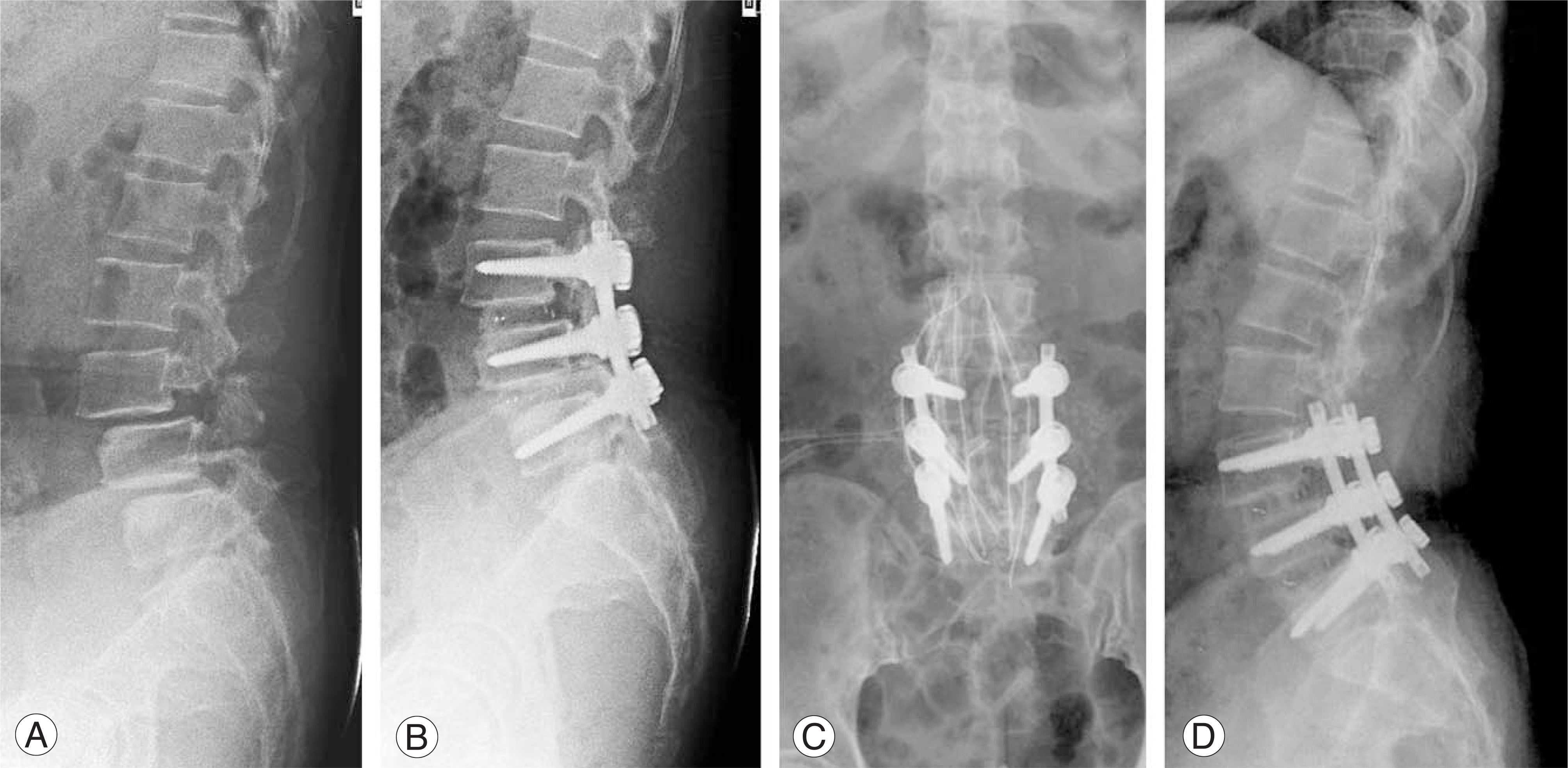 | Fig. 2.62 year-old female underwent the PLIF on L4-5-S1 due to spondylolisthesis (A, B). The deep infection occurred on postoperative 14 days, we took the two times debridement and antibiotic bead insertion (C). The last follow-up plain lateral radiogram shows bony fusion and no lower back pain and neurologic deficit clinically (D). |
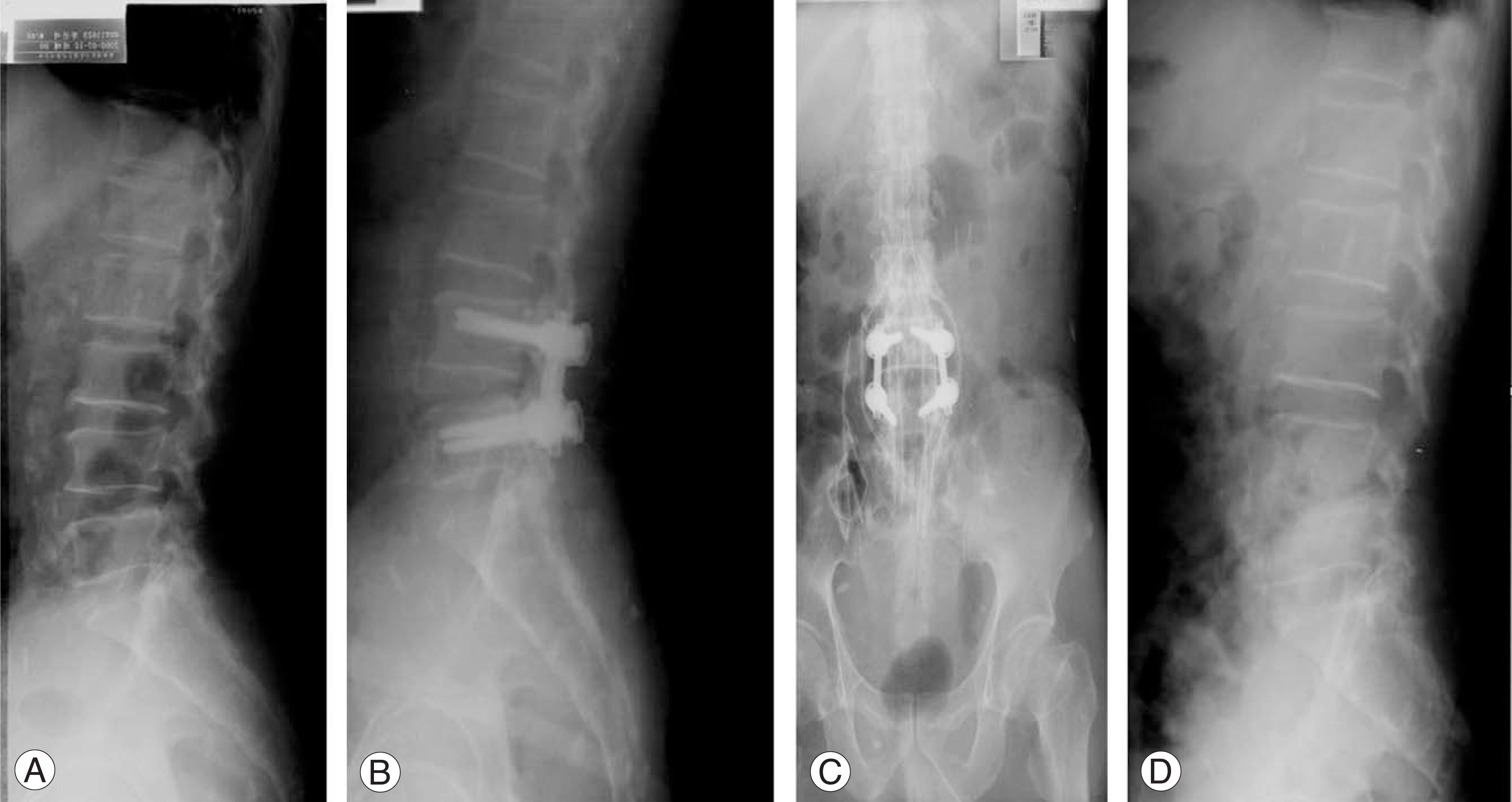 | Fig. 3.60 year-old male underwent the PLF on L4-5 due to spinal stenosis (A, B). The deep infection occurred on postoperative 26 days, we took the three times debridement and antibiotic bead insertion, but sustained the infection sign, and removed the implants (C). The last follow-up plain lateral radiogram shows bone loss and destruction of L4-5 disc space and moderate lower back and right leg pain clinically (D). |
Table 1.
Infecting Microorganism
Table 2.
The significant factors affecting the postoperative deep infection by multiple logistic regression test.
| Variables | B | S.E. | Wald | df | p-value |
|---|---|---|---|---|---|
| PLF | -2.115- | 1.100 | 3.698 | 1 | 0.054 |
| Blood loss amount | 3.675 | 1.121 | 10.7540 | 1 | 0.001 |
| OP time | 0.012 | 0.005 | 5.062 | 1 | 0.024 |
Table 3.
A comparison of mean value between implant retained and removal group.
 XML Download