

 PDF
PDF ePub
ePub Citation
 Print
Print


Abstract
Objectives
We analyzed the radiological and clinical results to verify the efficacy of anterior interbody fusion with using cages gradually increases in the treatment of cervical radiculopathy.
Summary of the Literature Review
Anterior cervical decompression and fusion is well accepted treatments for cervical radiculopathy. Performing anterior interbody fusion using cages has recently gradually increased to minimize the extent of surgery. While there are numerous reports on the primary stabilizing effects of the cervical cages, little is known about the subsidence behavior of such cages in vivo.
Materials and Methods
We retrospectively analyzed 38 patients with cervical disc herniation who underwent anterior decompression and interbody fusion with autoiliac bone graft and plate fixation (Group I, 21 patients) or who underwent with standalone cage (Group II, 17 patients). We statistically analyzed the changes of the cervical lordosis, the segmental lordosis, the vertebral body height, the fusion rate on the plain x-ray and the clinical results with using a pain visual analogue scale.
Results
All the cases were fused by 11.2±2.7 weeks after operation. The changes of the cervical lordosis and segmental lordosis show no statistically significant difference between the two groups (p=0.07, 0.66). The anterior and posterior vertebral heights of the fused segments of group II were more decreased than those of group I, but there was no statistically difference between the two groups (p=0.06, 0.30). The clinical results were not statistically difference between the two groups (p=0.64, 0.45).
Conclusions
Implantation of autoiliac cancellous bone impacted standalone cages or on a tricortical iliac crest autograft after anterior decompression was safe and reliable options for the treatment of cervical disc herniation that causes single level radiculopathy. Both procedures produced equally satisfying clinical and radiological results, leading to a high fusion rate and they maintained the intervertebral height.
Go to : 
REFERENCES
1). Robinson R, Smith G. Anterolateral cervical disc removal and interbody fusion for cervical disc herniation. Bull Johns Hopkins Hosp. 1955; 96:223–224.
2). Cloward R. The anterior approach for removal of ruptured cervical discs. J Neurosurg. 1958; 15:602–617.
3). Wilson DH, Campbell DD. Anterior cervical discectomy without bone graft. Report 71 cases. J Neurosurg. 1977; 47:551–555.
4). Savolainen S, Rinne J, Hernesniemi J. A prospective randomized study of anterior single-level cervical disc operations with long-term follow-up: surgical fusion is necessary. Neurosurgery. 1998; 43:51–55.
5). Barlocher C, Barth A, Krauss JK, Binggeli R, Seiler RW. Comparative evaluation of microdiscectomy only, autograft fusion, polymethylmethacrylate interposition, and threaded titanium cage fusion for treatment of single-level cervical disease: a prospective randomized study in 125 patients. Neurosurg Focus. 2002; 12:E4.
6). Park HJ, Shim YJ. Treatment of distractive flexion injury in lower cervical spine using anterior cervical fusion. J Korean Soc Spine Surg. 2007; 14:221–228.

7). Schnee C, Freese A, Weil RJ, Marcotte PJ. Analysis of harvest morbidity and radiographic outcome using autograft for anterior cervical fusion. Spine. 1997; 22:2222–2227.
8). Goffin J, Van Calenbergh F, van Loon J, et al. Intermediate follow-up after treatment of degenerative disc disease with the Bryan Cervical Disc Prosthesis: single-level. Spine. 2003; 28:2673–2678.
9). Bagby GW. Arthrodesis by the distraction-compression method using a stainless steel implant. Orhtopaedics. 1988; 11:931–934.
10). Ray CD. Threaded titanium cages for lumbar interbody fusions. Spine. 1997; 22:667–679.
11). Kuslich SD, Ulstrom CL, Griffith SL, Ahern JW, Dow-dle JD. The Bagby and Kuslich method of lumbar interbody fusion. History, techniques, and 2-year follow-up results of a United States prospective, multicenter trial. Spine. 1998; 23:1267–1279.
12). Brantigan JW, Steffee AD. A carbon fiber implant to aid interbody lumbar fusion. Two-year clinical results in the first 26 patients. Spine. 1993; 18:2106–2107.
13). Wilke HJ, Kettler A, Claes L. Stabilizing effect and sintering tendency of 3 different cages and bone cement for fusion of cervical vertebrae segments Orthopade. 2002; 31:472–480.
14). Furderer S, Schollhuber F, Rompe JD, Eysel P. Effect of design and implantation technique on risk of progressive sintering of various cervial vertebrae cages. Orthopade. 2002; 31:466–471.
15). Vavruch L, Hedlund R, Javid D, Leszniewski W, Shal-abi A. A prospective randomized comparison between the Cloward procedure and a carbon fiber cage in the cervical spine. A clinical and radiologic study. Spine. 2002; 27:1694–1701.
16). Gercek E, Arlet V, Delisle J, Marchesi D. Subsidence of standalone cervical cages in anterior interbody fusion: warning. Eur Spine J. 2003; 12:513–516.
17). Shimamoto N, Cunningham BW, Dmitriev AE, Minami A, MaAfee PC. Biomechamical evaluation of standalone interbody fusion cages in the cervical spine. Spine. 2001; 26:432–436.
18). Shono Y, McAfee PC, Cunningham BW, Brantigan JW. A biomechanical analysis of decompression and reconstruction methods in the cervical spine. Emphasis on a carbon-fiber-composite cages. J Bone Joint Surg. 1993; 75:1674–1684.
19). Curylo LJ, Lindsey RW, Doherty BJ, LeBlanc A. Segmental variations of bone mineral density in the cervical spine. Spine. 1996; 21:319–322.
20). Hollowell JP, Vollmer DG, Wilson CR, Pintar FA, Yoganandan N. Biomechanical analysis of thoracolumbar interbody constructs: how important is the endplate? Spine. 1996; 21:1032–1036.
21). Kettler A, Wilke HJ, Claes L. Effects of neck movements on stability and subsidence in cervical interbody fusion: an in vitro study. J Neurosurg. 2001; 94:97–107.
22). Wilke HJ, Kettler A, Goetz C, Claes L. Subsidence resulting from simulated postoperative neck movements: an in vitro investigation with a new cervical fusion cage. Spine. 2000; 25:2762–2770.
23). Frederic S, benedict R, Payer M. Implantation of an empty carbon fiber cage or a tricortical iliac crest autograft after cervical discectomy for single-level disc herniation: a prospective comparative study. J Neurosurg Spine. 2006; 4:292–299.
24). Bartels R, Donk R, Azn R. Height of cervical foramina after anterior discectomy and implantation of carbon fiber cage. J Neurosurg. 2001; 95:40–42.
25). Samandouras G, Shafafy M, Hamlyn PJ. A new anterior cervical instrumentation system combining an intradiscal cage with integrated plate: an early technical report. Spine. 2001; 26:1188–1192.
26). Hacker RJ, Cauthen JC, Gilbert TJ, Griffith SL. A prospective randomized multicenter clinical evaluation of an anterior cervical fusion cage. Spine. 2000; 25:2646–2655.
27). Agrillo U, Mastronardi L, Puzzilli F. Anterior cervical fusion with carbon cage containing coralline hydroxyapatite: preliminary observations in 45 consecutive cases of soft-disc herniation. J Neurosurg. 2002; 96:273–276.
28). van Limbeek J, Jacobs WC, Anderson PG, Pavlov PW. A systematic literature review to identify the best method for a single level anterior cervical interbody fusion. Eur Spine J. 2000; 9:129–136.
29). Boakye M, Mummaneni PV, Garrett M, Haid R. Anterior cervical discectomy and fusion involving a poly-etheretherketone spacer and bone moerphogenetic protein. J Neurosurg Spine. 2005; 2:521–525.
30). Hamburger C, Festenberg F, Uhl E. Ventral discectomy with pmma interbody fusion for cervical disc disease: long-term results in 249 patients. Spine. 2001; 26:249–255.
Go to :
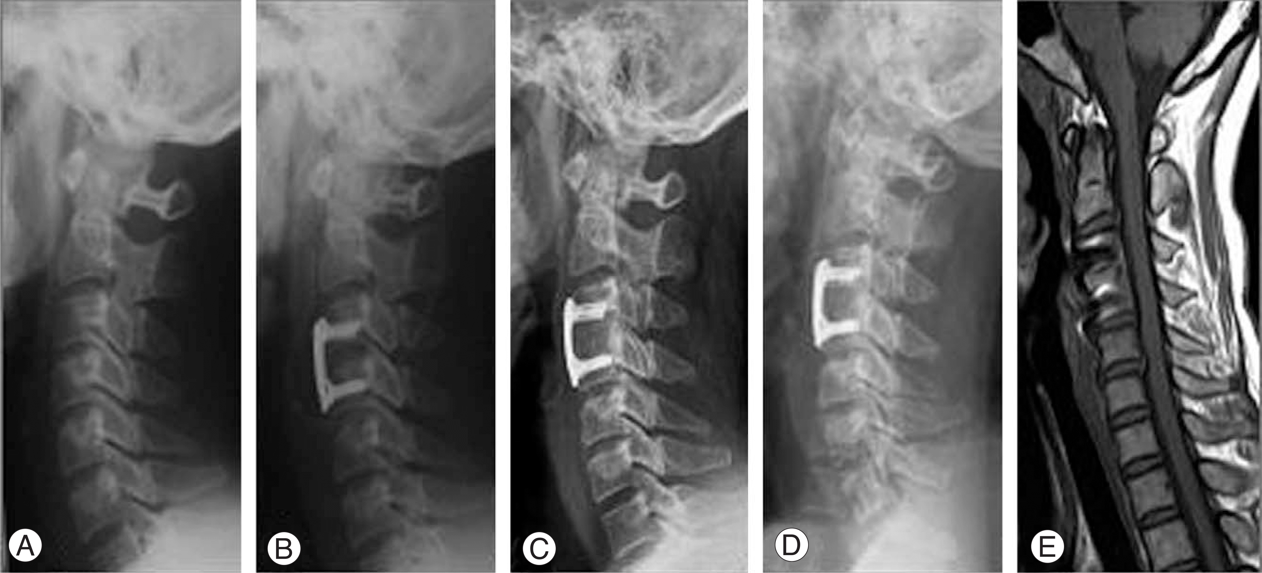 | Fig. 1.36-years-old female with cervical disc herniation on C3-4. (A) Preoperative lateral roentgenogram. (B)Lateral radiograph, immediately after surgery, shows anterior cervical fusion with autogenous iliac bone and cervical locking plate. (C) Lateral radiograph, 6years after surgery, shows the solid union of grafted bone. (D) Lateral roentgenogram of flexion/extention view that shows no motion at fused level. (E) Sagittal MRI, 6years after surgery, shows the solid union at fused level. |
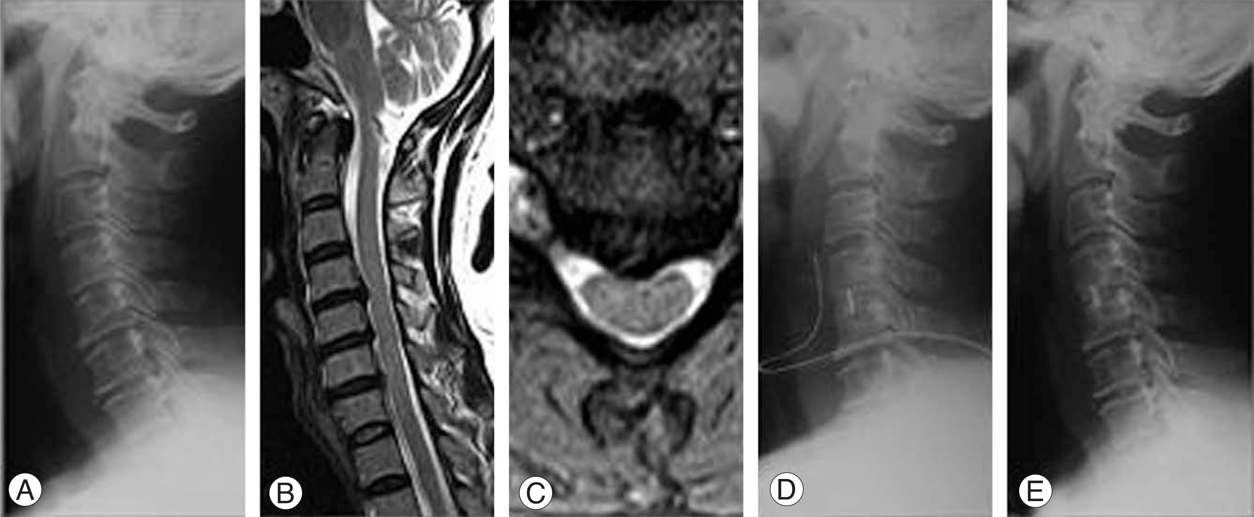 | Fig. 2.43-years-old female with cervical disc herniation on C4-5. (A) Preoperative lateral roentgenogram. (B), (C) T1 weighted sagittal and axial MRI image show a C4-5 disc herniation. (D) Lateral radiograph, immediately after surgery, show anterior cervical fusion with Solis PEEK cage that packed with cancellous iliac bone. (E) Lateral radiograph, 16 months after surgery, shows the solid union at fused level but disc height is decreased slightly than after surgery. |
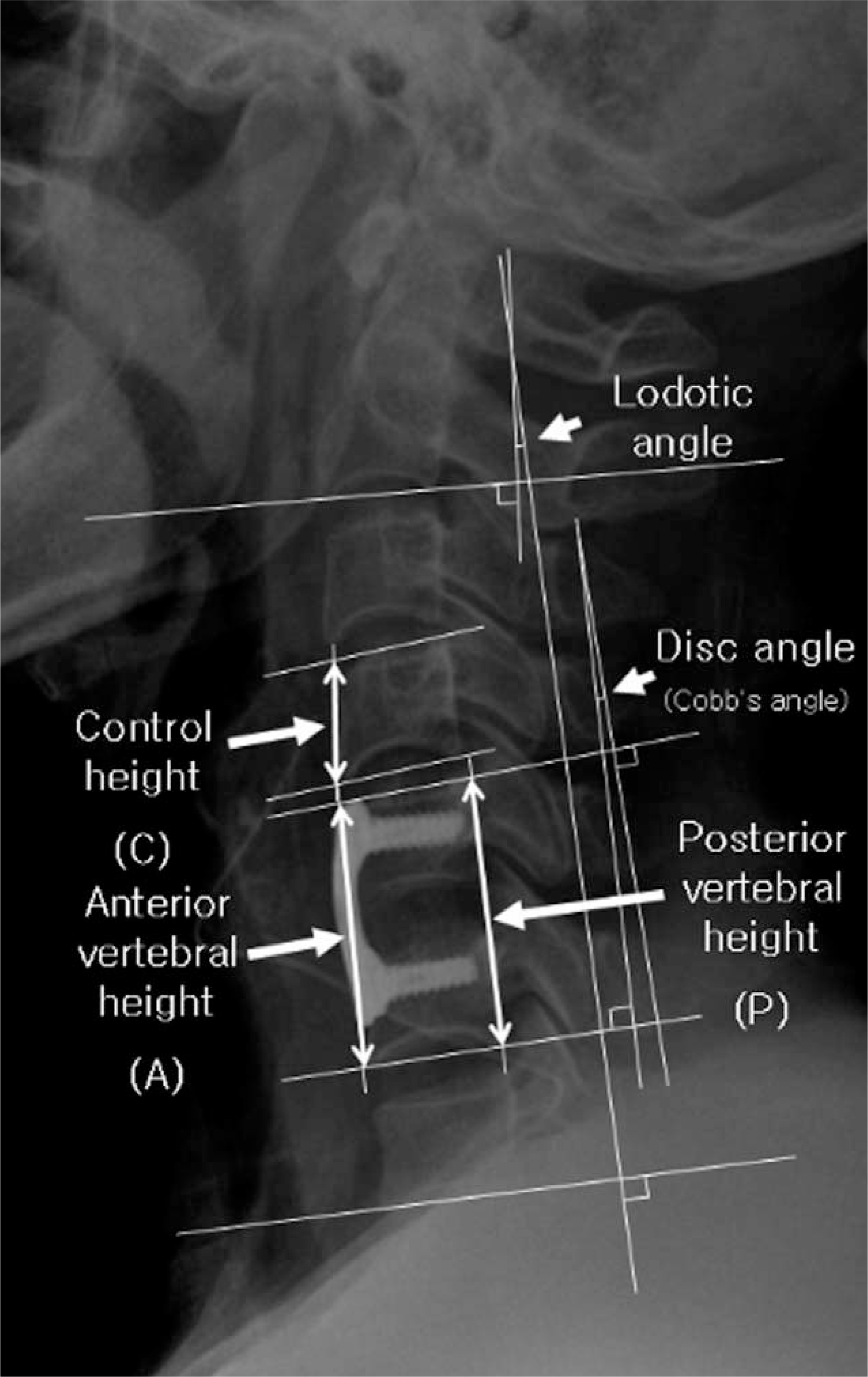
Table 1.
Demography of sampling errors
Table 2.
Radiological comparison between two groups
| Plate group (n=21) | Cage group (n=17) | P-value | ||
|---|---|---|---|---|
| Lordotic angle (®) | Preoperation | 13.7±7.4 | 12.5±8.0 | 0.658 |
| Postoperation | 8.5±5.3 | 11.9±6.4 | 0.090 | |
| Last Follow-up | 12.6±4.7 | 12.7±6.4 | 0.935 | |
| Δ(Last f/u-Post op) | 4.1±5.4 | 0.9±4.7 | 0.073 | |
| Disc angle (®) | Preoperation | 3.2±2.7 | 4.1±3.3 | 0.344 |
| Postoperation | 5.8±4.3 | 4.2±3.1 | 0.256 | |
| Last Follow-up | 3.6±2.9 | 2.7±3.6 | 0.384 | |
| Δ(Last f/u-Post op) | -2.1±3.8 | -1.6±3.8 | 0.658 | |
| Anterior body height (ABH∗, %) | Preoperation | 259.6±61.2 | 234.5±47.8 | 0.221 |
| Postoperation | 284.9±59.9 | 256.8±48.9 | 0.148 | |
| Last Follow-up | 277.2±55.9 | 236.9±36.6 | 0.022∗ | |
| Δ(Last f/u-Post op)® | -2.3±8.0 | -7.1±6.4 | 0.064 | |
| Posterior body height (PBH∗, %) | Preoperation | 260.9±62.5 | 231.6±48.4 | 0.143 |
| Postoperation | 275.0±57.6 | 247.4±44.3 | 0.134 | |
| Last Follow-up | 270.4±56.7 | 235.5±37.5 | 0.048∗ | |
| Δ(Last f/u-Post op)® | -1.3±9.2 | -4.4±7.0 | 0.297 |
 XML Download