

 PDF
PDF ePub
ePub Citation
 Print
Print


Abstract
– Abstract – Multidrug-resistant tuberculosis, resistant to at least isoniazid and rifampicin, continues to present a serious challenge to human health. However, there are no reports addressing multidrug-resistant tuberculous spondylitis in Korea. We report a case of multidrug-resistant tuberculous spondylitis at L2-L3 in a 30-year-old woman.
Go to : 
REFERENCES
1). Br Med Res Counc. Treatment of pulmonary tuberculosis with streptomycin and paraaminosalicylic acid. Br Med J. 1950; 2:1073–1085.
2). Bai GH. Anti-tuberculosis drug resistance in Korea. CDMR. 2005; 16:101–107.
3). Chaulet P, Boulahbal F, Grosset J. Surveillance of drug resistance for tuberculosis control: why and how? Tuber Lung Dis. 1995; 76:487–492.

4). Ahn JI, Oh HY, Rah JH, Kang KS. A clinical study of tuberculous spondylitis. J Korean Orthop Assoc. 1981; 16:300–310.
5). Pablos-Mendez A, Raviglione MC, Laszlo A, et al. World Health Organization-International Union against Tuberculosis and Lung Disease Working Group on Anti-Tuberculosis Drug Resistance Surveillance. N Engl J Med. 1998; 338:1641–1649.
6). Kim BJ, Lee HI, Lee DH, et al. The current status for Multidrug-resistant Tuberculosis in Korea. J of Tuberculosis and respiratory disease. 2006; 60:404–411.
7). Ahn JS, Lee JK, Jeon TS, Kwon YS, Kwak SK. Changes of kyphotic angle following operative treatment of tuberculous spondylitis. J Korean Soc Spine Surg. 2001; 8:148–155.
8). Martin NS. Tuberculosis of the spine. A study of the results of treatment during the last twenty-five years. J Bone Joint Surg Br. 1970; 52:613–628.
Go to :
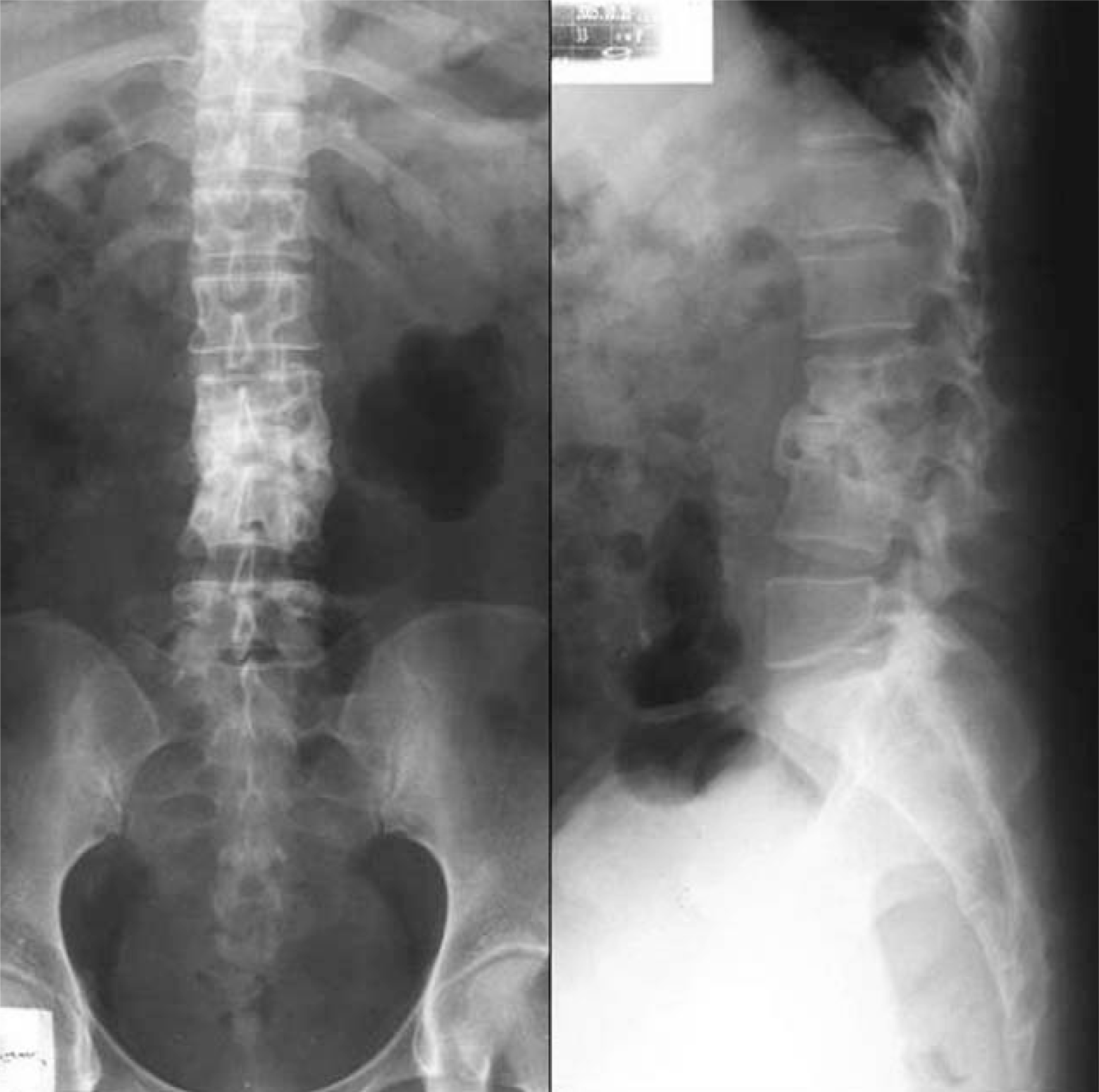
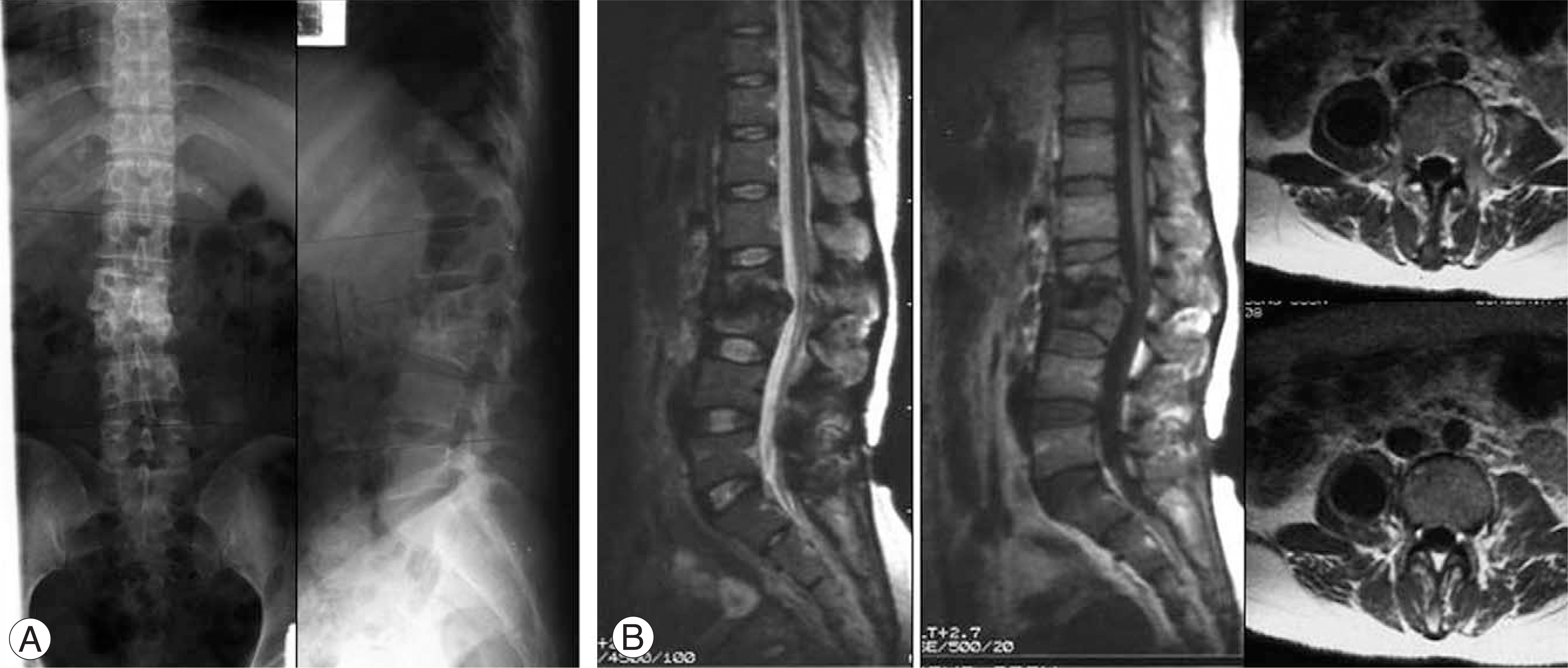 | Fig. 1.Preoperative radiographs and MR Imaging. (A) Radiographs show destruction of vertebral body and kyphotic deformity (Sagittal index: 40 degrees) at L2-3. (B) Sagittal T1 and T2-weighted imaging of L2-3 demonstrate destruction of vertebral bodies and compression of dural sacs. Axial T2-weighted images show the abscess formation at psoas muscle, right. |
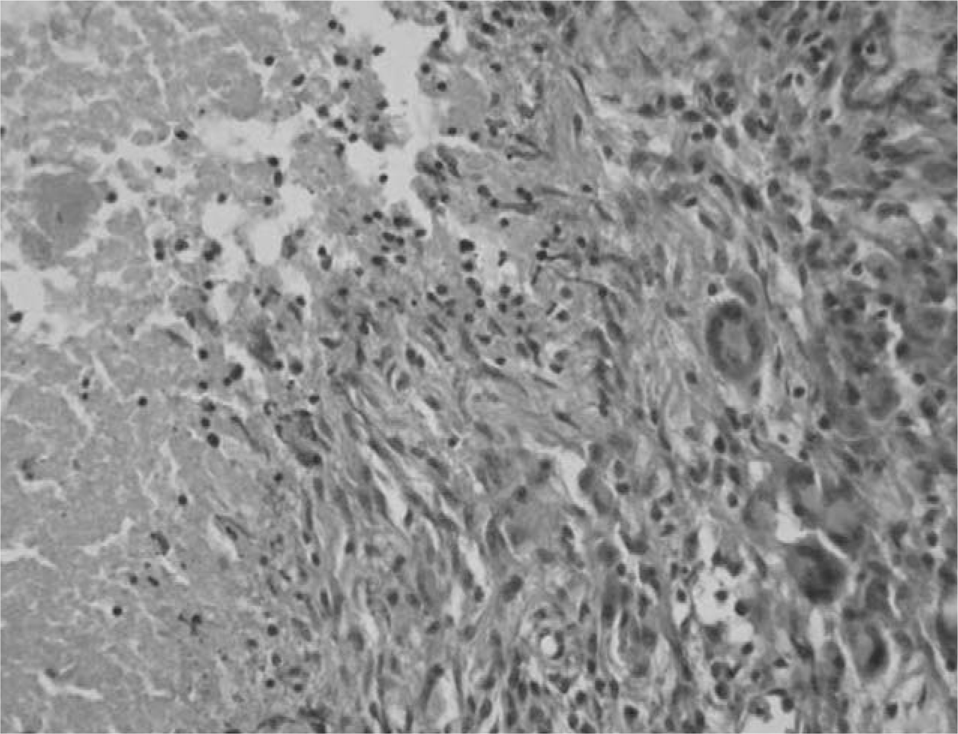 | Fig. 2.Histologic finding shows caseous necrosis with chronic granulated inflammation (H-E×100). |
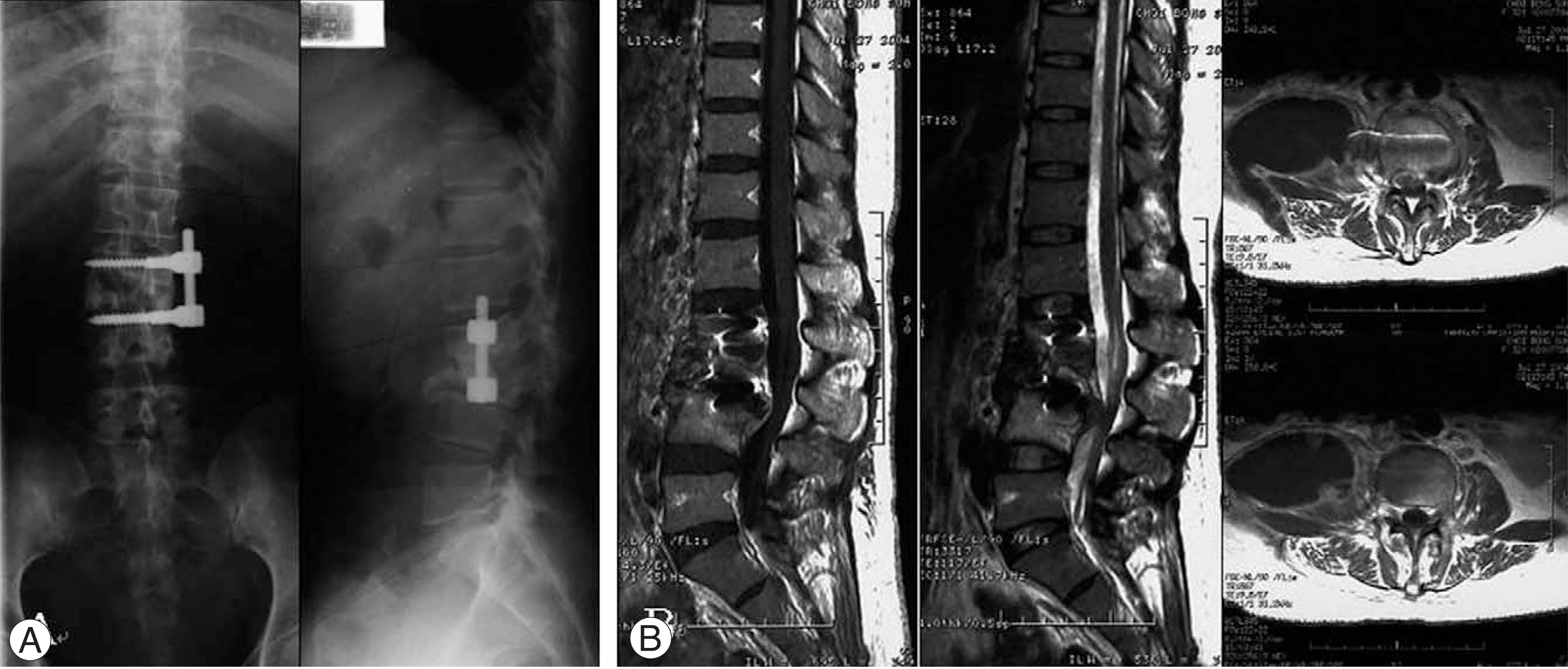 | Fig. 3.Radiographs and MR Imaging at 26months after the operation. (A) Radiographs show collapse of grafted bone, destruction of vertebral bodies and kyphotic deformity at L2-4. (B) Sagittal T1 and T2-weighted imaging of L2-4 demonstrate destruction of vertebral bodies and severe compression of dural sacs. Axial T2-weighted images show the large abscess formation at psoas muscle and fistula formation, right. |
 XML Download