Abstract
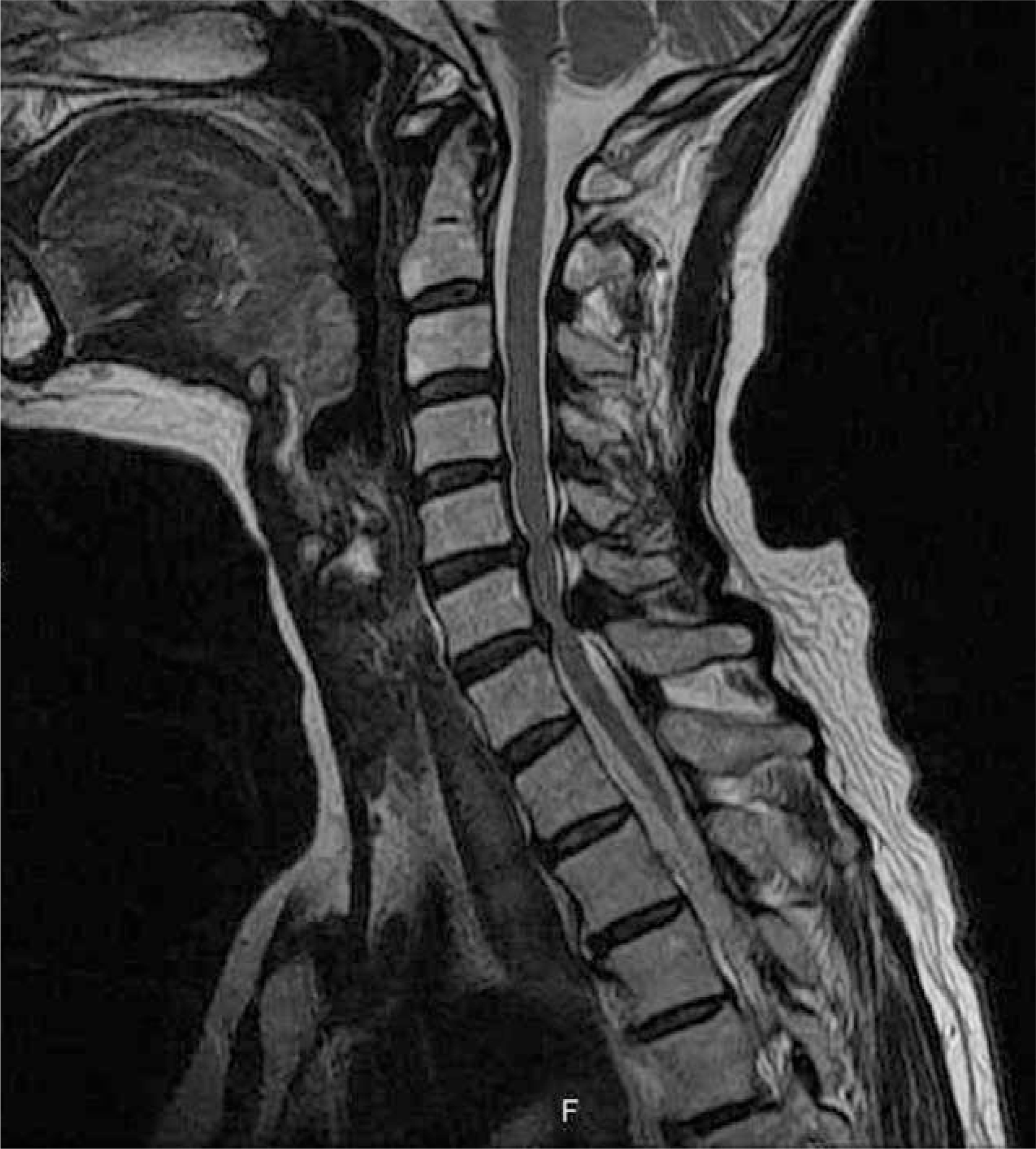
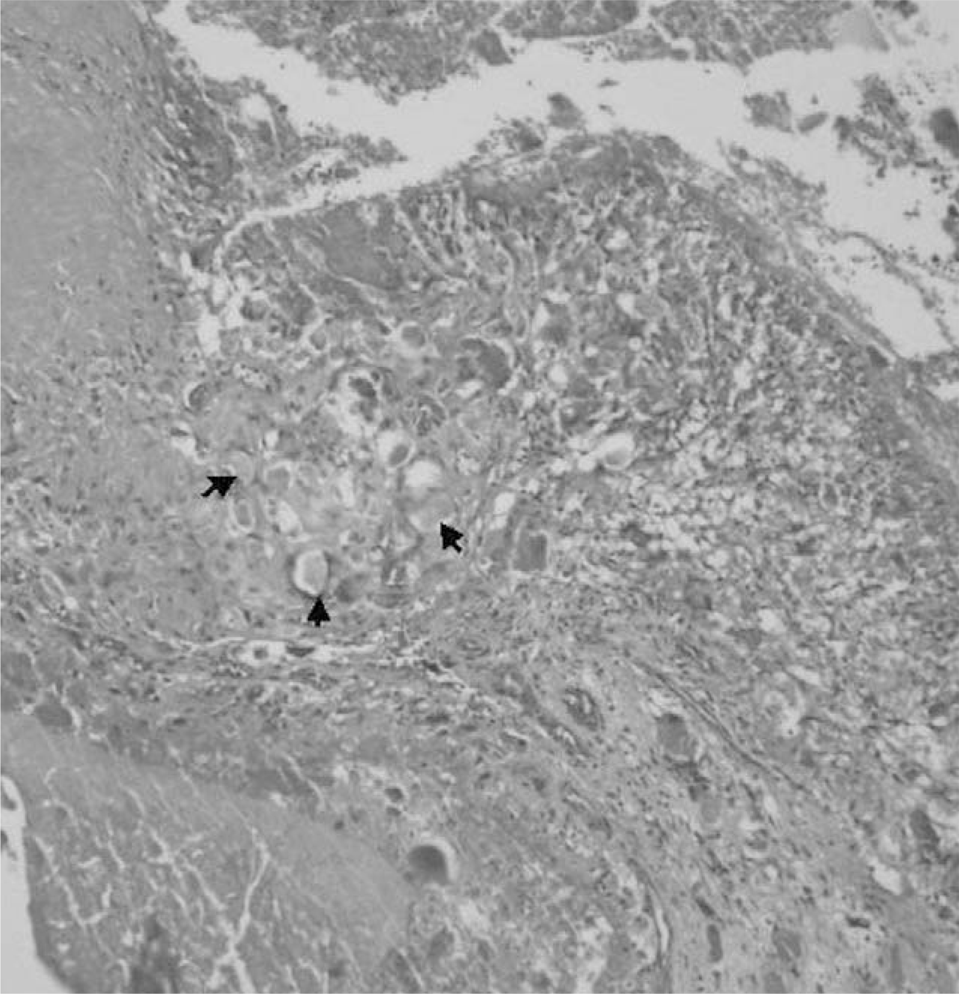
Calcium pyrophosphate dihydrate deposition disease (CPPD) is an inflammatory arthropathy that is defined by the deposition of CPPD crystals in articular and periarticular structures. The cervical ligamentum flavum is a rare location of CPPD deposition. A 65-year-old woman was admitted with complaints of neck pain and a tingling sensation and numbness below the xiphoid process for 2 months. Magnetic resonance (MR) imaging and computed tomography (CT) revealed compression of the spinal cord due to a nodular calcified mass in or attached to the ligamentum flavum at the C4-5, C5-6, or C6-7 level. The patient underwent a laminectomy at C4-5, C5-6, and C6-7, and resectioning of calcified extradural nodules that impinged on the cervical cord. The operation resulted in a resolution of neck pain and hypoesthesia, except in the feet. Histopathological examination of the excised specimen revealed rectangular CPPD crystals. Here, we report a case of compressive cervical spine due to CPPD deposition disease of the cervical spine and describe the literature relevant to CPPD deposition disease of the cervical spine.
Go to : 
REFERENCES
1). Ellman MH, Vazquez T, Ferguson L. Calcium pyrophosphate deposition in ligamentum flavum. Arthritis Rheum. 1978; 21:611–613.
2). Fye KH, Weinstein PR, Donald F. Compressive cervical myelopathy calcium pyrophosphate dihydrate deposition disease: Report of a case and review of the literature. Arch Intern Med. 1999; 159:189–193.
3). Joseph J, McGrath H. How to differentiate crystal-induced arthropathies. Geriatrics. 1995; 50:33–39.
4). Muthukumar N, Karuppaswamy U, Sankarasubbu B. Calcium pyrophosphate dihydrate deposition disease causing thoracic cord compression. Neurosurg. 2000; 46:222–225.
5). Kohn NN, Hughes RE, McCarty DJ, Faires JS. The significance of calcium pyrophosphate crystals in the syn-ovial fluid of arthritic patients, The “pseudogout syndrome”. Ann Intern Med. 1962; 56:738–745.
6). Fam AG. Calcium pyrophosphate crystal deposition disease and other crystal deposition disease. Curr Opin Rheumatol. 1995; 7:364–368.
8). Maigne JY, Ayral X, Guerin-Surville H. Frequency and size of ossifications in the caudal attachments of the ligamentum flavum of the thoracic spine. Surg Radiol Anat. 1992; 14:119–124.
9). Sato R, Takahashi M, Yamashita Y, et al. .:. Calcium crystal deposition disease in cervical ligamentum flavum, CT and MRI findings. J Comput Assist Tomogr. 1992; 16:352–355.
10). Sugimura H, Kakitsubata Y. MRI of ossification of the ligamentum flavum. J Comput Assist Tomogr. 1992; 16:73–76.
Go to : 
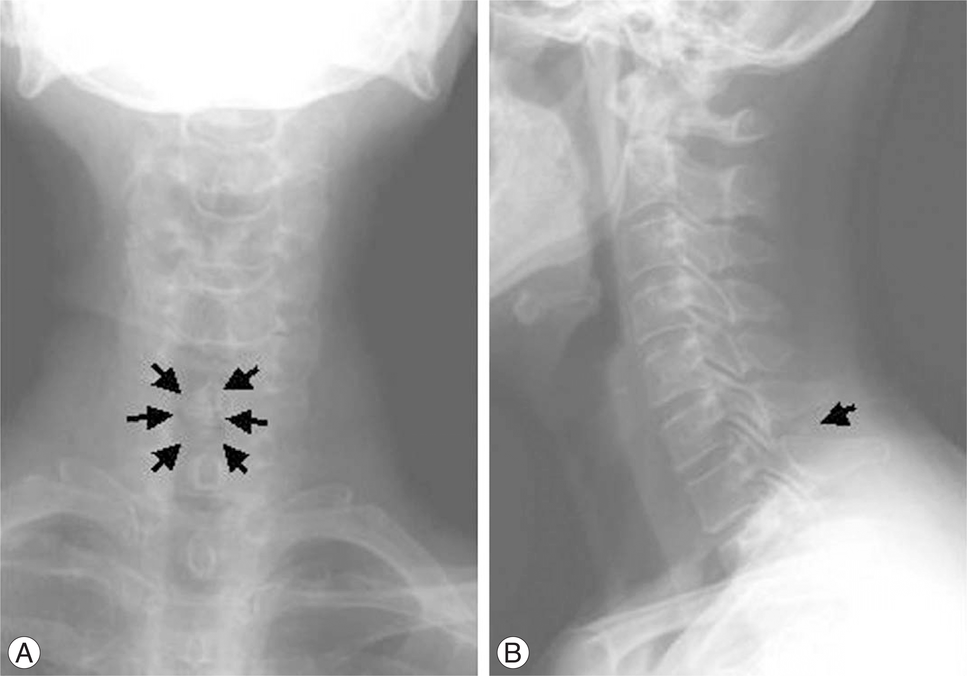
 | Fig. 1.Calcium pyrophosphate dihydrate (CPPD) crystal deposition disease in a 65-year-old woman with a history of neck pain and numbness below xiphoid process. Plain radiographs show dense and homogenous radiopaque deposits (arrows indicated) at C6-7 level in AP view (A) and lateral view (B). |
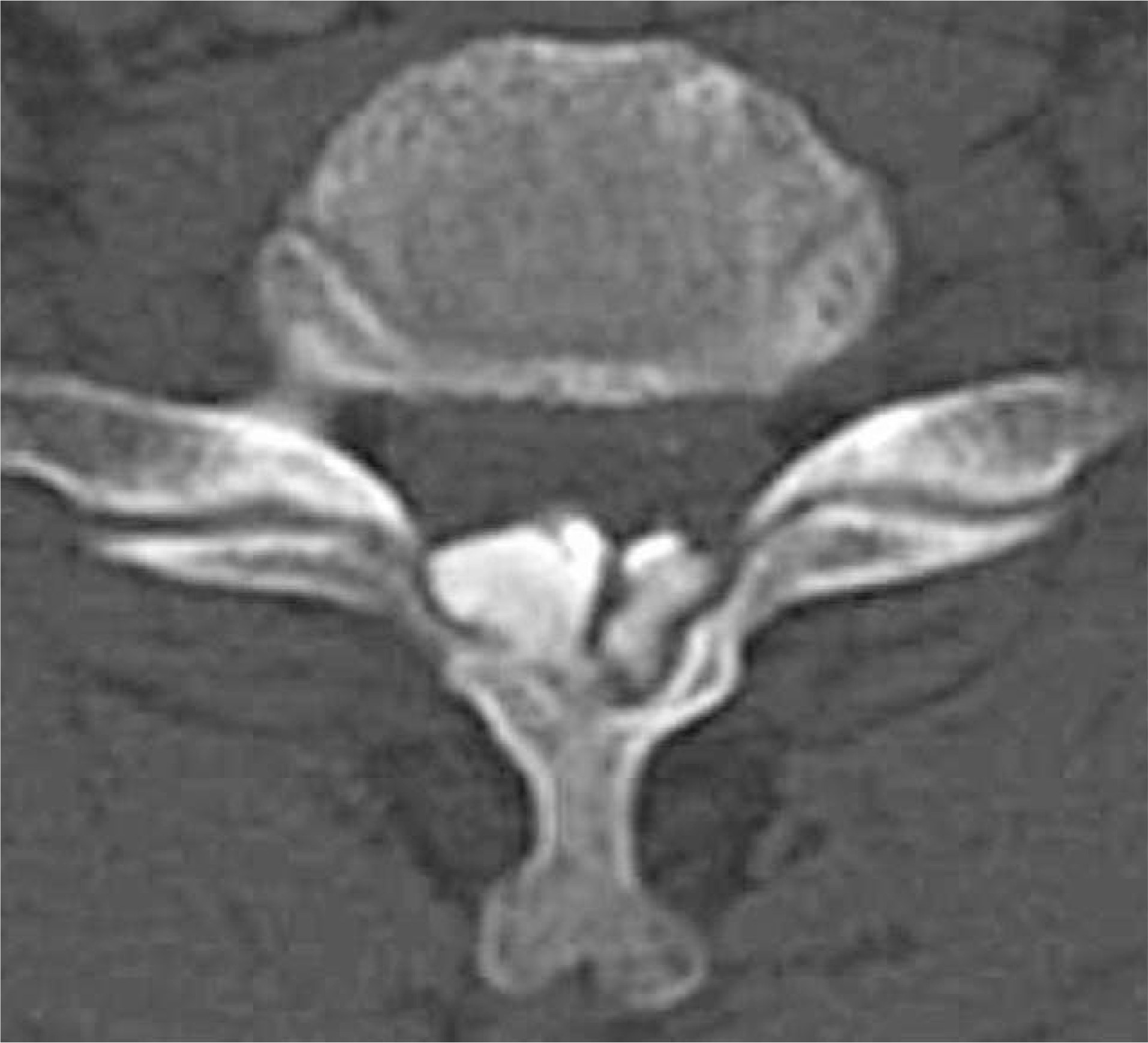
 | Fig. 2.Computed tomographic scan showing nodular calcified deposits in the ligamentum flavum at C6-7. |


 PDF
PDF ePub
ePub Citation
 Print
Print


 XML Download