Abstract
Objectives
To evaluate the efficacy of transpedicular bone graft and pedicle screw fixation in delayed collapse of osteoporotic vertebral fracture with claudication. Summary of Literature
Review
Delayed collapse of osteoporotic vertebral fracture may result in seemingly unrelenting back pain and neurologic deficits. Though there are many surgical options for such cases, comprehensive improvement of symptoms is uncertain.
Materials and Methods
Nineteen patients who underwent operation and were followed-up for more than 2 years were studied. The regional sagittal angle, restoration ratio of the vertebral body, standing sagittal balance, and additional fracture were assessed. Improvement of back and leg pain was assessed using 10 point Visual Analog Scales (VAS). The causes of sustained clinical symptoms were analyzed.
Results
The regional sagittal angle was corrected from 25.2±13.9。 to 12.4±10.4。(p=0.000). The vertebral body ratio was restored from 36±14.1% to 72±16.7% (p=0.000). Six cases were found to be neutral and 13 cases showed a positive sagittal balance. Additional fractures were found in 11 cases. The VAS value for leg pain was improved from 6.6±1.0 to 1.0±1.1 (p=0.000), while that for back pain was not improved (6.4±1.7 to 7.1±2.3, p=0.474). Positive sagittal balance was a significant risk factor (p=0.037, odds ratio=58.084) for sustained back pain.
Conclusion
For the treatment of delayed collapse of osteoporotic vertebral fracture with claudication, transpedicular bone graft and pedicle screw fixation was effective in improving claudication and restoring the vertebral body and regional sagittal angle. However, it was not capable of alleviating back pain. Positive sagittal balance was considered to be a cause of sustained back pain.
Go to : 
REFERENCES
1). Ito Y, Hasegawa Y, Toda K, Nakahara S. Pathogenesis and diagnosis of delayed vertebral collapse resulting from osteoporotic spinal fracture. Spine J. 2002; 2:101–106.

2). Kim KT, Suk KS, Kim JM, Lee SH. Delayed vertebral collapse with neurological deficits secondary to osteoporosis. Int Orthop. 2003; 27:65–69.

3). Shikata J, Yamamuro T, Iida H, Shimizu K, Yoshikawa J. Surgical treatment for paraplegia resulting from vertebral fractures in senile osteoporosis. Spine. 1990; 15:485–489.

4). Young WF, Brown D, Kendler A, Clements D. Delayed post-traumatic osteonecrosis of a vertebral body (Ku¨mmell's disease). Acta Orthop Belg. 2002; 68:13–19.
5). Halvorson TL, Kelley LA, Thomas KA, Whitecloud TS 3rd, Cook SD. Effects of bone mineral density on pedicle screw fixation. Spine. 1994; 19:2415–2420.

6). Alanay A, Acaroglu E, Yazici M, Aksoy C, Surat A. The effect of transpedicular intracorporeal grafting in the treatment of thoracolumbar burst fractures on canal remodeling. Eur Spine J. 2001; 10:512–516.

7). Kaminski A, Muller EJ, Muhr G. Burst fracture of the fifth lumbar vertebra: results of posterior internal fixation and transpedicular bone grafting. Eur Spine J. 2002; 11:435–440.
8). Knop C, Fabian HF, Bastian L, et al. .:. Fate of the transpedicular intervertebral bone graft after posterior stabilization of thoracolumbar fractures. Eur Spine J. 2002; 11:251–257.
9). Leferink VJ, Zimmerman KW, Veldhuis EF, ten Verg-ert EM, ten Duis HJ. Thoracolumbar spinal fractures: radiological results of transpedicular fixation combined with transpedicular cancellous bone graft and posterior fusion in 183 patients. Eur Spine J. 2001; 10:517–523.

10). Mirovsky Y, Anekstein Y, Shalmon E, Peer A. Vacuum clefts of the vertebral bodies. AJNR Am J Neuroradiol. 2005; 26:1634–1640.
Go to : 
Figures and Tables%
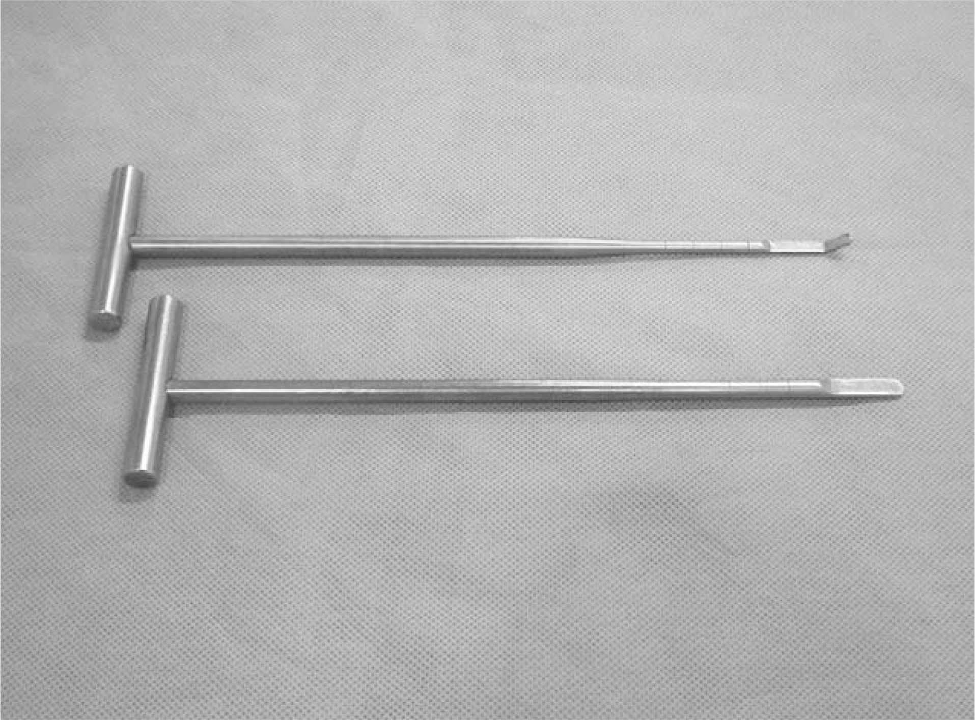
 | Fig. 1.Photographs of the pedicle reamer and end plate eleva-tor which are used for extension of interbody space. |
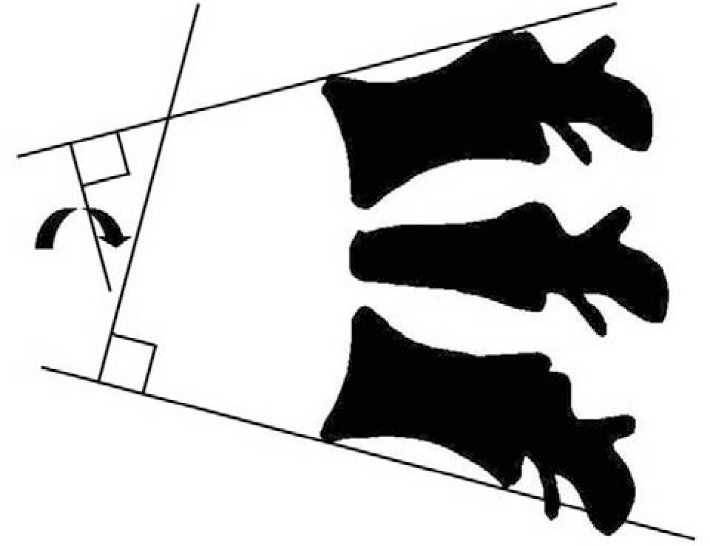
 | Fig. 2.Regional sagital angles were estimated by Cobb's method which is consist of upper end plate of upper vertebra and lower end plate of lower vertebra adjacent to collapsed vertebral body. |
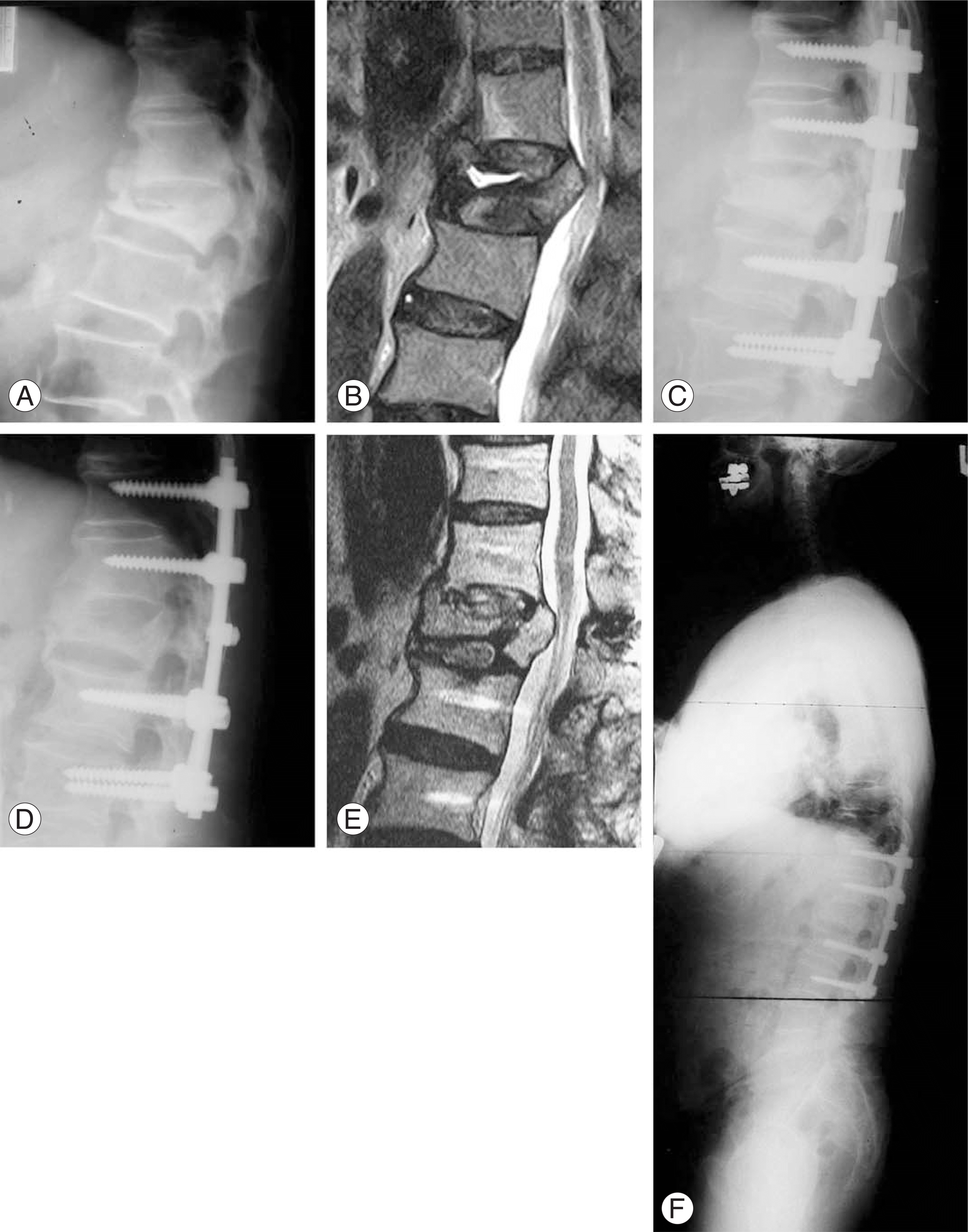
 | Fig. 3.These are images of 73 year old male patient with delayed collapse of osteporotic vertebral fracture with claudication. (A) Initial lateral radiograph shows cleft in L1 body. (B) Initial T2-weighted MRI film demonstrates not only vertebral body collapse but also neural canal encroachment. (C) Immediate postoperative lateral radiograph shows restoration of regional sagittal angle and body height. (D) Postoperative 31 months lateral radiograph shows maintenance of correction angle. (E) Postoperative 31 months T2-weighted MRI shows solid union of body and decompressed neural canal. (F) Postoperative 31 months whole spine standing lateral radiograph shows neutral sagittal balance. |


 PDF
PDF ePub
ePub Citation
 Print
Print


 XML Download