

 PDF
PDF ePub
ePub Citation
 Print
Print


Abstract
Study Design
This retrospective study was designed to evaluate treatment options for spinal stenosis with degenerative scoliosis.
Purpose
To evaluate the clinical outcomes based on the degree of spinal deformity for selective pedicle screw fixation with a long fusion for spinal stenosis with degenerative scoliosis.
Materials and Methods
We reviewed 54 cases performed from March 1996 to March 2006, and divided them into three groups based on osteophyte formation, pedicular rotation, and lateral transition. The three groups were analyzed for degree of correction of scoliotic and lordotic angle and bone fusion rate, as well as radiographically and clinically using the Kirkaldy-Willis questionnaire.
Results
Mild or moderate deformities (49 cases) were improved an average of 3 degrees of scoliotic angle, grade 1 of pedicular rotation, and 1 mm of lateral transition and were satisfied clinically. Severe deformities (5 cases) improved an average of 8 degrees of scoliotic angle, grade 2 of pedicular rotation, and 3 mm of lateral transition, but were clinically unsatisfactory. There was insignificant correction of the lordotic angle in all deformities and a fusion rate of 81.5% in mild-to-moderate deformities and 40% in severe deformities.
Go to : 
REFERENCES
1). Benner B, Ehni G. Degenerative lumbar scoliosis. Spine. 1979; 4:548–552.

2). Boos N, Marchesi D, Zuber K, Aebi M. Treatment of severe spondylolisthesis by reduction and pedicular fixation: A 4-6 year followup study. Spine. 1993; 18:1655–1661.
3). Chen SH, Mo Lin R, Chen HH, Tsai KJ. Biomechanical effects of polyaxial pedicle screw fixation on the lumbosacral segments with an anterior interbody cage sup-port. BMC Musculoskelet Disord. 2007 Mar 10; 8:28.
4). Dalenberg DD, Asher MA, Robinson R, Jayaraman G. The effect of stiff spinal implant and its loosening on bone mineral content in canines. Spine. 1993; 18:1862–1866.
5). Doherty JH. Complication of fusion in lumbar scoliosis. J Bone Joint Surg Am. 1973; 53:438.
6). Epstein JA, Epstein BS, Lavine LS. Surgical treatment of nerve root compression caused by scoliosis of the lumbar spine. J Neurosurg. 1974; 41:449–454.
7). Grubb SA, Lipscomb HJ. Diagnostic findings in painful adults scoliosis. Spine. 1992; 17:518–527.
8). Jutte PC, Castelein RM. Complications of pedicles screws in lumbar and lumbosacral fusions in 105 consecutive primary operations. Eur Spine J. 2002; 11:594–598.
9). Krag MH. Biomechanics of thoracolumbar spinal fixation. A review. Spine. 1990; 15:1216–1222.
10). Katz JN, Lipson SJ, Lew RA, et al. Lumbar laminectomy alone or with instrumented or noninstrumented arthrodesis in degenerative lumbar spinal stenosis, patient selection, costs and surgical outcomes. Spine. 1997; 22:1123–1131.
11). Kim YT, Lee CS, Lee HM, Shin SJ. Operative treatment of degenerative scoliosis. J Korean Soc Spine Surg. 2001; 8:491–496.
12). Kirkaldy-Willis WH, Wedge JH, Yong-Hing K, Tchang S, de Korompay V, Shannon R. Lumbar spinal nerve lateral entrapment. Clin Orthop. 1982; 169:171–178.
13). Kostuik JP, Bentivoglio J. The incidence of low back-pain in adult scoliosis. Spine. 1981; 6:268–273.
14). La Grone MO. Loss of lumbar lordosis, A complication of spinal fusion for scoliosis. Orthop Clin North Am. 1988; 19:383–393.
15). Lenke LG, Birdwell KH, Bullis D, Betz RR, Baldus C, Schoenecker PL. Results of in situ fusion for isthmic spondylolisthesis. J Spinal Disorder. 1992; 5:433–442.
16). Louis R. Fusion of the lumbar and sacral spine by internal fixation with screw plates. Clin Orthop. 1986; 203:18–33.
17). Moe JH, Denis F. The iatrogenic loss of lumbar lordosis, Orthop Trans. 1977; 1:131.
18). Nash CL, Moe JH. A study of vertebral rotation. J Bone joint Surg Am. 1969; 51:223–229.
19). Robin GC, Span Y, Steinberg R, Makin M, Menczel J. Scoliosis in the elderly: a follow up study. Spine. 1982; 7:355–359.
20). Simmons EH, Jackson RP. The management of nerve root entrapment syndromes associated with the collapsing scoliosis of idiopathic lumbar and thoracolumbar curves. Spine. 1979; 4:533–541.
21). Turner JA, Ersek M, Herron L, et al. Patient outcomes after lumbar spinal fusions. JAMA. 1992; 268:907–911.
22). Zdeblick TA. Laparoscopic spinal fusion. An international course on global. Orthop Clin North Am. 1998; 29:635–645.
23). Zucherman JF, Hsu K, Picetti G 3rd, White A, Wynne G, Taylor L. Clinical efficacy of spinal instrumentation in lumbar degenerative disc disease. Spine. 1992; 17:834–837.
Go to :
Figures and Tables%
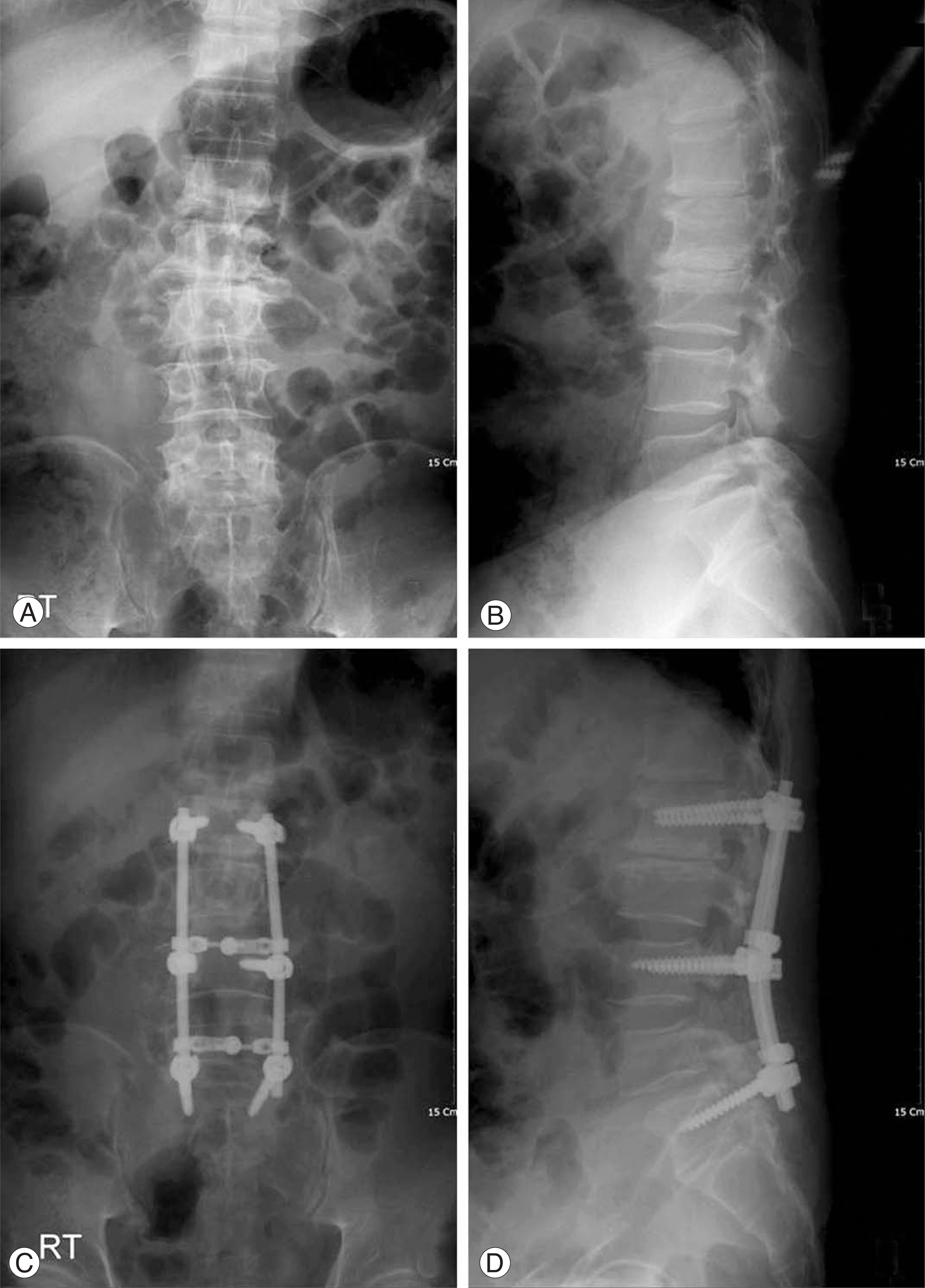 | Fig. 1.A 52-year-old female with degenerative scoliosis with mild spinal deformity. (A) Preoperative anteroposterior radiograph shows 11 degrees of scoliotic angle, grade 1 pedicular rotation and 2mm lateral transition. (B) Preoperative lateral radiograph. (C) Postoperative anteroposterior radiograph shows 2 degrees of scoliotic angle, grade 0 pedicular rotation and 1mm lateral transition. (D) Postoperative lateral radiograph. |
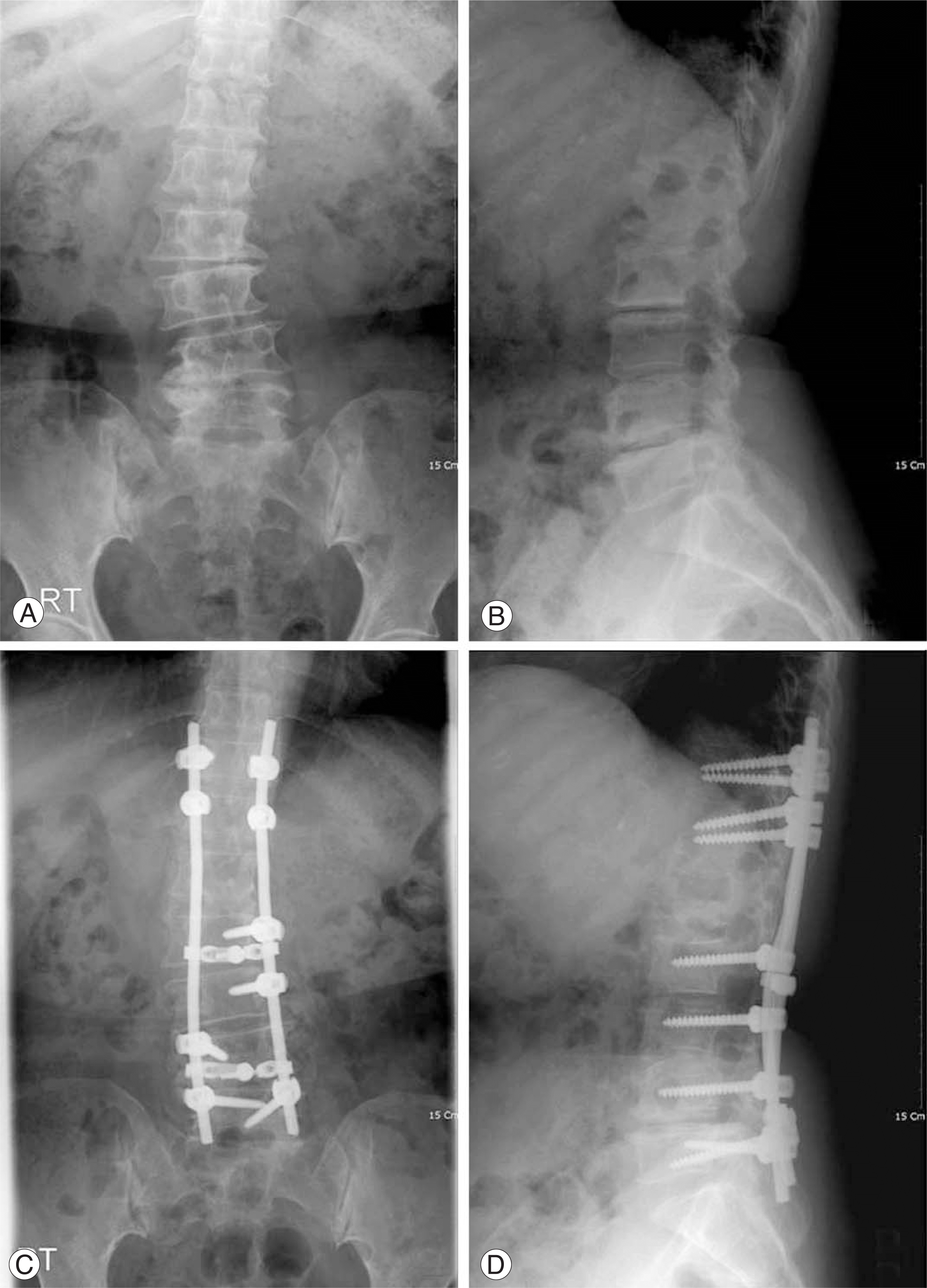 | Fig. 2.A 62-year-old female with degenerative scoliosis with severe spinal deformity. (A) Preoperative anteroposterior radiograph shows 26 degrees of scoliotic angle, grade 2 pedicular rotation and 5mm lateral transition. (B) Preoperative lateral radiograph. (C) Postoperative anteroposterior radiograph shows 11 degrees of scoliotic angle, grade 1 pedicular rotation and 2mm lateral transition. (D) Postoperative lateral radiograph. |
Table 1.
Kirkaldy-Willis Questionnaire
 XML Download