Abstract
Tumoral calcinosis is a rare disease involving the ectopic calcifications in the major juxtaarticular sites that was first described by Inclan Alberto in 1943. The etiology of tumoral calcinosis is still obscure. A disturbance of the phosphate metabolism in the kidney has been considered a major cause. However, some patients have no laboratory abnormalities. Tumoral calcinosis in the spine has not been reported in Korea. Recently, we encountered a case of tumoral calcinosis in the lumbar region. The clinical and pathological findings are discussed with a review of the relevant literature.
REFERENCES
2). Mitnick PD, Goldfarb S, Slatopolsky E, Lemann J Jr, Gray RW, Agus ZS. Calcium and phosphate metabolism in tumoral calcinosis. Ann Intern Med. 1980; 92:482–487.

3). Harkness JW, Perters HS. Tumoral calcinosis. A report 6 cases. J Bone and Joint Surg Am. 1967; 49:721–731.
5). Baldursson H, Evans EB, Dodge W, Jackson T. Tumoral calcinosis with hyperphosphatemia. J Bone and Joint Surg. 1969; 51A:960–964.

6). Lever WF, Schaumberg-Lever G. Histopathology of the skin. 6 th ed. Philadelphia: JB Lippincott Co;420. 1983.
7). Duret MH. Tumeurs multiples et singulieres des bourse sereuses. Bulletin de la Societe Anatomique de Paris. 1899; 74:725–731.
8). Teutschlaender O. Lipid calcinosis. Zieglers Beitr. 1947; 110:402–405.
10). Kirk TS, Simon MA. Tumoral calcinosis. Report of a case with successful medical management. J Bone and Joint Surg Am. 1981; 63:1167–1169.

11. Durant DM, Riley LH 3 rd, Burger PC, McCarthy EF. Tumoral calcinosis of the spine. A study of 21 cases. Spine. 2001; 26:1673–1679.
Fig. 2.
Lateral X-ray of lumbar spine shows scanty calcification (arrow) at L3~4 interspinous ligament area.
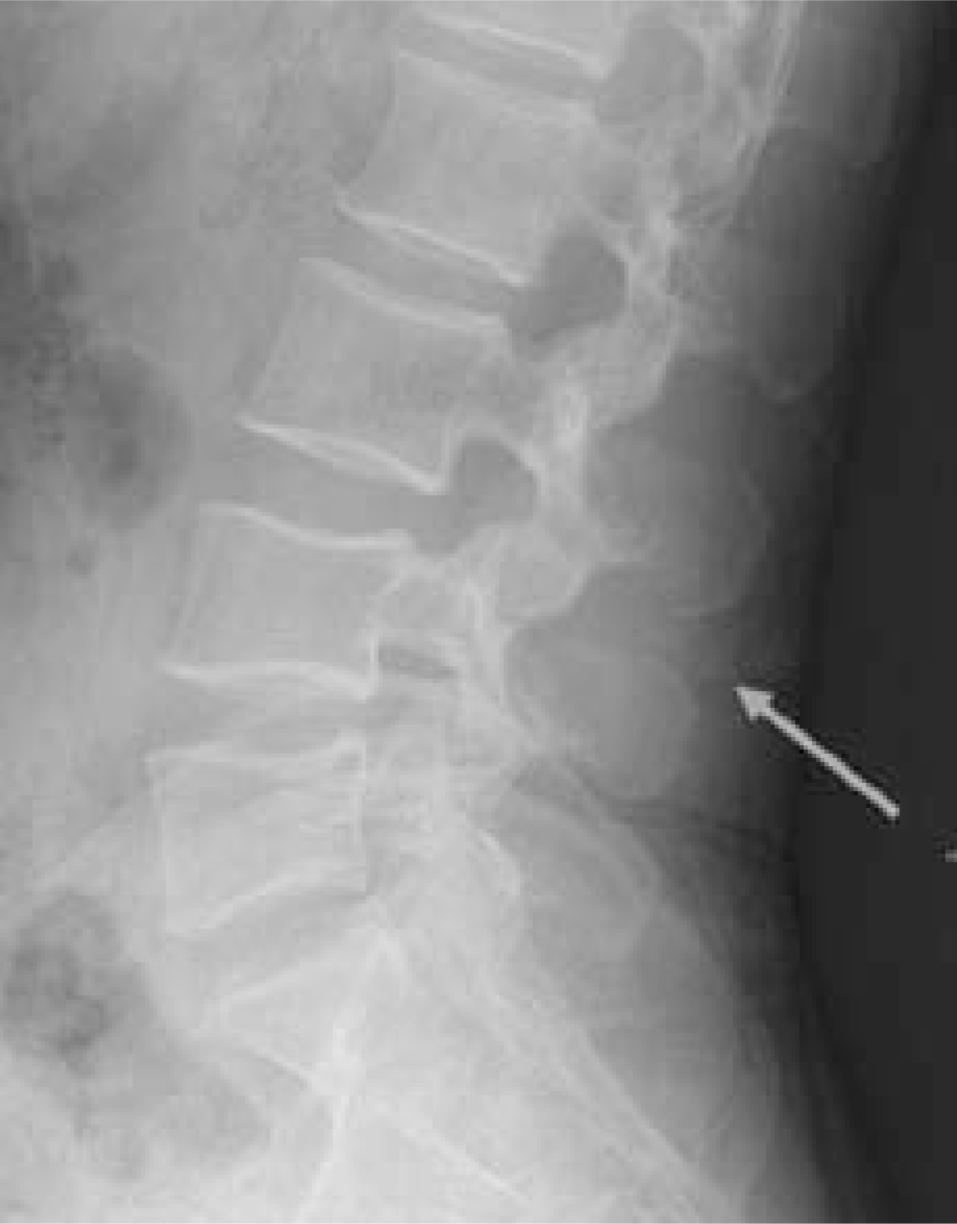
Fig. 3.
Sagittal images of MRI shows a mass at interspinous ligament area. T1-weighted and T2-weighted images show that heterogenous low signal change is surrounded by a rim of increased signal. (A) T1-weighted image, (B) T2-weighted image.
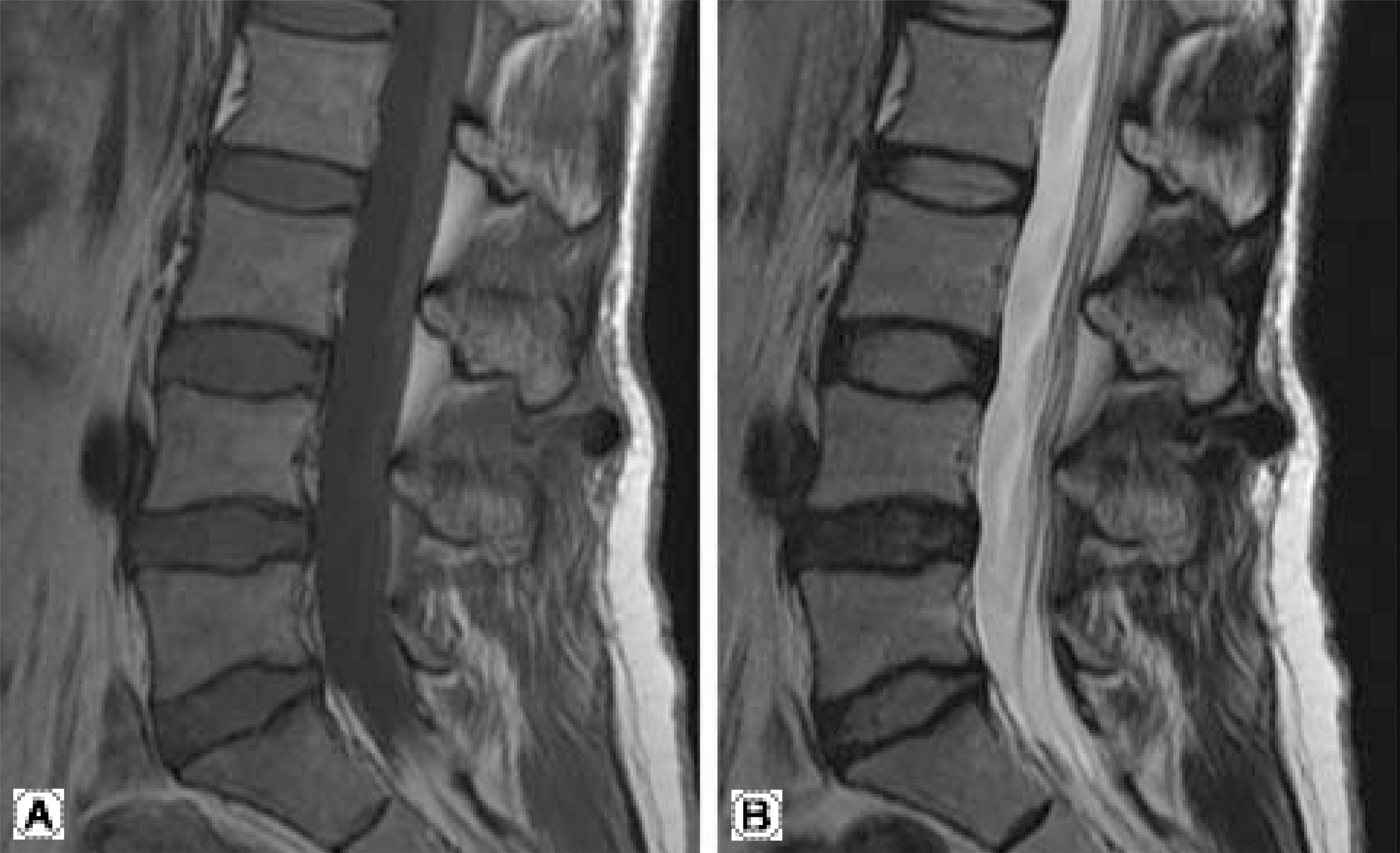
Fig. 4.
Axial images of MRI shows septum of increased signal at T1-weighted and T2-weighted image. T1 weighted image shows sedimentation sign (arrow). (A) T1-weighted image, (B) T2-weighted image.
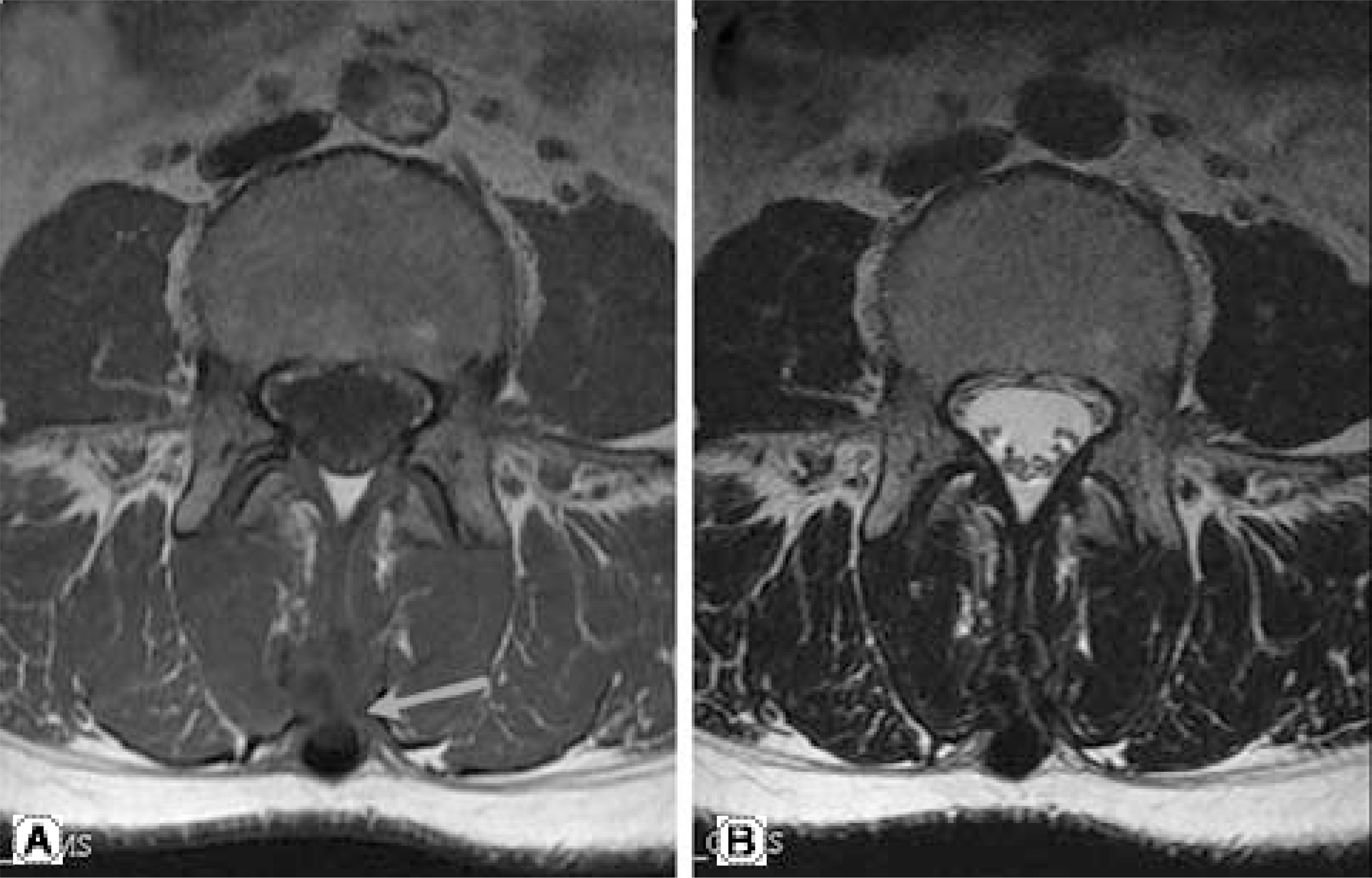
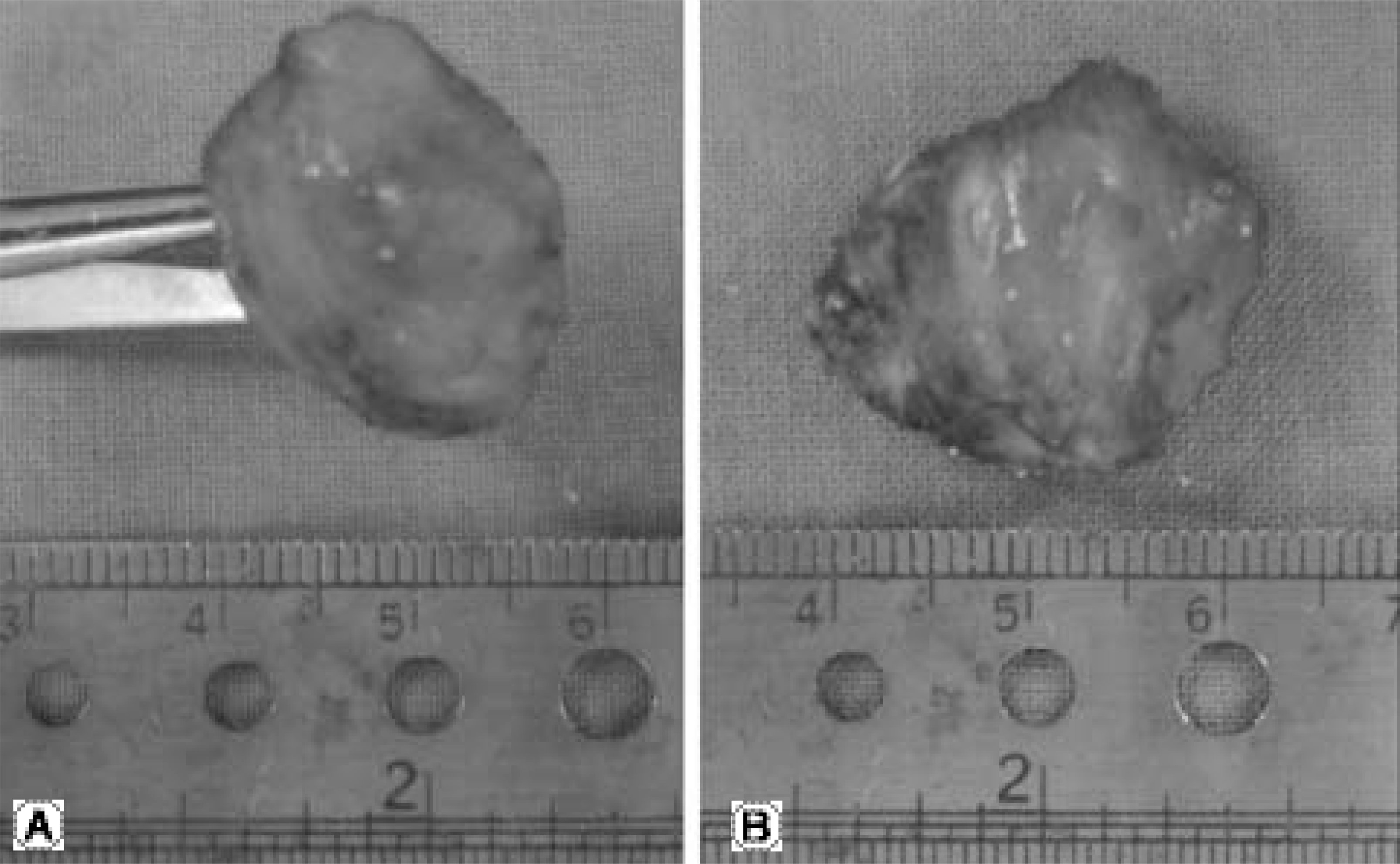
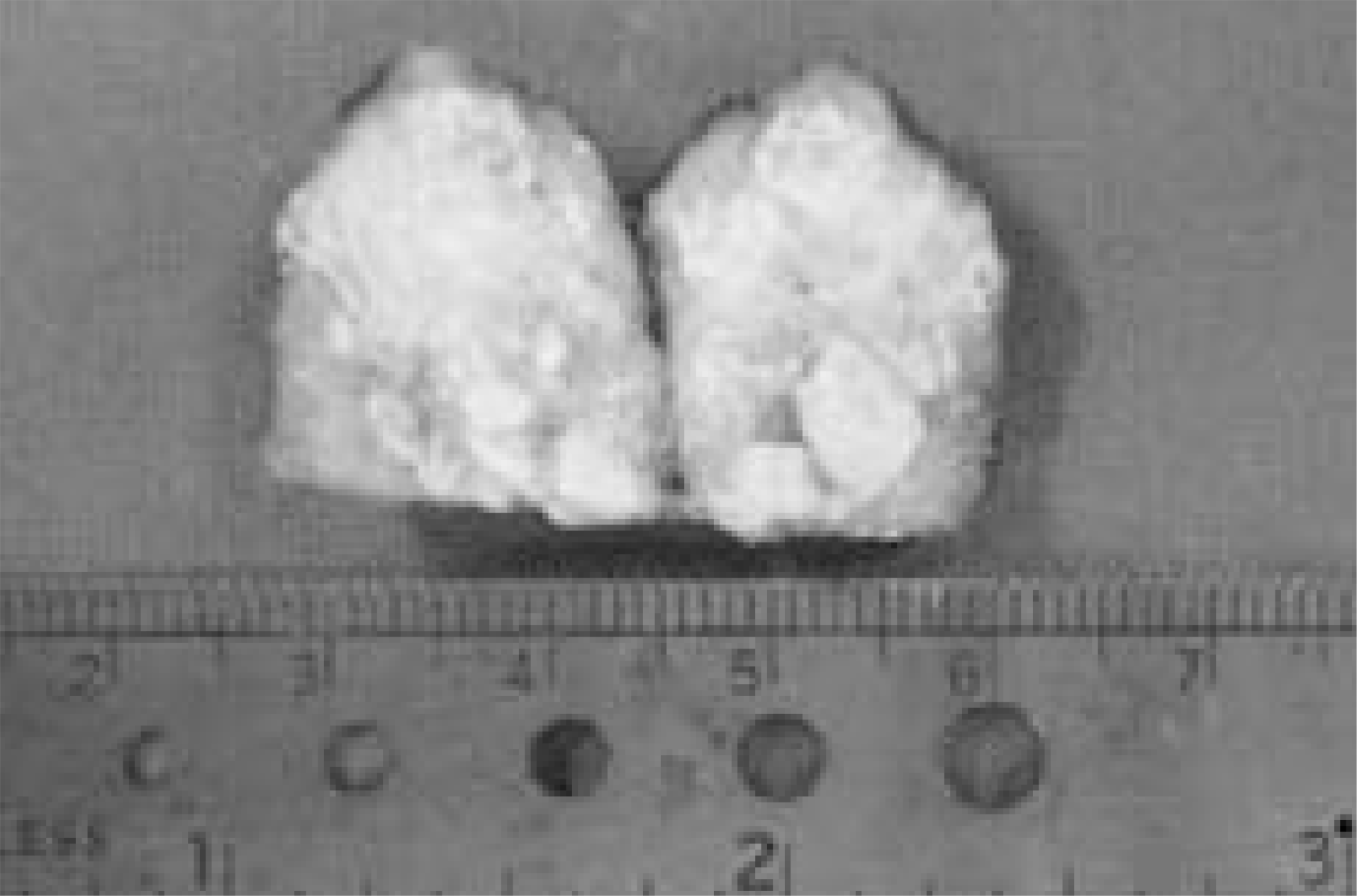
Fig. 5.
It is a photograph of the mass after excision. The excised specimen is measured 1.5×2.0×2.0 cm in size. (A) Axial view of the mass, (B) Sagittal view of the mass


 PDF
PDF ePub
ePub Citation
 Print
Print


 XML Download