

 PDF
PDF ePub
ePub Citation
 Print
Print


Abstract
Two percent of neurological complications after spine surgery for various reasons have been reported. Most are static or improve with time. We encountered two cases of newly developed, progressive neurological deficits with severe pain radiating along the exiting root after posterior decompression, adhesiolysis, posterior lumbar interbody fusion (PLIF) with a cage for spinal stenosis after previous lumbar spine surgery. When explored, the severely tightened and less movable, edematous exiting root was entrapped by a pedicle without evidence of pedicle violation, direct injury, epidural hematoma or iatrogenic foraminal stenosis. A wider decompression with a resection of the pedicle reduced the root course and made it more movable. Immediately, the severe radiating pain subsided and the neurological deficit recovered. A progressive neurological deficit after spinal surgery for spinal diseases with foraminal stenosis can develop as a result of the inordinate manipulation of the root, which may pro-voke root edema, root self-entrapment around a pedicle and local ischemia. An image test and exploration should be performed immediately in cases of progressive single root neurological deficits immediately after spine surgery. Total decompression, even with a pedicle resection, should be considered to resolve the neurological deficits.
Go to : 
REFERENCES
1). Mayfield FH. Complications of laminectomy. Clin Neurosurg. 23:435–439. 1975.

2). Stambough JL, Simeone FA. Neurogenic complications in spinal surgery. (in. Rothman RH, Simeone FA, editors. The spine, 4th ed. ,.Philadelphia: WB Saunders Co;p. 1724. 1999. .).
3). Ahn DK, Lee S. Neurologically symptomatic epidural hematoma after lumbar spine surgery. J Koreans Soc Spine Surg. 11:154–160. 2004.
4). Shin BJ. Lee JC. Intraoperative spinal nerve root injuries surgery for degenerative low back disease. J Korean Soc Spine Surg. 9:142–147. 2002.
5). Rydevik B, Brown MD, Lundborg G. Pathoanatomy and pathophysiology of nerve root compression. Spine. 9:7–15. 1984.
6). Henriques T, Olerud C, Petren-Mallmin M, Ahl T. Cauda equina syndrome as a postoperative complication in five patients operated for lumbar disc herniation. Spine. 26:293–297. 2001.
Go to :
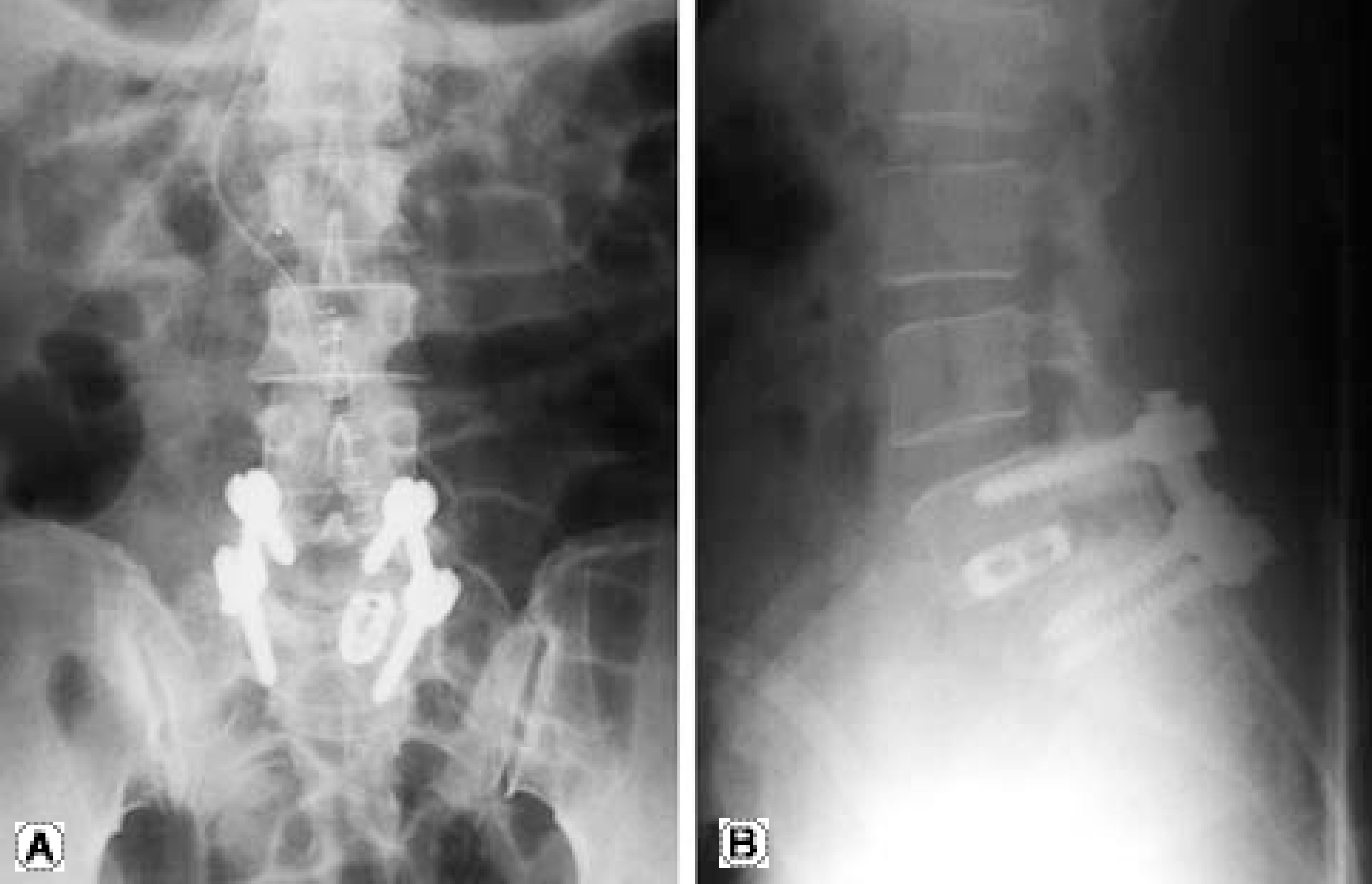 | Fig. 1.(A, B) After the first operation, plane radiographs showed that a right-sided cage violated upper end plate. |
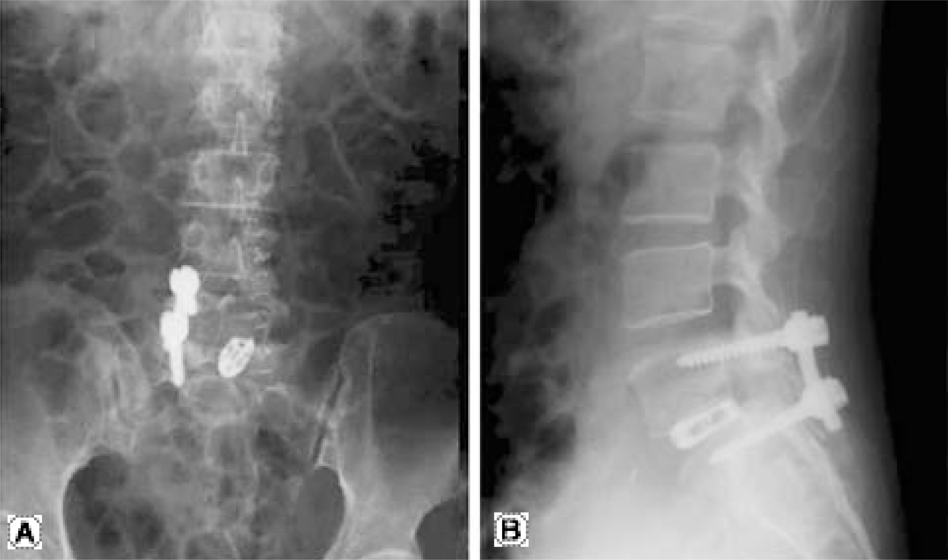 | Fig. 3.(A, B) After the third operation, plane radiographs showed that the L5 left-sided pedicle screw-removal and pedicle-resection was performed to decompress and intervene further ischemia of exiting root. |
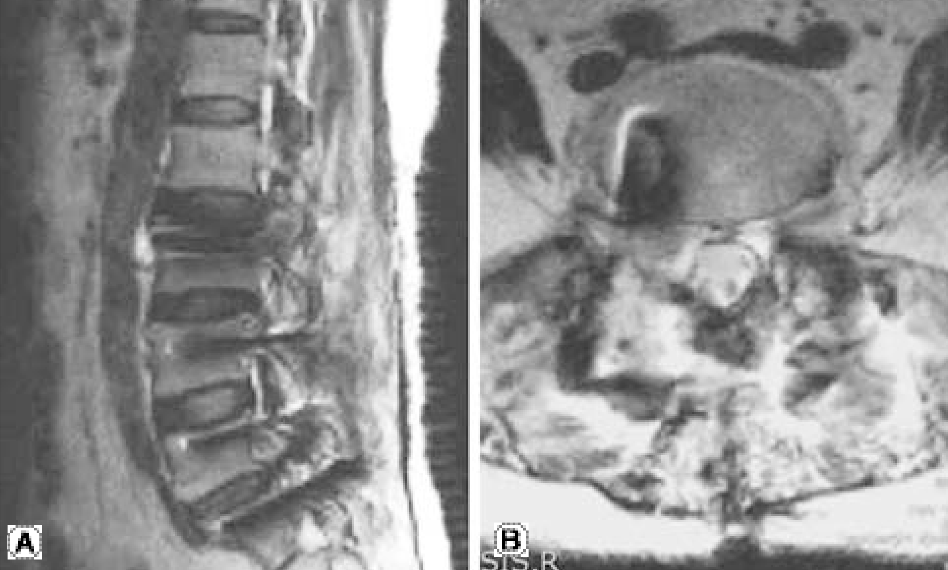
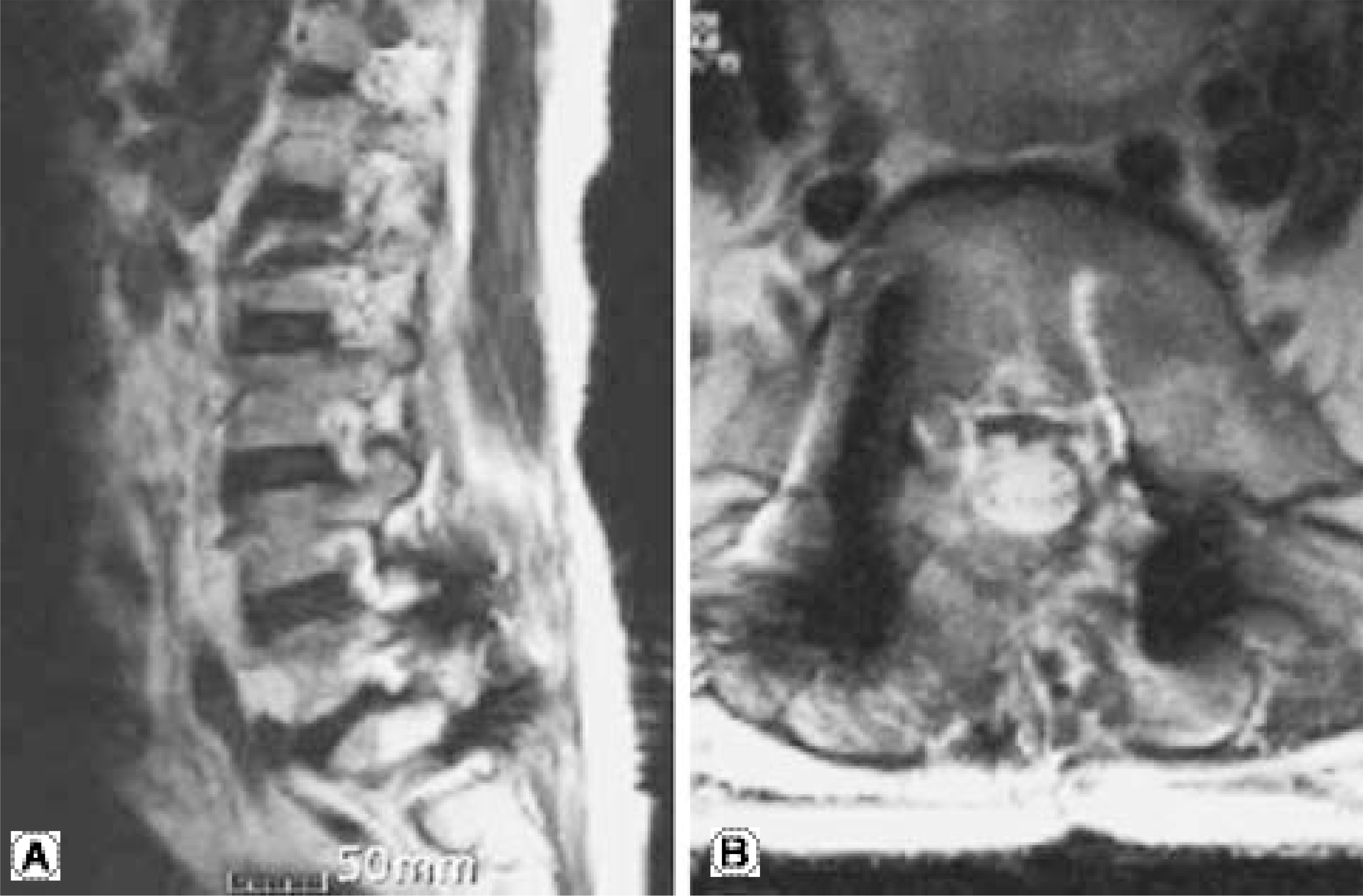
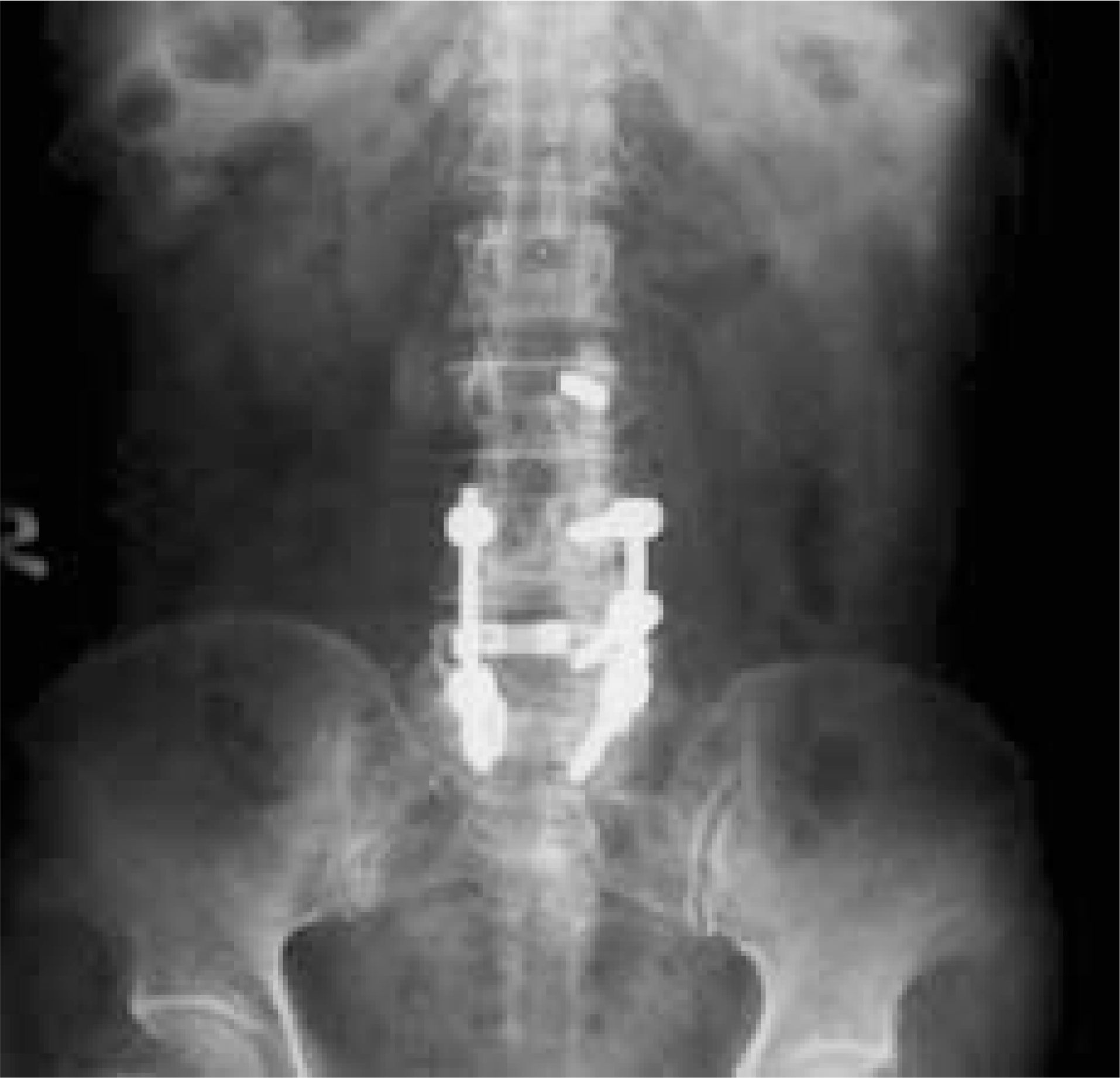 | Fig. 5.After the second operation, anteroposterior view showed that the L5 left-sided pedicle screw-removal and pedicle resection was performed. |
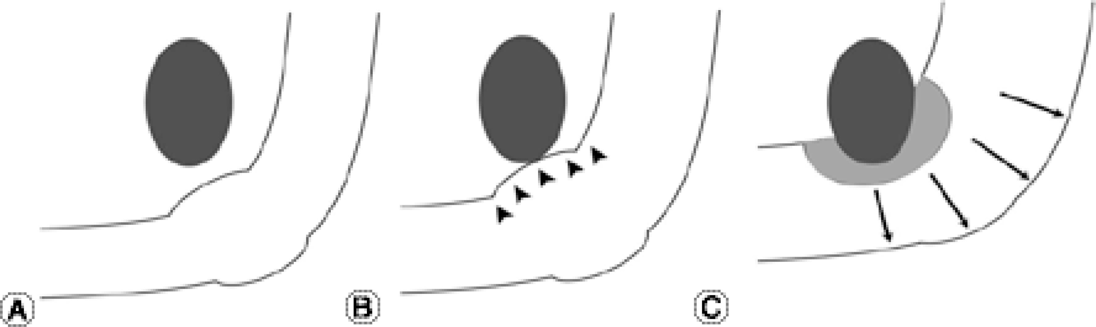 | Fig. 6.Hypothesis of progressive neurologic deficit after spine surgery (A) Normal: No compression of nerve root. (B) Swelling without impingement (as in “Battered root syndrome”): There might be pain and numbness, but not much edema enough to show definite neurologic complication or no motor weakness. (C) Impingement state : There might be severe pain and neurologic deficit. Stretching and edema of nerve root with subsequent venous congestion provoking local ischemia, caused severe pain and motor deficit. There should be need of surgical decompression immediately. |
 XML Download