Abstract
Objectives
To investigate the type of postsurgical spinal stenosis in patients who had undergone a primary laminectomy and discectomy for a herniated lumbar disc, and to evaluate the clinical outcomes of the revision operation.
Summary and Literature Review
Spinal stenosis occurs frequently after a laminectomy and discectomy. Facet joint arthritis, hypertrophy of the ligamentum flavum, iatrogenic instability, postsurgical scarring or any combination of these conditions can cause spinal stenosis.
Materials and Methods
Twenty-four patients, who had postsurgical spinal stenosis were reviewed. Patients with a simple recurrent disc herniation without a spinal stenosis were excluded. The mean age was 52.5 years (range 31~70). There were 19 males and 5 females. The primary discectomy were performed at L4-5 in 21 patients, L5-1 in 2 patients, and both L4-5 and L5-1 in 1 patient. The mean interval between the first discectomy and revision surgery was 11.6 years (range 2.7~40). The anatomical site of the spinal stenosis, combined herniated disc, height of the disc space, segmental instability, hypertrophy of facet joint and thickening of the ligamentum flavum in radiographs was evaluated. The clinical outcome was measured using the Oswestry disability index.
Results
Lateral spinal stenosis was observed in all patients. Eight patients showed both central and lateral stenosis. The lateral stenosis was caused by hypertrophy of the facet joint in 20 patients and a thickening of the ligamentum flavum in 8 patients. Nineteen patients showed herniated lumbar disc, including disc protrusion in 8 patients, disc extrusion in 9 patients, and disc sequestration in 2 patients. A loss of disc height was observed in 12 patients, segmental instability in 5 patients, and spondylolisthesis in 3 patients. All the patients received posterior decompression and posterolateral fusion with pedice screw instrumentation. Eighteen patients received a discectomy simultaneously. The average Oswestry score at the last visit was 24.4.
Go to : 
REFERENCES
1). Baba H, Chen Q, Kamitani K, Imura S, Tomita K. Revision surgery for lumbar disc herniation. Int Orthop. 1995; 19:98–102.

2). Homar CP, Peter D. Failed lumbar disc surgery. Clin Orthop Relat Res. 1982; 164:93–104.
4). Adams MA, Hutton WC. Prolapsed intervertebral disc. A hyperflexion injury. Spine. 1982; 7:184–191.
5). Campbell E, Whitfield RD. Certain reasons for failure following disc operations. NY State J Med. 1947; 47:2569–2572.
6). Amundsen T, Weber H, Lilleas F, Nordal HJ, Abdel-noor M, Magnaes B. Lumbar spinal stenosis clinical and radiologic features. Spine. 1995; 20:1178–1186.
7). Howard SA, Glover JM. Lumbar spinal stenosis: His-torical perspectives, classification and pathoanatomy. Seminars in spine surgery. 1994; 6-2:69–77.
8). Lee HM, Kim NH, Kim HJ, chung IH. Morphometric study of the lumbar spinal canal in the Korean population. Spine. 1995; 20(15):1967–1984.

9). John KE, Rosen I, Uden A. The natural course of lumbar spinal stenosis. Clin Orthop Relat Res. 1992; 279:82–6.
10). Scho¨nstro¨m N, Willen J. Imaging lumbar spinal stenosis. Radiol Clin North Am. 2001; 39:31–53.
11). Cantu RC. Neurologic athletic head and neck injury. The cervical spinal controversy. Clin Sports Med. 1998; 17:121–126.
12). Kim DY, Lee SH, Lee HY, et al. Validation of the Korean Version of the Oswestry Disability Index. Spine. 2005; 30:123–127.

13). Long DM, Filtzer DL, BenDebba M, Hendler NH. Clinical features of the failed-back syndrome. J Neurosurg. 1988; 69:61.

14). Suk SI, Lee JH, Min HJ, Kim HS, Ha CW, Park SE. Salvage procedure in failed low back surgery. J Korean Orthop Assoc. 1993; 28:1009–1016.

15). Kayaoglu CR, Calikoglu C, Binler S. Reoperation after lumbar disc surgery. J Int Med Res. 2003; 31:318–323.
17). Ivanic GM, Pink TP, Homann NC, Scheitza W, Goyal S. The post-discectomy syndrome. Arch Orthop Trauma Surg. 2001; 121:494–500.

18). Steven R, Harry N, Mirkovic S. Spinal stenosis. J Bone Joint Surg. Am. 1999; 81-A:572–586.
19). Breig A. Adverse mechanical tension in the central nervous system. An analysis of cause and effect. Stockholm: Almqvist & Wiksell International; New York: J. Wiley;1978.
20). Turner JA, Ers M, Herron L, et al. Patient outcomes after lumbar spinal fusions, JAMA. 1992; 268:907–911.
21). Katz JN, Lipson MSJ, Lew RA, et al. Lumbar laminectomy alone or with instrumented or noninstrumented arthrodesis in degenerative lumbar spinal stenosis patient selection, costs, and surgical outcomes, Spine. 1997; 22:1123–1131.
Go to : 
Figures and Tables%
 | Fig. 1.Plain lumbar spine AP and lateral view shows intervertebral disc space narrowing on L4-5 and L5-S1. |
 | Fig. 2.MRI image shows hypertrophy of facet joint and thickening of ligamentum flavum, causing lateral spinal stenosis. |
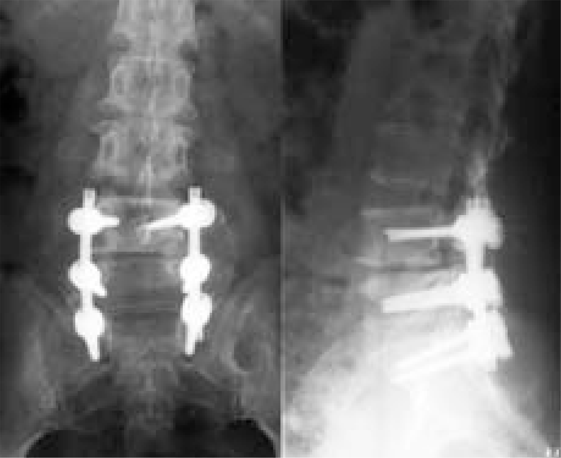
 | Fig. 3.1 year after posterolateral fusion with pedicle screw instrumentation, roentgenography shows good bony union. |
Table 1.
The etiologic factors of post-surgical spinal stenosis after discectomy for lumbar herniated disc


 PDF
PDF ePub
ePub Citation
 Print
Print


 XML Download