

 PDF
PDF ePub
ePub Citation
 Print
Print


Abstract
Objectives
To demonstrate the motile properties of the cartilage endplate (CE) chondrocytes and the effect of notochordal cells on this property.
Literature Review
Although previous in vivo studies have provided evidence for the migration of CE chondrocyte from hyaline CEs into the notochordal nucleus pulposus (NP), it is unclear if CE chondrocytes of the IVD actually have motile properties. In addition, the effect of notochordal cells on these properties has not been reported.
Materials and Methods
Notochordal cells and CE chondrocytes were harvested from three-month-old male Wistar rats and cultured separately. The motility was assayed in quadruplicate using a 48-well microchemotaxis chamber and a gelatin-coated 8-μm polycarbonate membrane filter. The control medium (serum-free culture medium), notochordal cells (4×, 2×, 1× and 0.5×10⁶) and concentrated conditioned medium (10-, 50-fold) where notochordal cells were cultured were loaded into the wells of the lower chamber, and CE chondrocytes were added to the wells of the upper chamber. At the end of the assays, the CE chondrocytes that migrated to the bottom side of the membrane filter were stained, counted, and compared.
REFERENCES
1). Buckwalter JA. Aging and degeneration of the human intervertebral disc. Spine. 1995; 20:1307–1314.

2). Kim KW, Lim TH, Kim JG, Jeong ST, Masuda K, An HS. The origin of chondrocytes in the nucleus pulposus and histologic findings associated with the transition of a notochordal nucleus pulposus to a fibrocartilaginous nucleus pulposus in intact rabbit intervertebral discs. Spine. 2003; 28:982–990.
3). Peacock A. Observations on the postnatal structure of the intervertebral disc in man. J Anat. 1952; 86:162–179.
4). Kim KW, Ha KY, Park JB, Woo YK, Chung HN, An HS. Expressions of Membrane-Type I Matrix Metallopro-teinase, Ki-67 Protein, and Type II Collagen by Chondrocytes Migrating from Cartilage Endplate into Nucleus Pulposus in Rat Intervertebral Discs: A Cartilage End-plate-Fracture Model Using an Intervertebral Disc Organ Culture. Spine. 2005; 30:1373–1378.
5). Singer SJ, Kupfer A. The directed migration of eukaryot-ic cells. Annu Rev Cell Biol. 1986; 2:337–365.
6). Aznavoorian S, Stracke ML, Krutzsch H, Schiffmann E, Liotta LA. Signal transduction for chemotaxis and haptotaxis by matrix molecules in tumor cells. J Cell Biol. 1990; 110:1427–1438.
7). Alini M, Li W, Markovic P, Aebi M, Spiro RC, Rough-ley PJ. The potential and limitations of a cell-seeded collagen/hyaluronan scaffold to engineer an intervertebral disc-like matrix. Spine. 2003; 28:446–454.
8). Gan JC, Ducheyne P, Vresilovic EJ, Shapiro IM. Intervertebral disc tissue engineering II: cultures of nucleus pulposus cells. Clin Orthop. 2003; 411:315–324.
9). Masuda K, Takegami K, An H, et al. Recombinant osteogenic protein-1 upregulates extracellular matrix metabolism by rabbit annulus fibrosus and nucleus pulposus cells cultured in alginate beads. J Orthop Res. 2003; 21:922–930.
10). Nam SW, Clair T, Campo CK, Lee HY, Liotta LA, Stracke ML. Autotaxin (ATX), a potent tumor motogen, augments invasive and metastatic potential of ras-trans-formed cells. Oncogene. 2000; 19:241–247.
11). Nam SW, Clair T, Schiffmann E, Liotta LA, Stracke ML. A sensitive screening assay for secreted motility-stimulating factors. Cell Motil Cytoskeleton. 2000; 46:279–284.
12). Hama K, Aoki J, Fukaya M, et al. Lysophosphatidic acid and autotaxin stimulate cell motility of neoplastic and non-neoplastic cells through LPA1. J Biol Chem. 2004; 279:17634–17639.
13). Kim KW, Kim YS, Ha KY, et al. An autocrine or paracrine Fas-mediated counterattack: a potential mechanism for apoptosis of notochordal cells in intact rat nucleus pulposus. Spine. 2005; 30:1247–1251.
Figures and Tables%
Fig. 1.
Schematic drawings for chemotaxis assays. Test materials (notochordal cells and concentrated conditioned medium) or control medium (α -MEM supplemented with 0.1% bovine serum albumin) is loaded in the lower chamber and cartilage endplate chondrocytes, in the upper chamber. The membrane filter separates the upper and lower chambers.
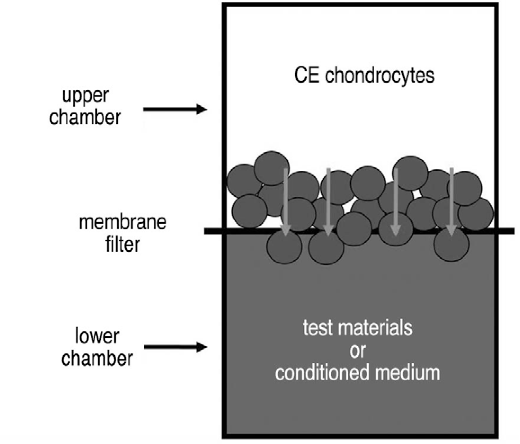
Fig. 2.
The effect of notochordal cells on the motility of cartilage endplate (CE) chondrocytes. (A), After chemotaxis assays, the membrane filter was stained with Diff-Quik reagents. On visual inspection, darker staining indicates more cell migration. (B-E), The numbers of notochordal cells loaded in the lower chamber were: (B) 4×10⁶, (C) 2×10⁶, (D) 1×10⁶, and (E) 0.5×10⁶. (F), For comparison, control medium (CM, α -MEM supplemented with 0.1% bovine serum albumin) was loaded in the lower chamber. The small round dots are the membrane pores. CE chondrocytes that migrated through the pores and attached to the bottom of the membrane are stained purple; deep purple indicates the nucleus of the CE chondrocyte (×400). The results of this experiment are summarized in Table 1.
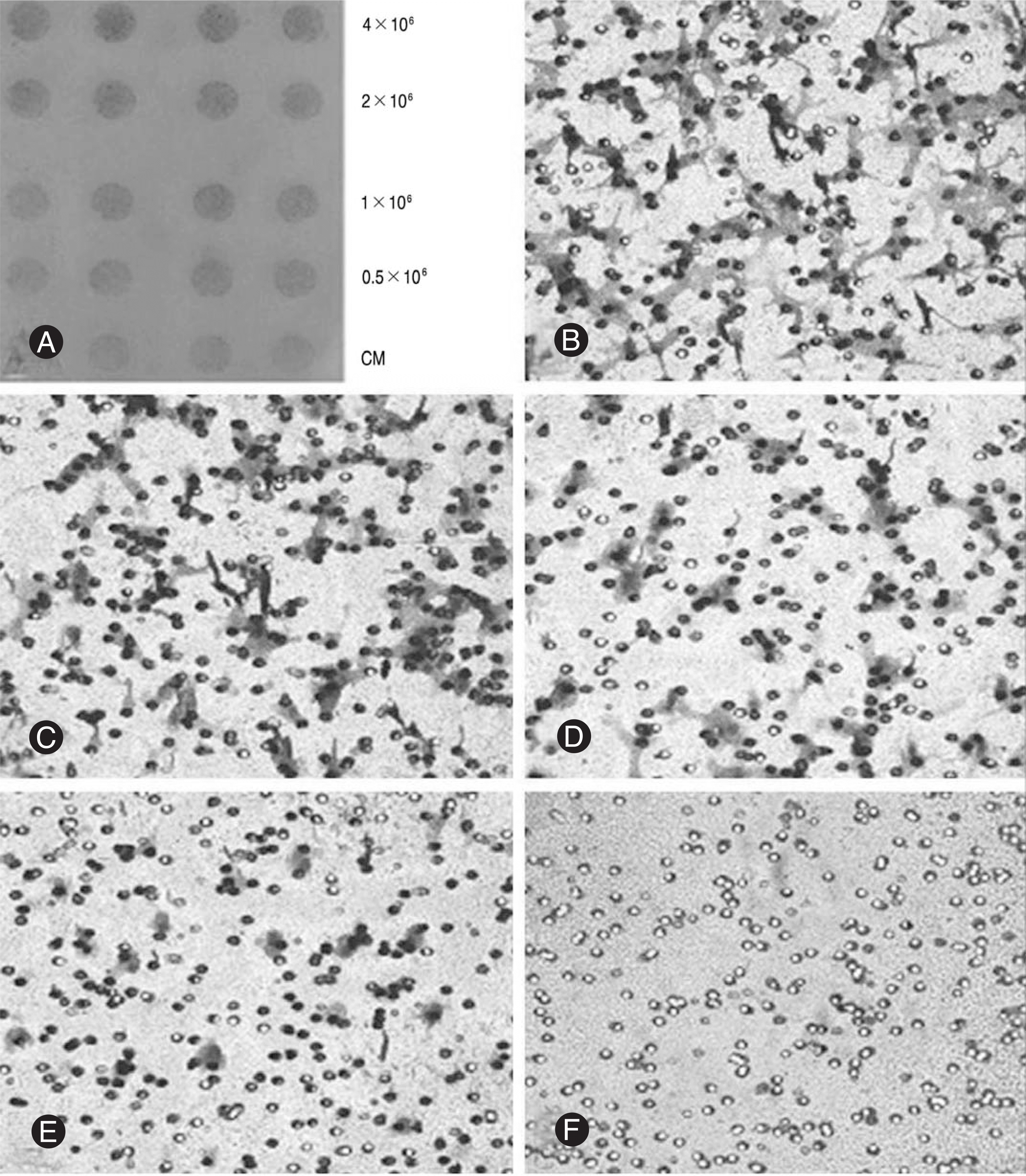
Fig. 3.
The effect of soluble factors produced by notochordal cells on the chemotactic motility of cartilage endplate chondrocytes. (A) visual inspection and (B-D) microscopic examinations (×400). Materials loaded in the lower chamber are (B) control medium (CM, α -MEM supplemented with 0.1% bovine serum albumin), (C) 50-fold concentrated conditioned medium and (D) 10-fold concentrated conditioned medium. The results of this experiment are summarized in Table 2. CCM indicates concentrated conditioned medium.
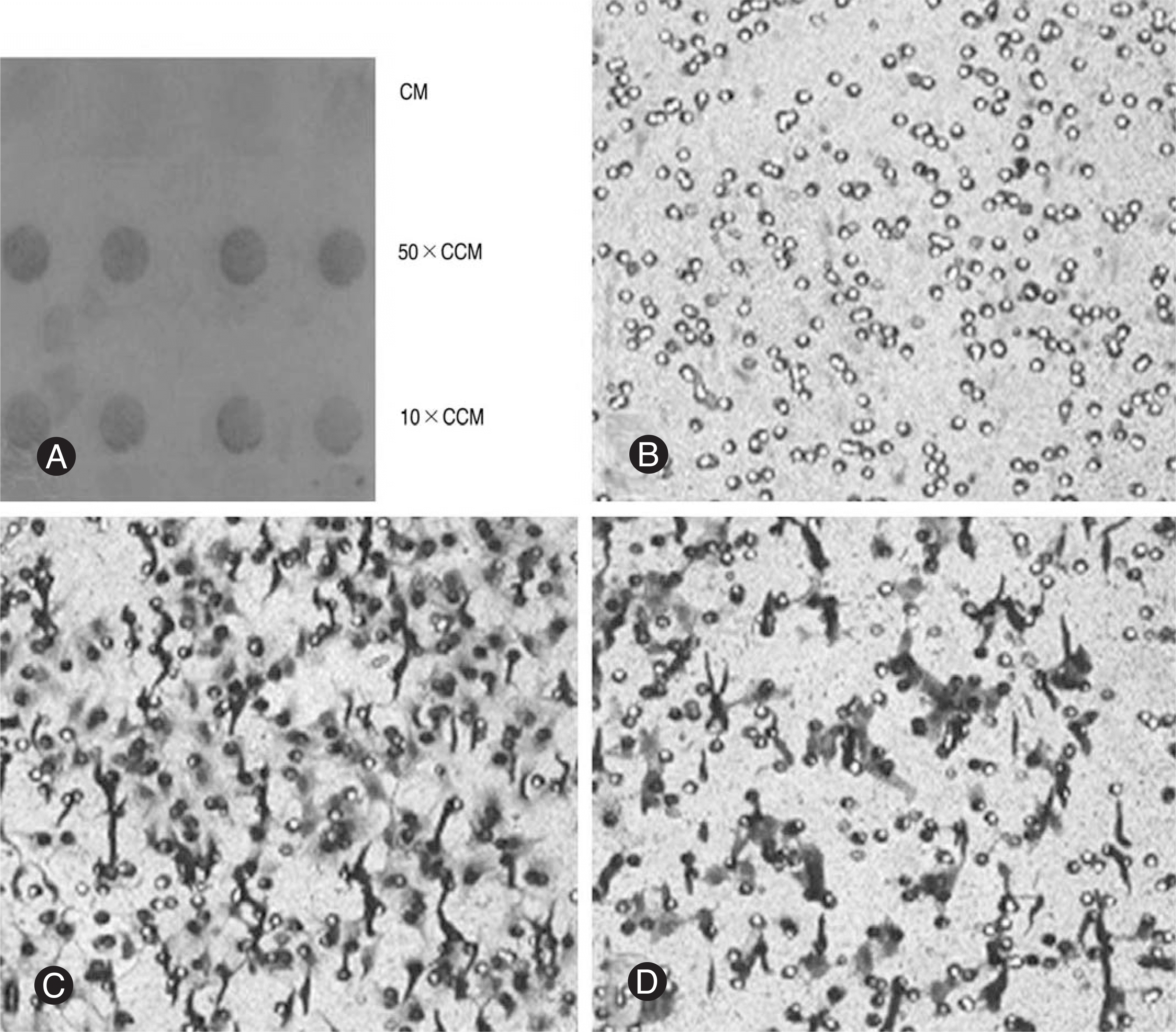
Table 1.
The effect of notochordal cells on the motility of cartilage endplate (CE) chondrocytes
| Materials Loaded in the Lower Chamber | Number of Migrated CE Chondrocytes (Mean±SD)* |
|---|---|
| Control Medium | 8.5±3.4 |
| Notochordal Cells (Number) | |
| 0.4×10⁶ | 91.5±21.0 |
| 0.2×10⁶ | 69.3±15.7 |
| 0.1×10⁶ | 46.8±14.5 |
| 0.5×10⁶ | 42.0±12.0 |
Table 2.
The effect of soluble factors produced by notochordal cells on the chemotactic motility of cartilage endplate (CE) chondrocytes
| Materials Loaded in the Lower Chamber | Number of Migrated Chondrocytes (Mean±SD)* |
|---|---|
| Control Medium | 08.5±3.4 |
| Concentrated Conditioned Medium | |
| 50-fold | 218.3±14.1 |
| 10-fold | 146.5±12.6 |
 XML Download