Abstract
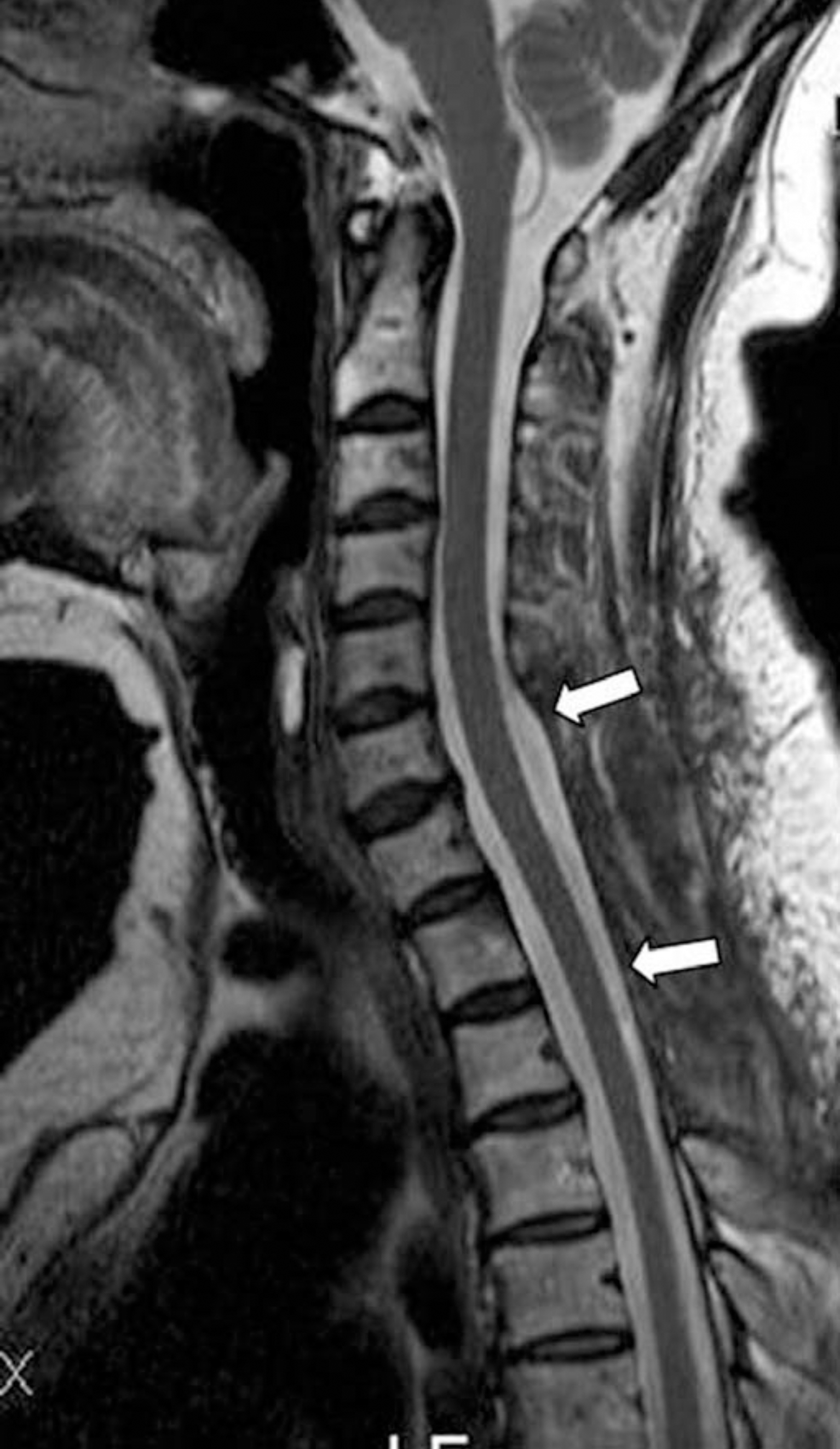
Spinal epidural hematomas can often result from a spinal tap, trauma, pregnancy, bleeding diathesis, vascular malformations, hypertension, etc. However, a spontaneous spinal epidural hematomas (SSEH) without any risk factors are relatively rare clinical entities and the clinical suspicion is very difficult in an acute setting. The outcome for patients with SSEH usually is determined by the speed of the diagnosis and the initiation of the appropriate treatment. We present a good surgical outcome of a rare case of acute SSEH without any risk factors. The patient presented initially with paresis of both upper and lower extremi-ties, upper thoracic and neck pain and mild headache. We report the diagnosis and treatment method of SSEH in this case with a review of the relevant literature.
REFERENCES
1). Betty RM, Winston KR. Spontaneous cervical epidural hematoma. A consideration of etiology. J Neurology. 1984; 61:143–148.
2). Ravid S, Schneider S, Maytal J. Spontaneous spinal epidural hematoma: an uncommon presentation of a rare disease. Childs Nerv Syst. 2002; 18:345–347.

3). Groen RJ, van Alphen HA. Operative treatment of spontaneous spinal epidural hematomas: a study of the factors determining postoperative outcome. Neurosurgery. 1997; 39:494–504.

4). Fukui MB, Swarnkar AS, Williams RL. Acute spontaneous spinal epidural hematomas: Am J Neuroradiol. 1999; 20:1365–1372.
5). Holtas S, Heiling M, Lonntoft M. Spontaneous spinal epidural hematoma: findings at MR imaging and clinical correlation. Radiology. 1996; 199:409–413.

6). Muthukumar N. Chronic spontaneous spinal epidural hematomas ?a rare cause of cervical myelopathy. Eur Spine J. 2003; 12(1):100–103.
7). Serizawa Y, Ohshiro K, Tanaka K, Tamaki S, Matsuu-ra K, Uchihara T. Spontaneous resolution of an acute spontaneous spinal epidural hematoma without neurological deficits. Intern Med. 1995; 34(10):992–994.

8). Shin JJ, Kuh SU, Cho YE. Surgical management of spontaneous spinal epidural hematomas. Eur Spine J. 2006; 15(6):998–1004.
9). Hogan QH: Lumbar epidural anatomy. A new look by cryomicrotome section. Anesthesiology. 1991; 75:767–775.
Fig. 1.
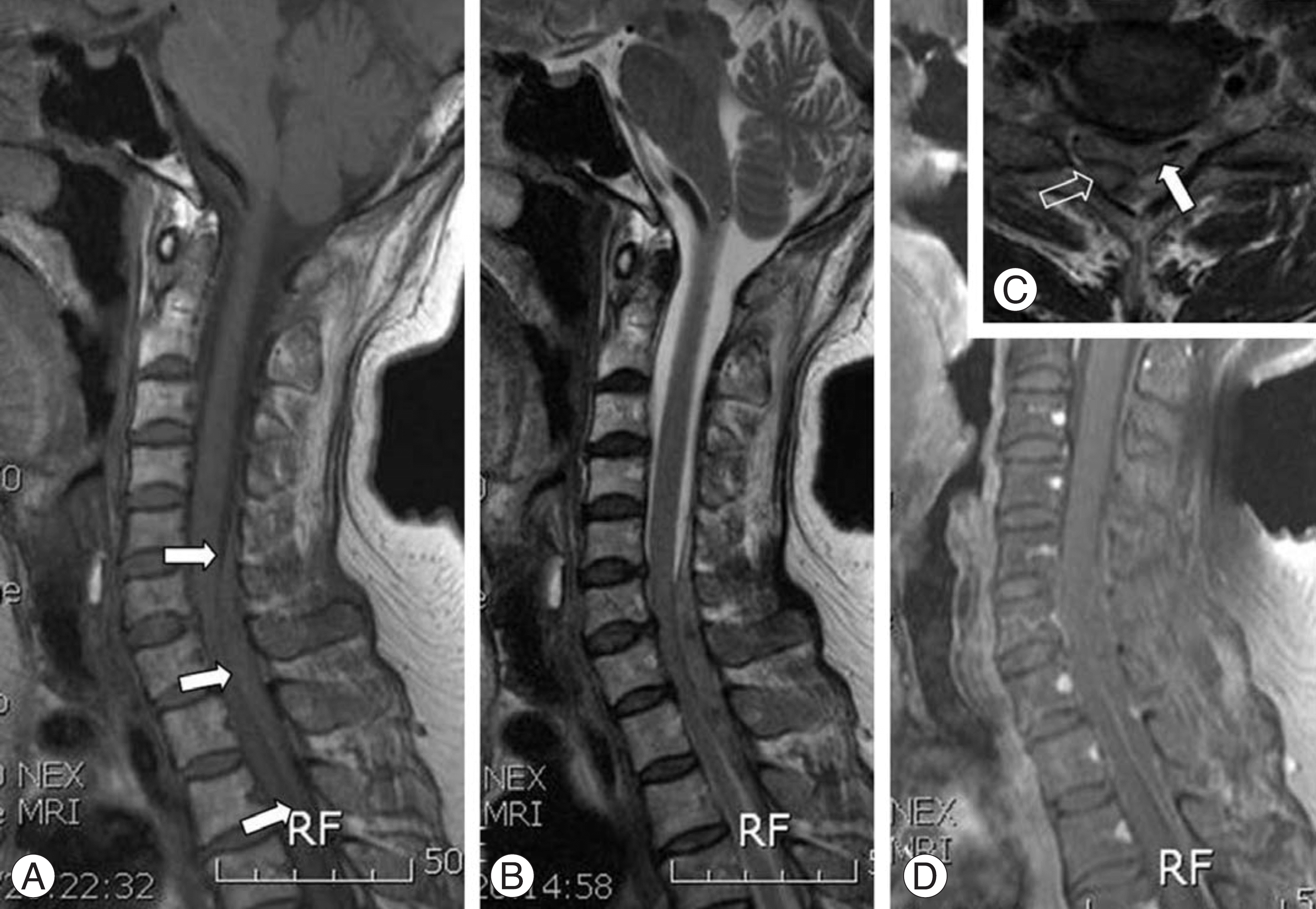
The cervicothoracic spine magnetic resonance imaging show epidural spaceoccupying lesion (arrows) extending from C5 to T2. The lesion is isointense to the spinal cord on the T1-weighted image (A), a little hyperintense or mixed signal than spinal cord on the T2-weighted image (B) and poorly enhanced (C). On the axial T2-weight image (D), the hematoma (blank arrow) was located on dorsal portion to the cord (arrow) is severely compressed.


 PDF
PDF ePub
ePub Citation
 Print
Print


 XML Download