Abstract
Study Design
The difference between the improvement after operation depending on the surgical method on patients with a spinal canal stenosis without instability were examined retrospectively.
Objectives
To determine the difference between the improvement after surgery depending on the operative method on patients with spinal canal stenosis without an instability by using a retrospective study.
Summary and Literature review
There is a The clinical difference between simple decompression and fusion using instrument after decompression.
Material and methods
Sixty-six patients, who were diagnosed with pure spinal canal stenosis without instability and treated with surgery from October 2002 to April 2004 and were available for a follow up at least for 1 year were examined. There were 22 examples of decompression only and 22 examples of fusion using instruments. The change in postoperative pain was scaled using a visual analogue scale (VAS), and the functional disability in everyday life was clinically compared with the Korean ODI (KODI) and Lower Back Outcome Score for back pain (LBOS).
Results
20 male and 24 females were examined, and the mean age was 61(45~76) years. the Million Visual Analogue Scale (MVAS) showed improvement in 28.3% of group A with decompression, and the everyday life disability scale using the Korean ODI and Lower back outcome score for back pain (LBOS) showed a improvement of 16.1% (KODI) and 18.4% (LBOS). Group B each showed 18.0% improvement using the VAS. The Korean ODI and Lower Back Outcome Score for back pain each improved by 18.0% (KODI) and 17.3% (LBOS) in two groups showing no statistically significant difference.
Go to : 
REFERENCES
1). Hanley EN, Philips ED, Kostuik JP. The adult spine, Principles and practice. New York: Raven Press;p. 1821–1831. 1991.
2). Amundsen T, Weber H, Lilleas F, et al. Lumbar spinal stenosis: Clinical and radiologic features. Spine. 1995; 20:1178–1186.
3). Herno A, Saari T, Suomalainen O, et al. The degree of decompressive relief and its relation to clinical outcome in patients undergoing surgery for lumbar spinal stenosis. Spine. 1999; 24:1010–1014.

4). Javid MJ, Hadar EJ. Longterm followup review of patients who underwent laminectomy for lumbar stenosis: A prospective study. J Neurosurg. 1998; 89:1–7.

5). Fairbanks JC, Pynsent PB. The Oswestry disability index, Spine. 2000; 25:2940–2953.
6). Grob D, Humke T, Dvorak J. Degenerative lumbar spondylolisthesis: Decompression with and without arthrodesis. J bone Joint Surg. 1995; 77:1036–1041.
7). Katz Jn, Lipson SJ, Lew RA, et al. Lumbar laminectomy alone or with instrumented or noninstrumented arthrodesis in degenerative lumbar spinal stenosis: Patient selection, costs and surgical outcomes. Spine. 1997; 22:1123–1131.
8). Turner JA, Ersek M, Herron L, et al. Patient outcomes after lumbar spinal fusions. JAMA. 1992; 268:907–911.

9). Mardjetko SM, Connolly PJ, Shott S. Degenerative lumbar spondylolisthesis: A meta-analysis of the literature, 1970-1993. Spine. 1994; 19:2256–2265.
10). Fischgrund JS, Mackay M, Herkowitz HN, et al. Degenerative lumbar spondylolisthesis with spinal stenosis: A prospective, randomized study comparing decompressive laminectomy and arthrodesis with and without spinal instrumentation. Spine. 1997; 22:2807–2812.
Go to : 
Figures and Tables%
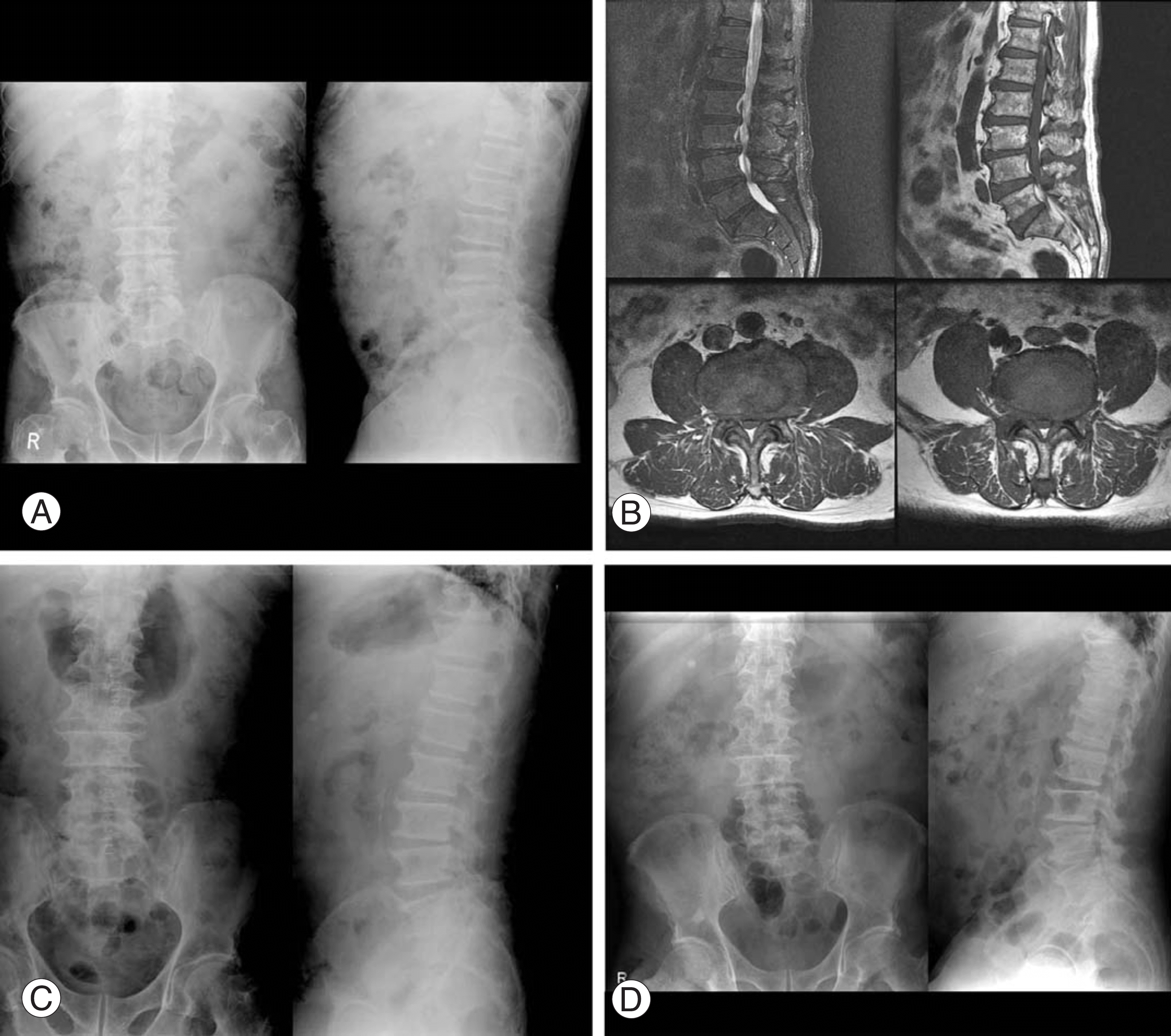
 | Fig. 1.(A) A 79 years old man sustained both lower leg pain 3 month ago. Initial radiograph showed general osteophytes and foraminal narrowing on L1-S1. (B) Preoperative MRI finding. Showing both foraminal stenosis, hypertrophy of ligamentum flavum and facet joint on L3-4-5-S1. (C) We made decompression with foraminotomy on L1-L5. (D) Postoperative radiograph at 1 year showed spinal alignment maintained well and not any instability. |
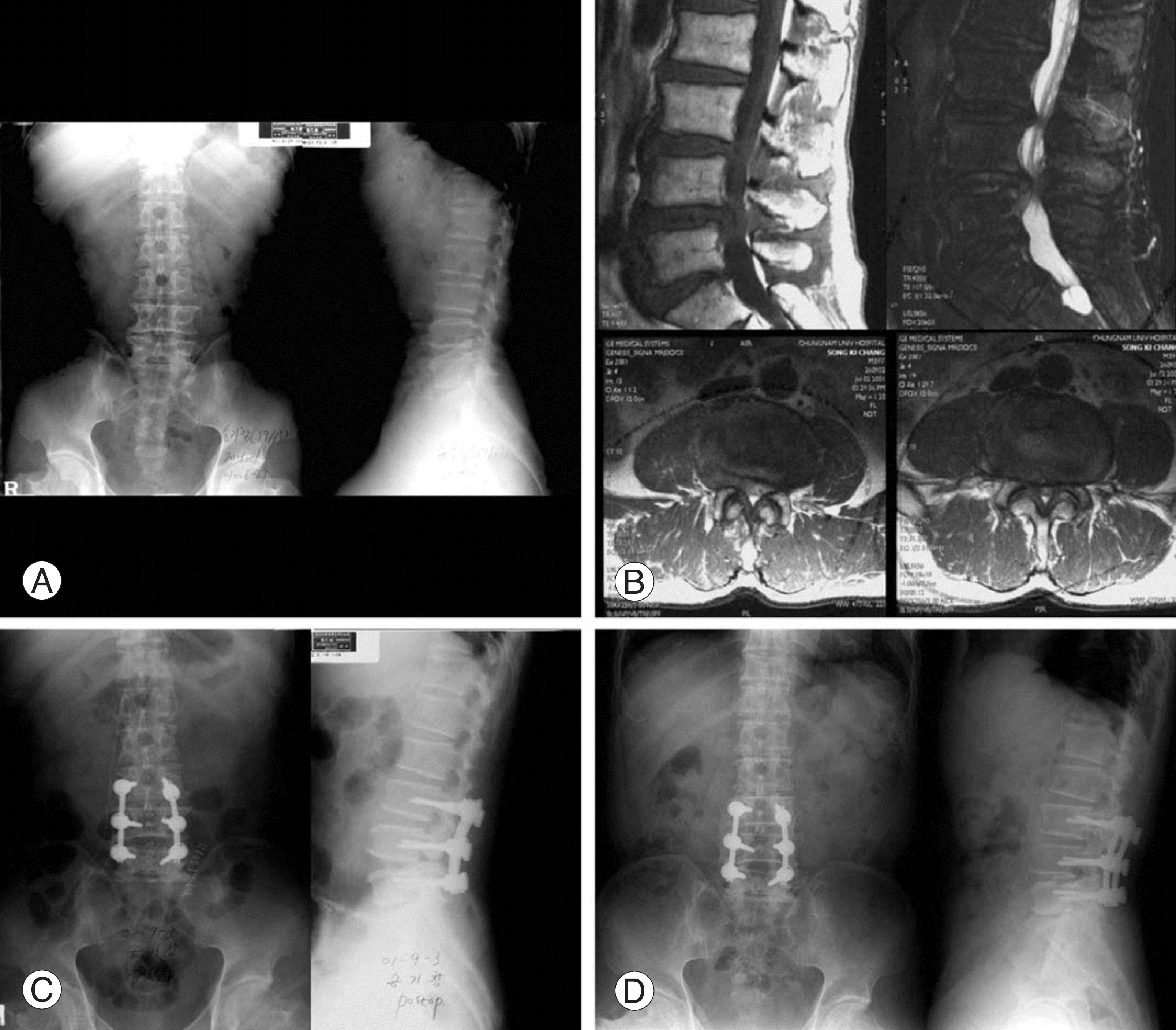
 | Fig. 2.(A) A 64 years old man sustained both lower leg pain 1 year ago. Preoperative radiograph showed moderate degenerative spondylosis and foraminal narrowing on L3-4-5. (B) Preoperative MRI showed lumbar disc extrusion and both foraminal stenosis on L3-4, L4-5. (C) We made decompression and fusion with instruments and autogenous iliac bone graft on L3-4, L4-5. (D) Postoperative radiograph at 3 year showed solid union on L3-4, L4-5 and maintained bone grafts well. |
Table 1.
Data of patients
Table 2.
Postoperative results on Group A and Group B (Oswestry disability index).
| Group | preop. ODI* | postop. ODI | Improvement (%) |
|---|---|---|---|
| A | 27.7 | 15.5 | 29.2 |
| B | 27.1 | 16.6 | 31.8 |
| Aver. | 27.7 | 15.9 | 25.8 |
Table 3.
Postoperative results on Group A and Group B (Lower Back Outcome Score for back).
| Group | preop LBO* score | postop LBO score | Improvement (%) |
|---|---|---|---|
| A | 28.4 | 42.0 | 18.1 |
| B | 30 | 42 | 15.8 |
| Aver. | 28.7 | 42.1 | 17.9 |


 PDF
PDF ePub
ePub Citation
 Print
Print


 XML Download