

 PDF
PDF ePub
ePub Citation
 Print
Print


Abstract
Objective
To categorize and analyze clinical results of degenerative lumbar deformity patients according to the degree of scoliosis and kyphosis.
Summary and Literature Review
A degenerative spinal deformity is classified into a coronal and sagittal deformity. There are some reports about treatment according to each classification but the classification is sometimes inappropriate and the treatment can vary.
Materials and Methods
From June 1998 to June 2003, 79 patients, who were diagnosed with a degenerative lumbar deformity and underwent conservative or operative treatment, were studied retrospectively. Group I had scoliosis ranging from 10 to 20。, and group II had scoliosis �20。 Each group was subdivided into A, B, and C according to the lordosis, group A �30。, group B between 20 to 30。, and group C �20。. Scoliosis and lordosis were measured from the radiographs and the clinical results were evaluated using the Kirkaldy-Willis criteria and VAS score before and after surgery.
Results
In group I, 9 cases underwent surgery and 11 cases underwent conservative treatment, without any significant differences in the clinical results. In group II, 35 cases underwent surgery and 24 cases underwent conservative treatment. Excellent results were obtained in 18(51.4%) cases, good in 15(42.9%) and 2(5.7%) were below fair. The average VAS score in group II given conservative treatment 8.9 preoperatively and 6.5 at the final follow up. Tn group II given surgical treatment the average VAS score was 9.2 preoperatively and 4.1 at the final follow up. There was a significant difference in the outcome (P�0.05).
Go to : 
REFERENCES
1). Glassman SD, Berven S, Bridwell K, Horton W, Dimar JR. Correlation of radiographic parameters and clinical symptoms in adult scoliosis. Spine. 2005; 30:682–688.

2). Keller TS, Colloca CJ, Harrison DE, Harrison DD, Janik TJ. Influence of spine morphology on intervertebral disc loads and stresses in asymptomatic adults: Impli-cations for the ideal spine. Spine J. 2005; 5:297–309.
3). Legaye J, Duval-Beaupere G. Sagittal plane alignment of the spine and gravity: A radiological and clinical evaluation. Acta Orthop Belg. 2005; 71:213–220.
4). Kirkaldy-Willis WH, Paine KW, Cauchoix J, McIvor G. Lumbar spinal stenosis. Clin Orthop Relat Res. 1974; 99:30–50.
5). Bradford DS. Adult scoliosis. Current concepts of treatment. Clin Orthop Relat Res. 1988; 229:70–87.
6). Lee CS, Kim YT, Kim EG. Clinical study of lumbar degenerative kyphosis. J Korean Soc Spine Surg. 1997; 4:27–35.
7). Perennou D, Marcelli C, Herisson C, Simon L. Adult lumbar scoliosis. Epidemiologic aspects in a low-back pain population. Spine. 1994; 19:123–128.
8). Takemitsu Y, Harada Y, Iwahara T, Miyamoto M, Miyatake Y. Lumbar degenerative kyphosis. Clinical, radiological and epidemiological studies. Spine. 1988; 13:1317–1326.
9). Abei M. Correction of degenerative scoliosis of the lumbar spine. A preliminary report. Clin Orthop Relat Res. 1988; 232:80–86.
10). Grubb SA, Lipscomb HJ. Diagnostic findings in painful adult scoliosis. Spine. 1992; 17:518–527.
11). Grubb SA, Lipscomb HJ, Suh PB. Results of surgical treatment of painful adult scoliosis. Spine. 1994; 19:1619–1627.
12). Bernhardt M. Normal spinal anatomy. Normal sagittal plane alignment. (in. Bridwell KH, Dewald RL, editors. The textbook of spinal surgery 2nded. Philadelphia: Lippin-cott-Raven;p. 185–191. 1997.
13). Moon MS, Lee KS, Lim CI, Kim YB, Lee HS. A clinical study of degenerative lumbar scoliosis. Yonenobu K, editor. Lumbar fusion and stabilization. Springer-Verlag;p. 98–112. 1992.
14). Pritchett JW, Bortel DT. Degenerative symptomatic lumbar scoliosis. Spine. 1993; 18:700–703.
15). Toyama Y. Surgical management of degenerative lumbar scoliosis. Yonenobu K, editor. Lumbar fusion and stabilization. Springer-Verlag;113-134. 1992.
16). Kostuik JP. Recent advances in the treatment of painful adult scoliosis. Clin Orthop Relat Res. 1980; 147:238–252.
17). Lee JK, Yang JY, Kim KC. Surgical treatment of degenerative lumbar scoliosis with multiple spinal stenosis. J Korean Soc Spine Surg. 2002; 9:197–203.
18). Kostuik JP, Israel J, Hall JE. Scoliosis surgery in adult. Clin Orthop Relat Res. 1973; 93:225–234.
Go to :
Figures and Tables%
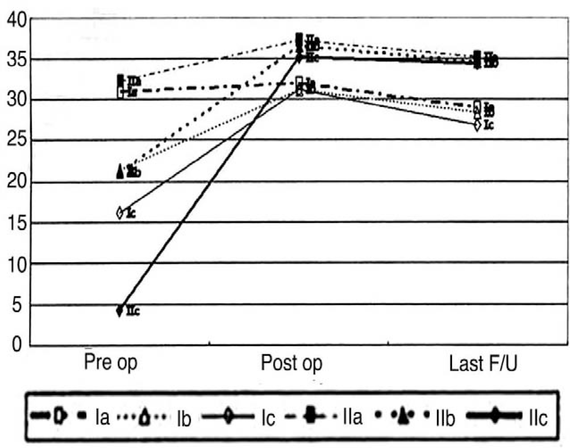 | Fig. 1.Correction of kyphotic angle of the operative group. IIC group show maximal correction. |
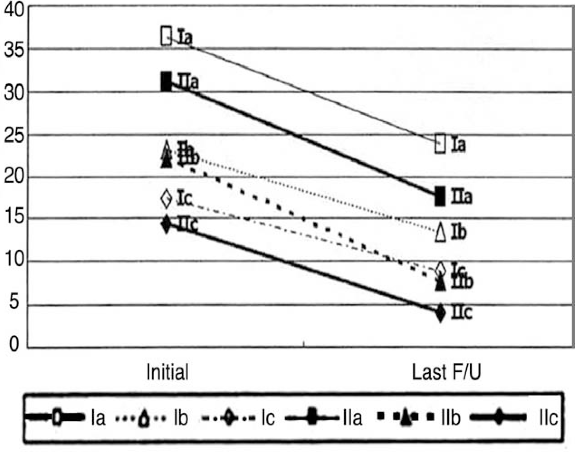 | Fig. 2.Changes of kyphotic angle of the nonoperative group. IIA group had progressed to the IIC group level and IIB group also had progressed to the IIC group level. |
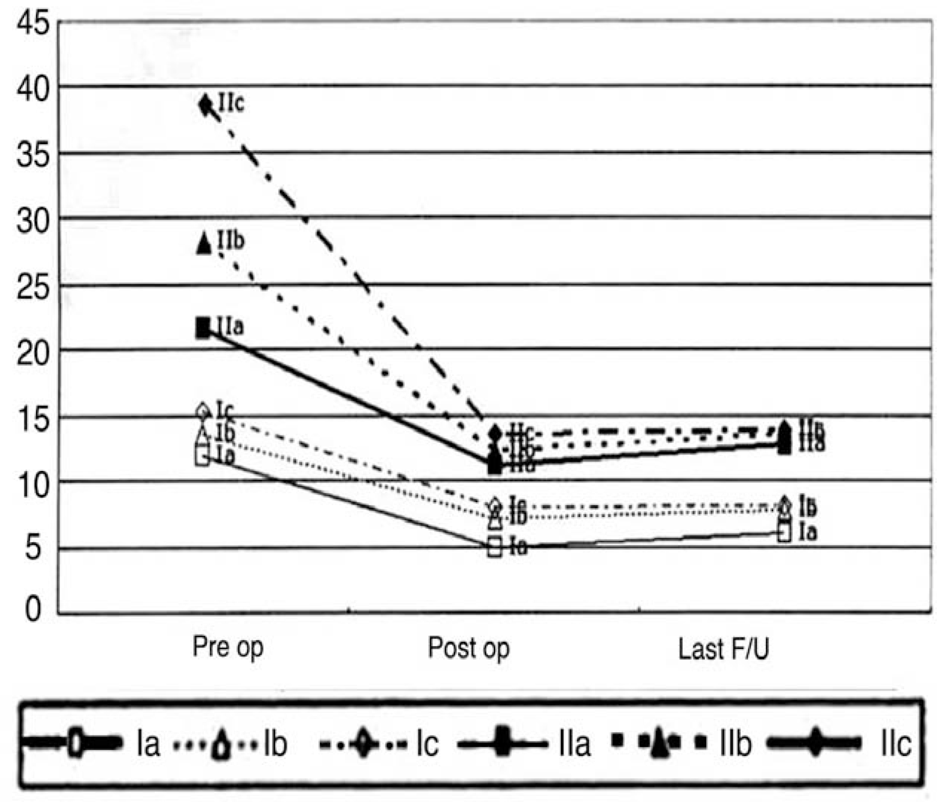 | Fig. 3.Correctioin of scoliotic angle of the operative group. IIC group show maximal correction. |
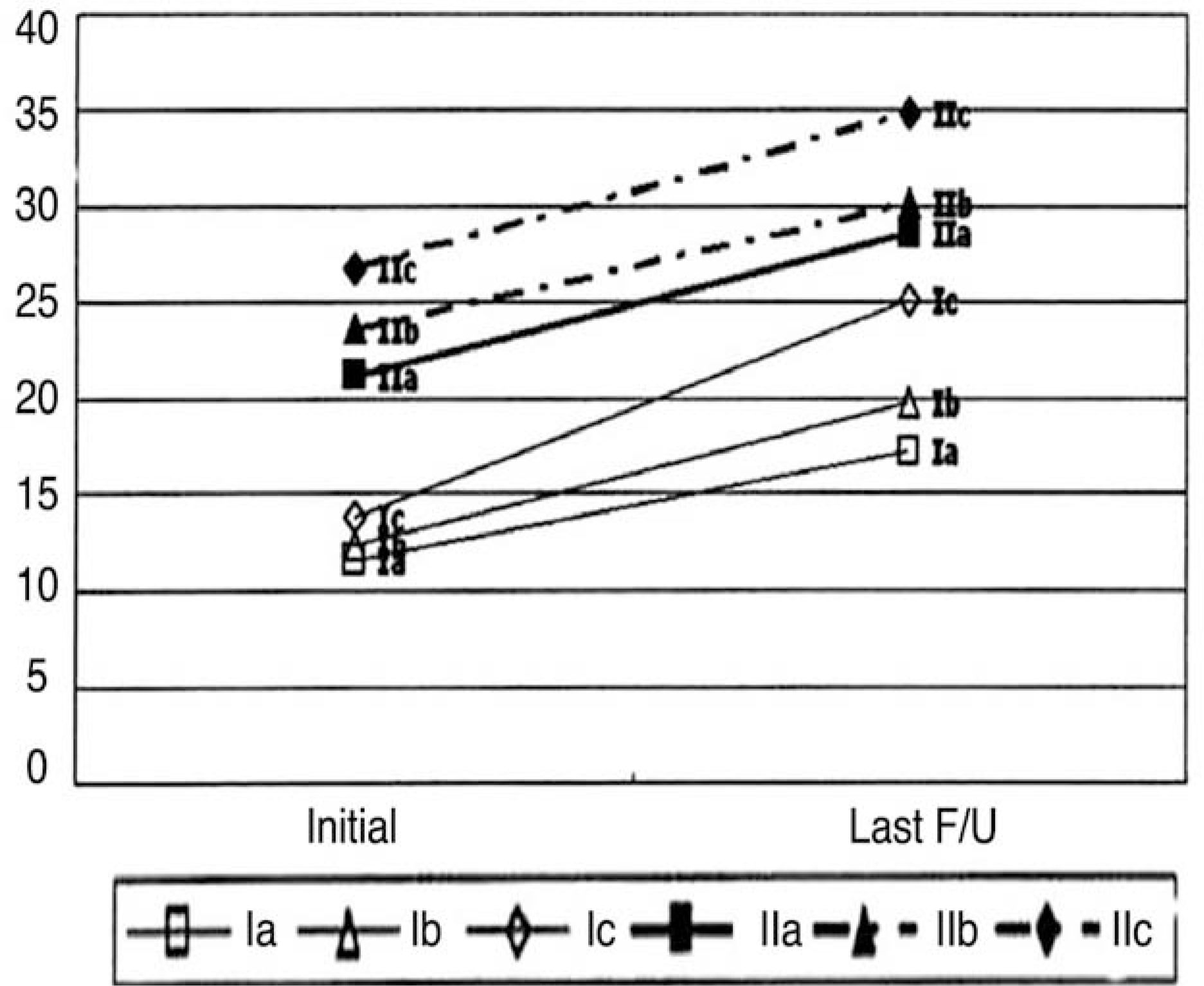 | Fig. 4.Changes of scoliotic angle of the nonoperative group. IC group had progressed to the II group level. |
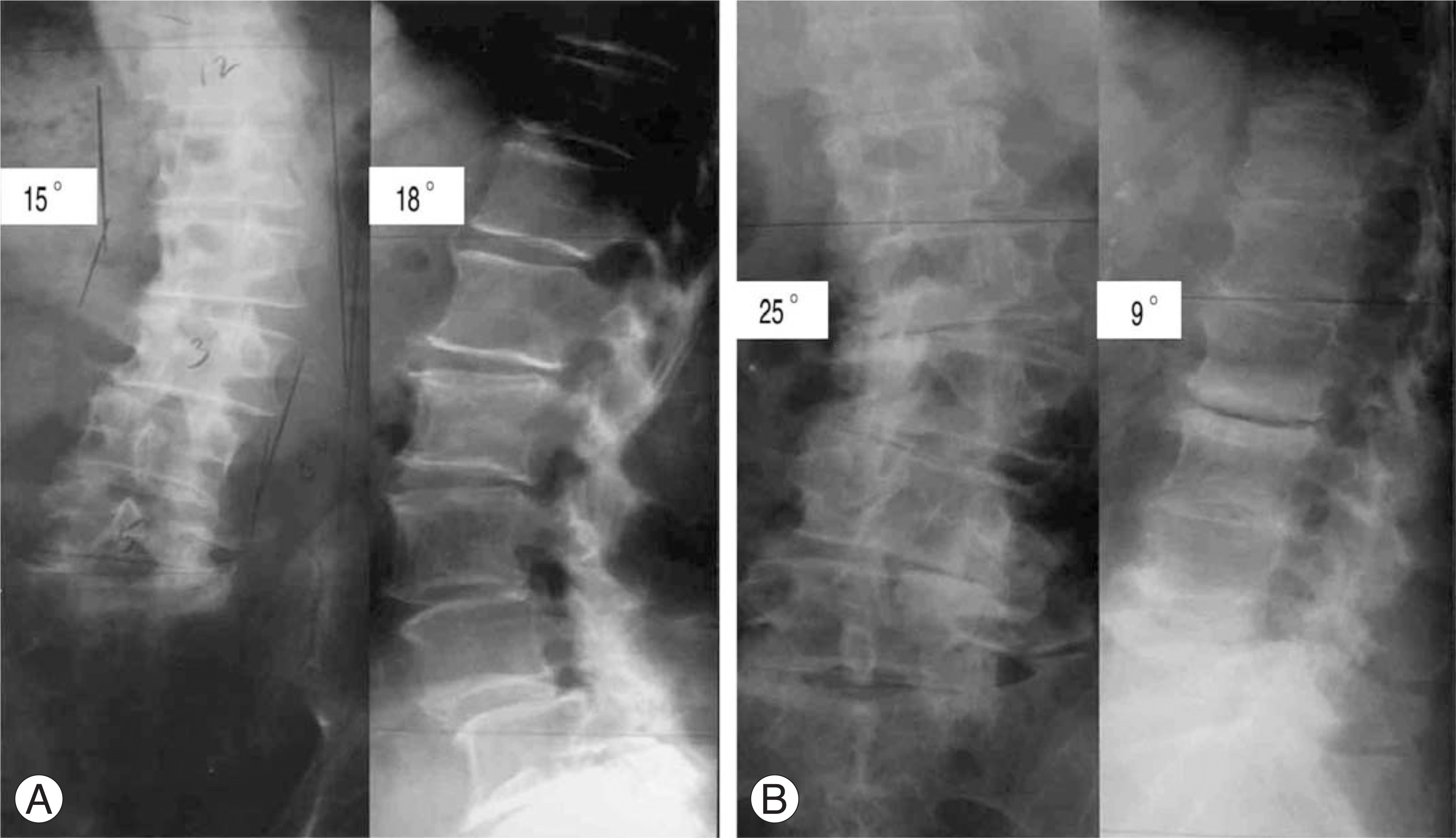 | Fig. 5.The case of conservative Treatment. (A) Initial AP and lateral radiographs. The Cobb's angle was 15 degree and lordotic angle was 18 degree and this case was classified into group IC. (B) The radiographs of 2 years follow up. The Cobb's angle increased up to 25 degree and lordotic angle decreased to 9 degree. |
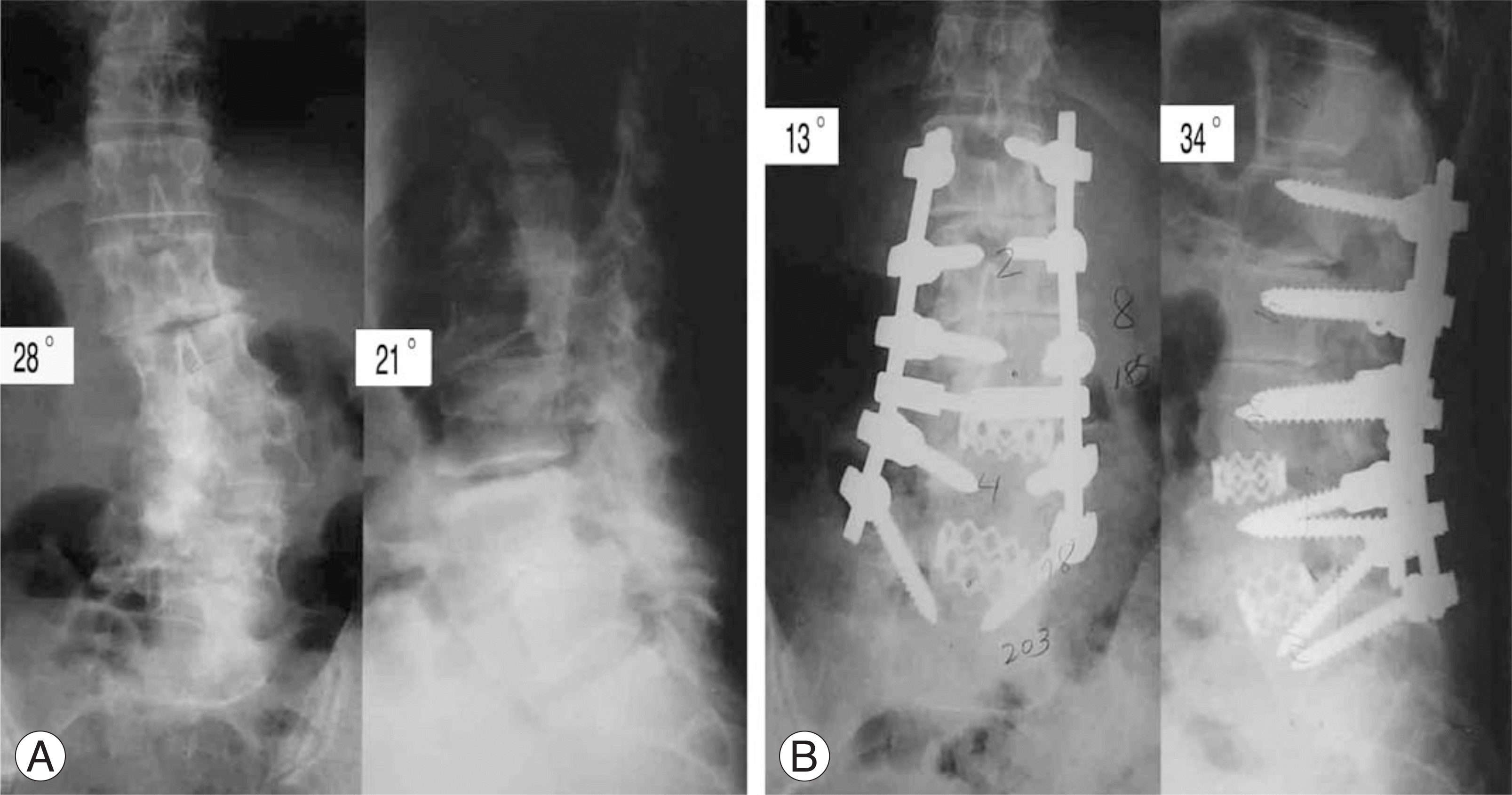 | Fig. 6.The case of long spinal fusion. (A) Initial AP and lateral radiographs. The Cobb's angle was 28 degree and lordotic angle was 21 degree and this case was classified into group IIB. (B) The radiographs of 2 years follow up. The Cobb's angle decreased to 13 degree and lordotic angle increased to 34 degree. |
Table 1.
Data of Each Group
| Group | I | II | ||||
|---|---|---|---|---|---|---|
| Subgroup | A | B | C | A | B | C |
| Conservative Tx. | 3 | 4 | 4 | 12 | 5 | 7 |
| Operative Tx. | 1 | 3 | 5 | 14 | 4 | 17 |
Table 2.
Correction of Kyphotic Angle of the Operative Group
| Group | Pre-operation | Postoperation | Last follow up | |
|---|---|---|---|---|
| A | 31 | 32 | 29 | |
| I | B | 21.5 | 31.2 | 28.4 |
| C | 16.2 | 31.3 | 26.9 | |
| A | 32.3 | 37.3 | 35.1 | |
| II | B | 21.1 | 36.5 | 34.6 |
| C | 4.2 | 35.1 | 34.4 |
Table 3.
Changes of Kyphotic Angle of the Nonoperative Group
| Group | Initial | Last follow up | |
|---|---|---|---|
| A | 36.3 | 23.8 | |
| I | B | 23.1 | 13.5 |
| C | 17.4 | 8.9 | |
| A | 31.1 | 17.5 | |
| II | B | 22.1 | 7.8 |
| C | 14.3 | 4.1 |
Table 4.
Correction of Scoliotic Angle of the Operative Group
| Group | Pre-operation | Postoperation | Last follow up | |
|---|---|---|---|---|
| A | 12 | 5 | 6 | |
| I | B | 13.5 | 7.2 | 7.8 |
| C | 15.3 | 7.9 | 8.1 | |
| A | 21.6 | 11.2 | 12.8 | |
| II | B | 28.2 | 12.2 | 13.5 |
| C | 38.7 | 13.5 | 13.9 |
 XML Download