Abstract
Objectives
To evaluate the usefulness of postoperative suction drainage tip cultures as a method of predicting the development of deep wound infections after thoracolumbar surgery using pedicle screws.
Summary of Literature Review
The primary diagnostic elements of postoperative spinal infections are a high degree of clinical suspicion by the surgeon combined with aspiration and culture of the suspected infection sites.
Materials and Methods
We analyzed the results of cultures on postoperative suction drainage tips from a total of 471thoracolumbar surgery cases. We calculated the sensitivity, specificity, and predictive value and investigated the isolated pathogens. In addition, we performed quantitative analyses of serum C-reactive protein using Turbidimetry.
Results
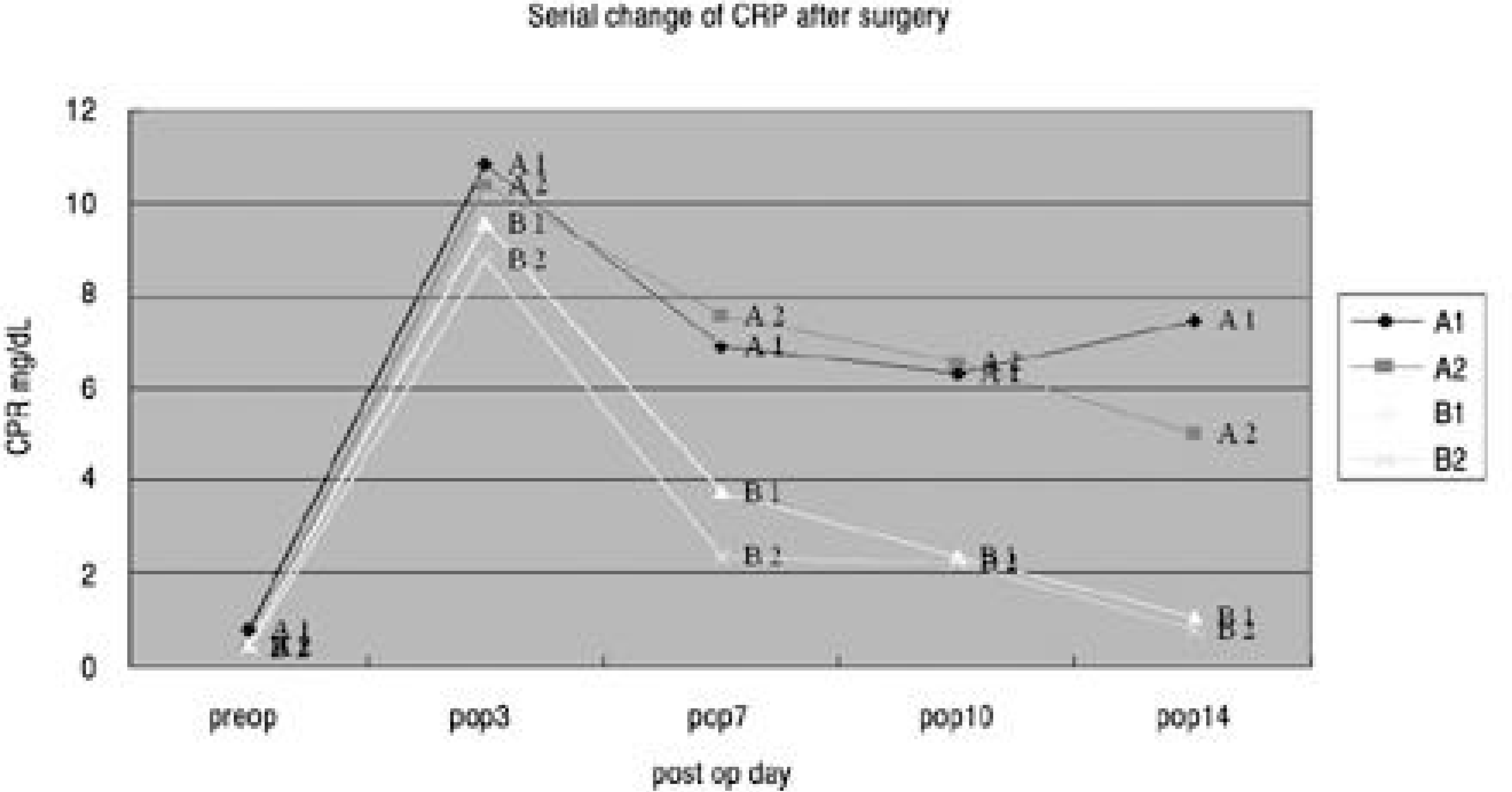
The postoperative infection rate was 4.0%. The most common isolated pathogen of the true positive cases was staphylococcus aureus, which was found in 3 cases (methicillin- resistant staphylococcus aureus in 2 cases); and that of the false positive cases was coagulase- negative staphylococcus in 5 cases. The sensitivity of the suction drainage tip culture was 52.6%, the specificity was 96.3%, the positive predictive value was 37.0%, and the negative predictive value was 98.0%. In cases of C-reactive protein, true positive and false negative cases followed the same course, where the CRP decreased slowly for the first week but remained elevated persistently at the 14th postoperative day.
Conclusions
Culture of the suction drainage tips could not predict the development of postoperative deep wound infections, but it had more significance in the exclusion of deep wound infections. We concluded that careful observation for other signs of deep wound infections are necessary when a clinically significant pathogens are isolated.
Go to : 
REFERENCES
1). Leah YC, Rolando MP, Juhn RD, Steven DG, John RJ. Perioperative complications of posterior lumbar decompression and arthrodesis in older adults. J Bone Joint Surg Am. 2003; 85(11):2089–2092.
2). Gepstein R, Eismont FJ. Postoperative spine infection. Garfin SE, editor. Complication of Spine Surgery. Baltimore: Williams & Wikins Co.;1989. p. 302–322.
3). Jutte PC, Castelein RM. Complications of pedicle screws in lumbar and lumbosacral fusions in 105 cansecutive primary operations, Eur spine J. 2002; 11:594–598.
4). Keller RB, Pappas AM. Infection after spinal fusion using internal fixation instrumentation, Orthop Clin North Am. 1972; 3:99–111.
5). Kim YM, Won CH, Choi ES, Seo JB, Lee HS, Cho BG. Management of deep infection after posterior spinal instrumentation with prolonged suction drainage. J Kor Spine Surg. 2001; 8:504–512.

6). Rah JH, Park HJ, Ahn JI. A clinical study of postoperative infection in posterior spinal surgery with pedicle screw system J Kor Orthop. Asso. 1994; 29:994–1003.
7). Bridwell KH, Dewald RL. The textbook of spinal surgery. 2nd ed.spinal infections-postoperative infections. philadelphia-new york: Lippincott-Raven publishers;II:p. 2171–2177. 1997.
8). Shim DM, Kim TK, Song HH, Shim YS, Lee SH, Song JH. Quantitation of C-reactive protein levels and erythro -cyte sedimentation rate after spinal surgery. J Kor Spine Surg. 1998; 5-1:33–39.
9). Lonstein J, Winter R, Moe JH. Wound infection with Harrington instrumentation and spine fusion for scoliosis. Clin Orthop. 1973; 284:99–108.

10). Gaine WJ, Ramamohan NR, Hussein NA, Hullin MG, McCreath SW. Wound infection in hip and knee arthroplasty. J Bone Joint Surg Br. 2000; 82:561–565.

11). Massie JB, Heller JG, Abitbol JJ, Mcpherson D, Garfin SR. Postoperative posterior spinal wound infection. Clin Orthop. 1992; 284:99–108.
12). Fizgerald RH, Nolan DR, Ilstrup DM, Vanscoy RE, Washington JA, Coventry MB. Deep wound sepsis following total hip arthroplasty. J Bone Joint Surg Am. 1977; 59:847–855.
13). Harwood DA, Ribbins SG, Zawadsky JP. The predictive value of intraoperative and postoperative cultures in patients undergoing total hip arthroplasty. Orthop Trans. 1989; 13:60–61.
14). Dietz FR, Koontz FP. The importance of positive bacterial cultures of specimens obtained during clean orthopaedic operation. J Bone Joint Surg Am. 1991; 73:1200–1207.
15). Cho MR, Kim C, Son JW, Kim JD. Identification of bacteria in postoperative infections after orthopaedic surgery. J. of Kor Orthop. Assoc. 2003; 38:771–775.
16). Fitzgerald RH, Peterson LF, Washington JA. Bacterial colonization of wounds and sepssis in total hip arthroplasty. J Bone Joint Surg Am. 1973; 55:1242–1250.
17). Kim EH, Song IS. Deep wound infection after lumbar spine fusion with pedicular screw fixation J Kor Spine Surg. 2000; 7:535–543.
18). Thomas DB, Michael TM. Biology of microorganism. 5th ed.Prentice hall inc;p. 749–750. 1988.
19). Cheo SS, Kim DG, Kim IS, et al. Preventive medicine and public health. 2nd ed.Preventive medicine pubassoci. Gye chuck publisher;p. 391–395. 2001.
20). Jeon JH, Son HS, Park BJ, Shin HL. Statistical training for clinician. 1st ed.Jin young publisher;p. 207–230. 2002.
21). Jason W, Michael K, Rovert D. C-reactive protein level after total hip and total knee replacement J Bone Joint Surg Br. 1998; 80:909–911.
22). Kim BJ, Lim Y, Lee JH, Jeon TH. The clinical significance of CRP for the postoperative infection in orthopedic surgery J Kor Orthop. Assoc. 1992; 27-4:1074–1082.
Go to : 
 | Fig. 1.Serial change of CRP before and after thoracolumbar surgery.(A) Deep wound infection after thoracolumar surgery A1: true positive - suction drainage tip culture positive A2: false negative - suction drainage tip culture negative (B) No infection after thoracolumar surgery B1: false positive - suction drainage tip culture positive B2: true negative - suction drainage tip culture negative |
Table 1.
Patient characteristics
| Deep infection (14cases) | No infection (457cases) | |||
|---|---|---|---|---|
| Disease | Spinal stenosis | 6 cases | Spinal stenosis | 266 cases |
| Spondylolisthesis | 61 cases | |||
| DLS∗ | 2 cases | Idiopathic scoliosis | 16 cases | |
| Idiopathic adolscoliosis | 2 cases | DLS∗ | 15 cases | |
| LDK# | 15 cases | |||
| Thoracic myelopathy | 5 cases | |||
| Trauma | Distractive flexion injury | 1 case | Distractive flexion injury | 37 cases |
| Unstable burst fracture | 1 case | Unstable burst fracture | 26 cases | |
| Revision | FBSS& | 2 cases | FBSS& | 16 cases |
| fusion level | 4.21 | 3.11 | ||
Table 2.
Organisms from suction drainage tip culture vs wound culture in deep wound infection patients
| Suction drainage tip culture | Wound culture | ||
|---|---|---|---|
| Organism | No. | Organism | No. |
| Staphylococcus aureus (MRSA)# | 3 (2) | MRSA | 7 |
| Staphylococcus epidermidis | 3 | Enterococcus sp | 6 |
| Enterococcus sp | 2 | Staphylococcus epidermidis (MRSE)∗ | 7 (2) |
| G(+) rod | 1 | Pseudomonas sp. | 2 |
| Micrococcus | 1 | Acinetobactor calcoaceticus | 2 |
| baumannii compelx | |||
| Micrococcus | 1 | ||
Table 3.
Organisms from positive suction drainage tip culture in uncomplicated group
| Organism | No. |
|---|---|
| Coagulase negative staphylococcus | 5 |
| Acinetobactor sp. | 3 |
| Enterococcus sp. | 1 |
| Pseudomonas sp. | 1 |
| Staphylococcus aureus | 1 |
| Staphylococcus viridans | 1 |
| G(+) rod | 1 |
| G(+) cocci | 1 |
| G(-) rod | 1 |
Table 4.
The sensitivity, the specificity, the predictive value for suction drainage tip culture
| Infection | ||||
|---|---|---|---|---|
| + | - | |||
| suction | ||||
| drainage | + | 10 | 17 | |
| tip | - | 4 | 440 | |
| culture | ||||
| Specificity | 96.28% | |||
| Sensitivity | 52.63% | |||
| Predictive value | Positive | 37.03% | ||
| Negative | 97.99% |
Table 5.
Serial change of CRP before and after thoracolumbar surgery
| Group No. | preOP | POP 3rd day | POP 7th day | POP 10th day | POP 14th day | |
|---|---|---|---|---|---|---|
| A | A1 | 0.78 | 10.82 | 6.89 | 6.32 | 7.46 |
| A2 | 0.41 | 10.37 | 7.54 | 6.53 | 5.03 | |
| B | B1 | 0.45 | 09.56 | 3.74 | 2.34 | 1.06 |
| B2 | 0.35 | 08.78 | 2.36 | 2.24 | 0.77 | |
A: Deep wound infection after thoracolumar surgery A1: true positive - suction drainage tip culture positive A2: false negative - suction drainage tip culture negative B: No infection after thoracolumar surgery B1: false positive - suction drainage tip culture positive B2: true negative - suction drainage tip culture negative


 PDF
PDF ePub
ePub Citation
 Print
Print


 XML Download