Abstract
Symptomatic epidural varix presenting with radiculopathy is extremely rare. The most common misdiagnosis is reported as a sequestrated prolapsed nucleus pulposus in the preoperative evaluation. The method of evaluating enhanced MRI studies improved the efficacy of discovery and treatment of this condition. We experienced 6 cases of epidural varices that were diag-nosed with T1 fat suppressed post-gadolinium enhanced MRI studies and we present the operative findings.
Go to : 
REFERENCES
01). Gumbel U., Pia HW., Vogelsang H. Lumbosacrale Gefassanomalien als Ursache von Ischialgien. Acta Neu-rochir. 1969. 20:131–51.
02). Cahan LD., Higashida RT., Halbach VV., Hieshima GB. Variants of radiculomeningeal vascular malformation of the spine, J Neurosurg. 1987. 66:333.
03). Hanley EN Jr., Howord BH., Brigham CD., Chapman TM., Guilford WB., Coumas JM. Lumbar epidural varix as a cause of radiculopathy. Spine. 1994. 19:2122–6.

04). Ivanovici F. Urine retention: an isolated sign of some spinal cord disorder. J Urol. 1970. 104:284–6.
05). Dickman CA., Zabramski JM., Sonntag VK., Coons S. Myelopathy due to epidural varicose veins of the cervi-cothoracic junction: case report. J Neurosurg. 1970. 69:940–1.
06). Asamoto S., Sugiyama H., Doi H., Nagao T., Ida M., Mat-sumoto K. Spinal epidural varices. No shinkei Geka. 1999. 27:911–3.
07). Bapat M., Metkar U. Epidural varix at the cervicotho-racic junction: unusual cause of quadriplegia: a case report. Spine. 2006. 31:E88–90.
08). Wong CH., Thng PL., Thoo FL., Low CO. Symptomatic spinal epidural varices presenting with nerve impinge-ment: report of the cases and review of the literature. Spine. 2003. 28:E347–350.
09). Demaeral P., Petre C., Wilms G., Plets C. Sciatica caused by a dilated epidural vein: MR findings. Eur radiol. 1999. 9:113–4.
10). Zimmerman GA., Weingarten K., Lavyne MH. Symptomatic lumbar epidural varices. Report of 2 cases. J Neurosurg. 1994. 80:914–8.
Go to : 
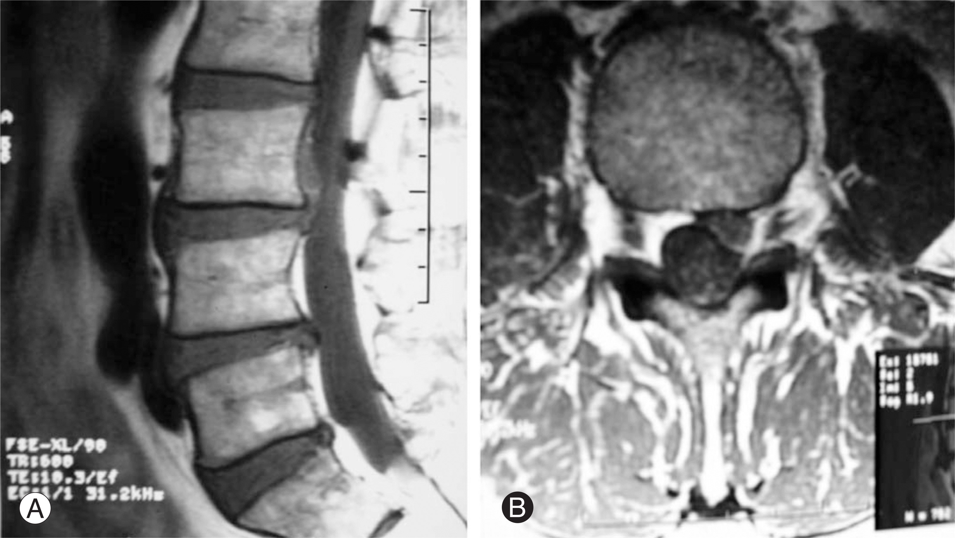
 | Fig. 1.(A, B) T1 sagittal and axial image, L3 body posterior, intermediate signal 0.5×1CM thecal sac compressing fusiform mass. |
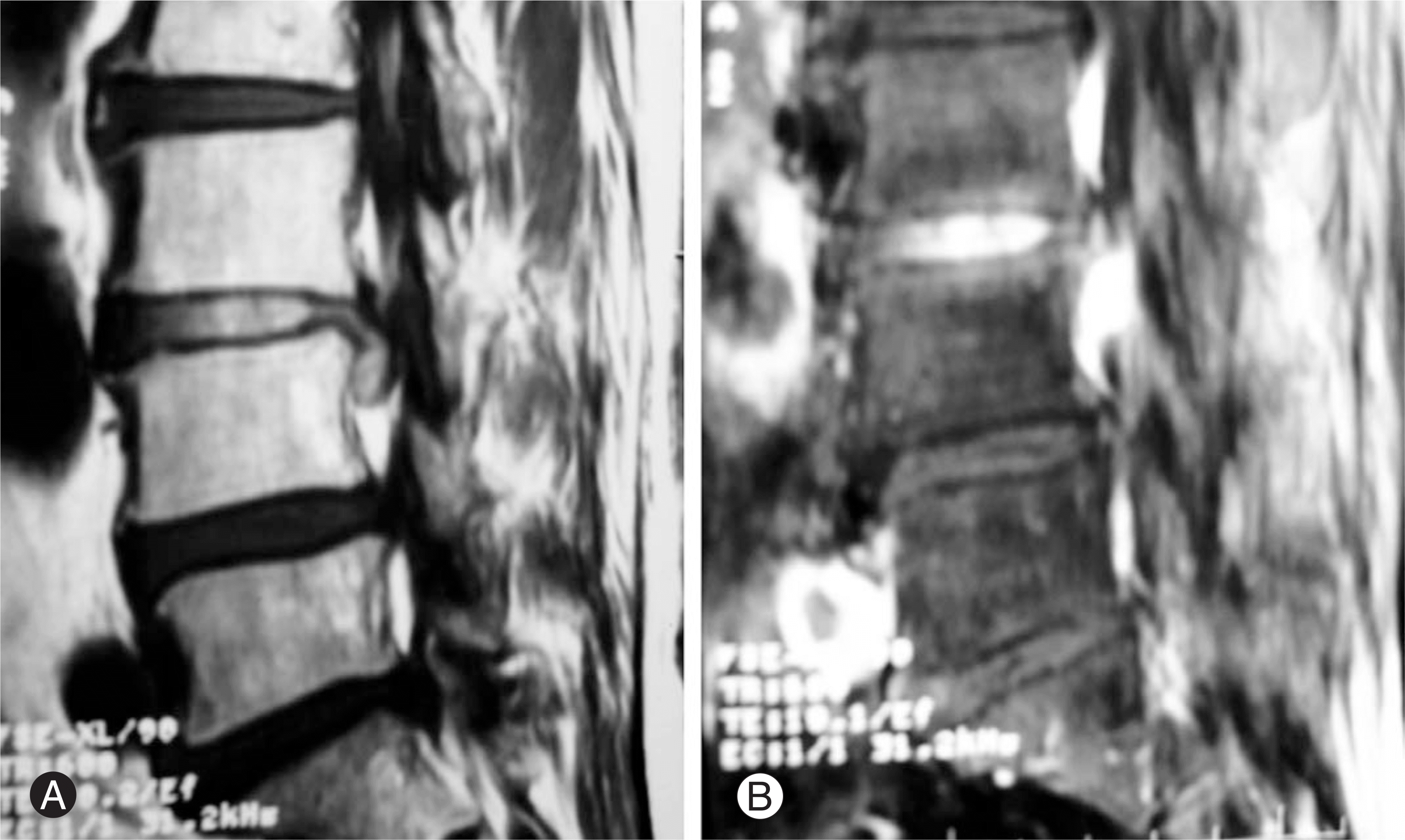
 | Fig. 2.(A) T1 sagittal image, L3/4, intermediate signal extruded, caudal migrated mass. (B) T1 fat-suppresed post-gadolini-um enhanced sagittal image, enhanced mass with serpiginous dilated vein. |
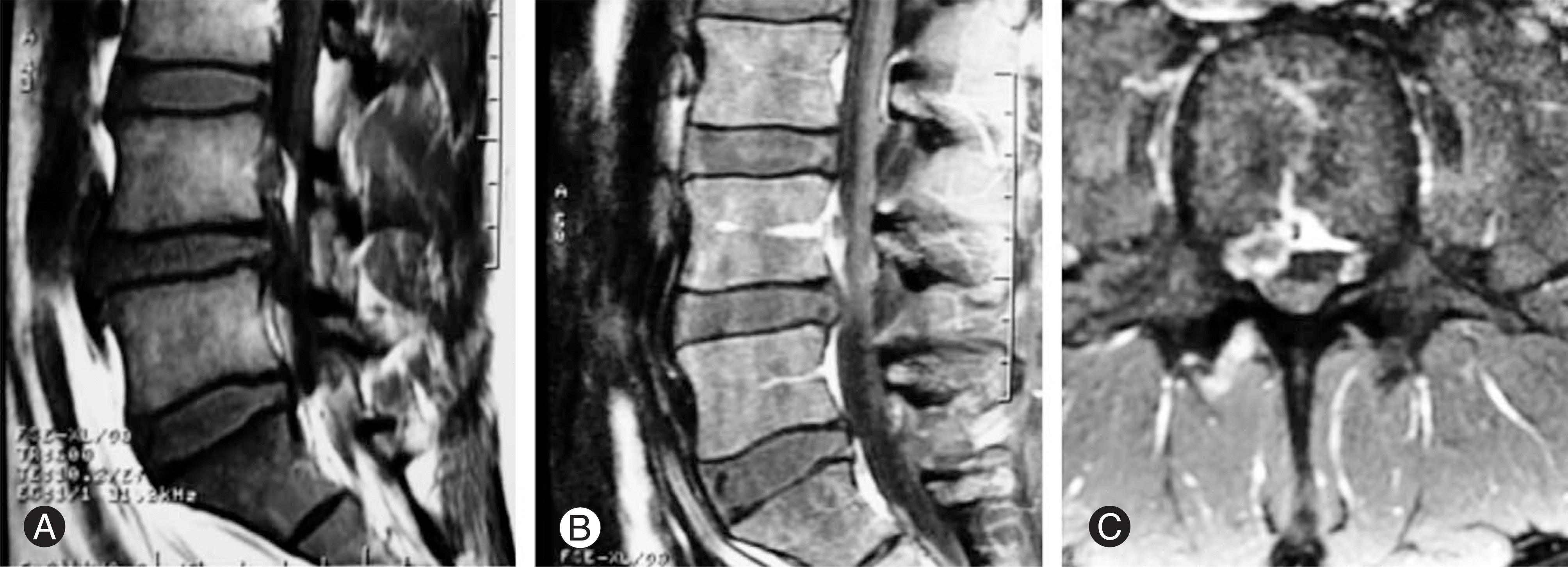
 | Fig. 3.(A) T1 sagittal image, L4 body posterior, intermediate signal fusiform mass, L4/5 disc protrusion. (B) T1 fat-supp-resed post-gadolinium enhanced sagittal image, L4 body posterior enhanced around the mass. (C) T1 enhanced axial image, Rt paracentral disc-like intermediate signal. At the central cephalad direction, low signal mass with enhanced, communicating L4 body. Serpiginous dilated vein, prominent lumbar segmental veins was seen. |


 PDF
PDF ePub
ePub Citation
 Print
Print


 XML Download