

 PDF
PDF ePub
ePub Citation
 Print
Print


Abstract
Objectives
The purpose of this study was to determine whether F-18 FDG-PET had value in distinguishing between vertebral pathologic fractures and osteoporotic compression fractures.
Summary of Literature Review
There were many reports in the literature about vertebral pathologic disease studied with F-18 FDG-PET, but few about the distinction between pathologic and benign causes in fractured vertebrae.
Materials and Methods
Twenty-nine patients with vertebral fractures that did not result from major trauma, who were admitted to our hospital from December 2002 to May 2004, were included in this study; and all of them were evaluated with MRI and F-18 FDG-PET. Their final diagnoses were confirmed by biopsy (n=12) or clinical follow-up (n=17). There were 18 cases of vertebral compression fractures, 11 cases of pathologic fractures (4 cases of tumor lesions and 7 cases of pyogenic spondylitis). F-18 FDG-PET images of those patients were interpreted as vertebral compression fractures or pathologic fractures by one nuclear medicine specialist and one radiology specialist without any clinical or radiologic information. The sensitivity and specificity of MRI and F-18 FDG-PET for the diagnosis of vertebral pathologic fractures were calculated and compared.
Results
Twenty-four (82.8 %) of 29 cases demonstrated a coincidence between MRI and F-18 FDG-PET interpretations. The sensitivity of F-18 FDG-PET for the diagnosis of vertebral pathologic fractures was 90.9 % and the specificity was 88.9 %. The sensitivity of MRI was 81.8% and the specificity was 83.3%. F-18 FDG-PET demonstrated a higher sensitivity and specificity, and these were statistically insignificant differences.
Go to : 
REFERENCES
01). Robert WB., James DH. Rockwood and Green' s fractures in adults,. 5th ed.Philadelphia: Lippincott Williams & Wilkins;p. 557–578. 2001.
02). Kim DH., Silber JS., Albert TJ. Osteoporotic vertebral compression fracture. Instructional Course Lectures. 52. Rosemont IL: American academy of orthopaedic surgeons;p. 541–550. 2003.
03). Daldrup-Link HE., Franzius C., Link TM., Laukamp D., Sciuk J., Jurgens H., Schober O., Rummeny EJ. Whole-body MR imaging for detection of bone metastases in chil-dren and young adults: comparison with skeletal scintigraphy and FDG-PET. Am J Roentgenol. 2001. 177:229–236.
04). Carrage EJ. The clinical use of magnetic resonance imaging in pyogenic vertebral osteomyelitis. Spine. 1997. 22(7):780–785.

05). Boutin RD. Musculoskeletal imaging update, part II: update on imaging orthopedic infections. Ortho Clin North Am. 1998. 29(1):41–66.
06). Vaccaro AR., Betz RR., Zeidman SM. Principle and practice of spinal surgery. Philadelphia: Mosby;p. 165–222. 2003.
07). Seong KH., Byun WM., Lee JK., Cho JH., Cho KH., Hwang MS., Park BH., Suh KJ. Differentiation between Osteoporotic and Metastatic Vertebral Compression Fractures by MRI. J Kor Rad Soc. 1997. 37(3):529–534.
08). Ghanem N., Uhl M., Brink I., Schafer O., Kelly T., Moser E., Langer M. Diagnostic value of MRI in comparison to scintigraphy, PET, MS-CT and PET/CT for detection of metastasis of bone. Eur J Radiology. 2005. 55:41–55.
09). Gustav K. Clinical Molecular Anatomic Imaging. Philadelphia: Lippincott Williams & Wilkins;p. 251–270. 412-428. 2003.
10). Bombardieri E., Crippa F. The increasing impact of PET in the diagnostic work-up of cancer, Nuclear medicine annual 2002. Philadelphia: Lippincott Williams & Wilkins;p. 75–121. 2002.
11). Schmitz A., Risse JH., Textor J., Zander D., Biersack HJ., Schmitt O., Palmedo H. FDG-PET findings of vertebral compression fractures in osteoporosis: preliminary results. Osteoposos Int. 2002. 13:755–761.
12). Chung JK. Mechanisms of Glucose Uptake in Cancer Tissue. Kor J Nucl Med. 1999 Feb. 33(1):1–10.
13). Metser U., Metser U., Lerman H., Blank A., Lievshitz G., Bokstein F., Even-Sapir E. Malignant involvement of the spine; assessment by 18F-FDG PET/CT. J Nucl Med. 2004. 45(2):279–284.
14). Bohdiewicz PJ., Wong CY., Kondas D., Gaskill M., Dworkin HJ. High predictive value of F-18 FDG PET patterns of the spine for metastasis or benign lesions with good agreement between readers. Clin Nucl Med. 2003. 28(12):966–970.
15). Seo JG., Oh WH., Kim TH., Kim BT. A preliminary Analysis of FDG-PET Findings in Musculoskeletal Tumors. J Kor Bone and Joint Tumor Soc? 1996. 2(2):186–193.
16). Yamada S., Kubota K., Kubota R., Ido T., Tamahashi N. High accumulation of fluorine 18-fluorodeoxyglucose in turpentine-induced inflammatory tissue. J Nucl Med. 1995. 36:1301–1306.
17). Stumpe KD., Dazzi H., Schaffner A., von Schulthess GK. Infection imaging using whole-body FDG-PET. Eur J Nucl Med. 2000. 27(7):822–832.
18). Gratz S., Do¨ rner J., Fischer U., Behr TM., Behe M., Altenvoerde G., Meller J., Grabbe E., Becker W. 18 F-FDG hybrid PET in patients with suspected spondylitis. Eur J Nucl Med. 2002. 29(4):516–523.
19). Schmitz A., Risse JH., Gru¨ nwald F., Gassel F., Biersack HJ., Schmitt O. Fluorine-18 fluorodeoxyglucose postron emission tomography findings in spondylodiscitis: preliminary results. Eur Spine J. 2001. 10:534–539.
Go to :
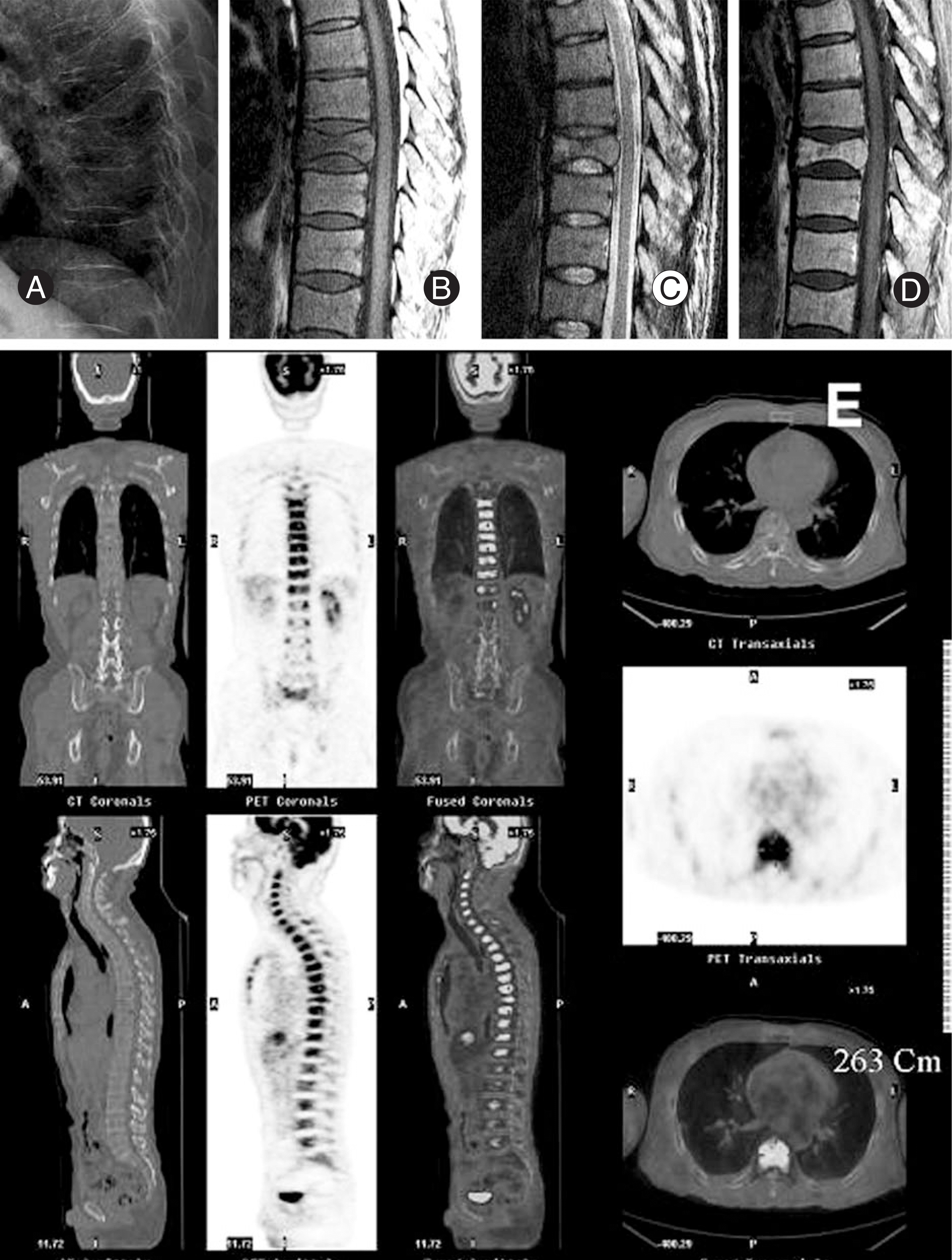 | Fig. 1.51-year old male with T8 fracture, histopathologically diagnosed as multiple myeloma. MRI shows round-protruded posterior cortex and destructed pattern rather than fractured, F-18 PET shows multiple homogeneous uptake stronger than liver. (A: plain X-ray, B: T1WI MRI, C: T2WI MRI, D: T1WI enhanced MRI, E: PET/CT) |
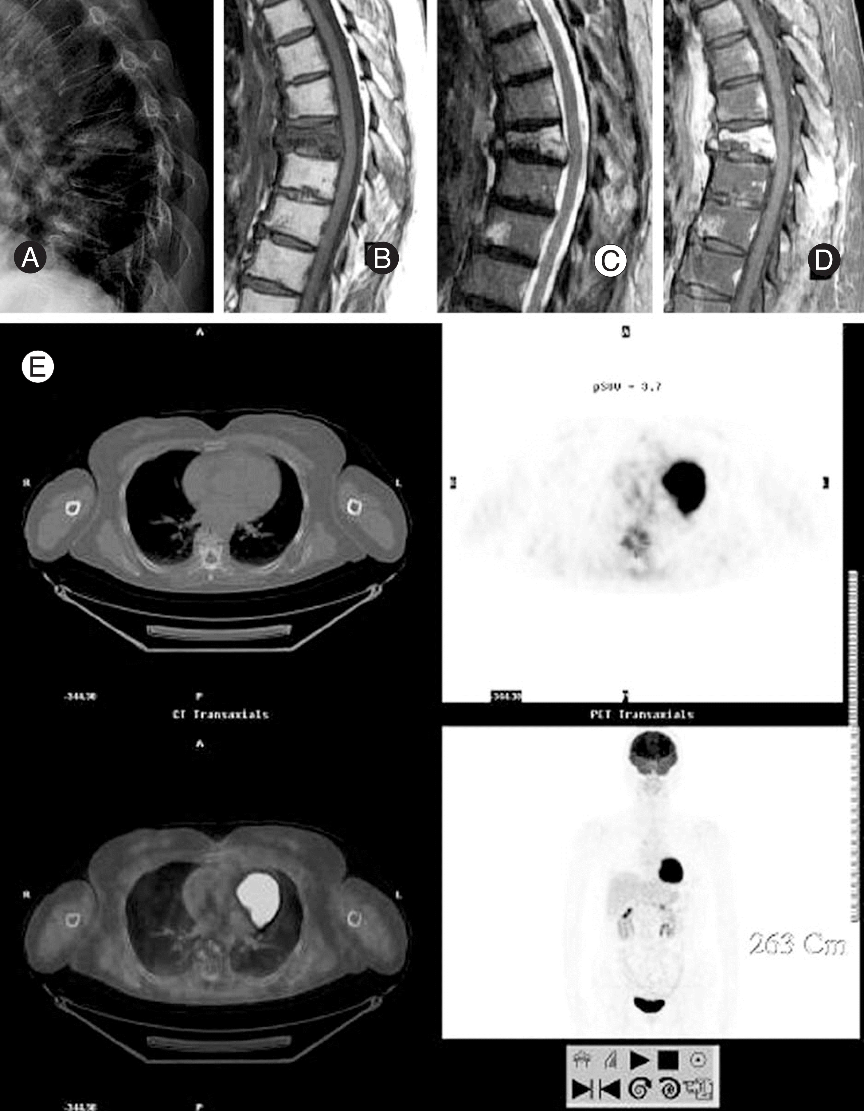 | Fig. 2.63-year old female with T7 fracture, clinically diagnosed as vertebral compression fracture. MRI shows fractured pattern rather than destructed with marrow edema, F-18 PET shows heterogeneous uptake weaker than liver. (A: plain X-ray, B: T1EI MRI, C: T2EI MRI, D: T1EI enhanced MRI, E: PET/CT) |
Table 1.
Patients data
| Patient | t Sex | Age | Level | MRI | SUV∗ | PET (1)∗∗ | PET (2)∗∗∗ | Bx.# | F/U (mo)## | Final diagnosis |
|---|---|---|---|---|---|---|---|---|---|---|
| 01 | F | 66 | T12,L1,2,3 | tumor | - | pathologic | pathologic | + | multiple myeloma | |
| 02 | M | 72 | T10,11 | benign | - | pathologic | benign | + | spondylitis | |
| 03 | F | 81 | T11,L1 | benign | - | benign | benign | - | 35 | VCF### |
| 04 | F | 63 | T8 | benign | - | benign | benign | - | 12 | VCF |
| 05 | M | 57 | C7 | metastatis | - | pathologic | pathologic | + | metatasis | |
| 06 | F | 81 | L4 | benign | - | benign | benign | - | 13 | VFC |
| 07 | M | 52 | L5,S1 | spondylitis | - | pathologic | pathologic | + | spondylitis | |
| 08 | F | 73 | T12,L1 | benign | - | benign | benign | - | 17 | VCF |
| 09 | M | 71 | T11,12 | benign | - | benign | benign | + | VCF | |
| 10 | M | 52 | L1,2 | benign | - | benign | benign | - | 10 | VCF |
| 11 | F | 83 | T12 | benign | - | benign | benign | - | 18 | VCF |
| 12 | F | 78 | L1 | benign | - | benign | benign | - | 10 | VCF |
| 13 | M | 68 | L1 | benign | - | benign | benign | - | 14 | VCF |
| 14 | F | 62 | L1 | benign | - | benign | benign | - | 25 | VCF |
| 15 | F | 71 | T12,L1 | benign | - | benign | benign | - | 18 | VCF |
| 16 | M | 76 | L3 | benign | - | benign | benign | - | 06 | VCF |
| 17 | F | 72 | L1 | equivocal | - | pathologic | benign | + | VCF | |
| 18 | M | 37 | L2,3 | benign | - | pathologic | pathologic | + | spondylitis | |
| 19 | F | 80 | L2,3 | metastatis | - | pathologic | pathologic | + | VCF | |
| 20 | F | 83 | L4,5 | spondylitis | - | benign | benign | - | 21 | spondylitis |
| 21 | F | 46 | L1,2,3,4,5 | metastasis | 5.96 | pathologic | pathologic | - | 10 | metastasis |
| 22 | F | 62 | T4,6,7,8,9 | benign | 1.97 | benign | benign | - | 08 | VFC |
| 23 | M | 51 | T8 | tumor | 5.23 | pathologic | pathologic | + | multiple myeloma | |
| 24 | F | 63 | T7 | benign | 3.20 | benign | benign | - | 06 | VCF |
| 25 | F | 61 | T7 | metastasis | 2.71 | benign | benign | + | VCF | |
| 26 | M | 67 | L4,5 | spondylitis | 3.08 | pathologic | pathologic | - | 07 | spondylitis |
| 27 | M | 66 | L3 | benign | 3.19 | benign | benign | - | 08 | VCF |
| 28 | F | 63 | L5,S1 | spondylitis | 3.60 | pathologic | pathologic | + | spondylitis | |
| 29 | M | 57 | L5,S1 | spondylitis | 5.41 | pathologic | pathologic | + | spondylitis |
Table 2.
Results
 XML Download