Abstract
Osteoid osteomas occur most commonly in the lumbar spine. However, they rarely occur in the sacrum, and there is no report of such a case in Korea. We report a case of osteoid osteoma in the sacrum in a 15-year-old boy who was treated by a surgical excision with a satisfactory outcome. Although unusual, an osteoid osteoma should be considered when making a differential diagnosis of benign tumors in the sacral areas.
REFERENCES
01). Dahlin DC., Unni KK. Bone tumors: general aspects and data on 8,543 cases. 4th ed.Springfield, CC Thomas;1986. p. 88–101.
02). Enneking WF. Musculoskeletal Tumor Surgery. New York, C Livingstone;1986. p. 49–66.
03). Maiuri F., Signorelli C., Lavano A., Gambardella A., Simari R., D' Andrea F. Osteoid osteomas of the spine. Surg Neurol. 1986. 25(4):375–380.

04). Sypert GW. Osteoid osteoma and osteoblastoma of the spine. Sundaresan N, Schmidek HH, Schiller AL, Rosenthal DI, editors. Tumors of spine,. Philadelphia: WB Sanders;1990. p. 117–127.
05). Frassica FJ., Waltrip RL., Sponseller PD., Ma LD., McCarthy EF Jr. Clinicopathologic features and treat-ment of osteoid osteoma and osteoblastoma in children and adolescents. Orthop Clin North Am. 1996. 27(3):559–574.

06). Bilchik T., Heyman S., Siegel A., Alavi A. Osteoid osteoma: the role of radionuclide bone imaging, conventional radiography and computed tomography in its manage-ment. J Nucl Med. 1992. 33(2):269–271.
07). Pettine KA., Klassen RA. Osteoid-osteoma and osteoblastoma of the spine. J Bone Joint Surg. 1986. 68(3):354–361.

08). Poey C., Clement JL., Baunin C, et al. Percutaneous extraction of an osteoid osteoma of the lumbar spine under CT guidance. J Comput Assist Tomogr. 1991. 15(6):1056–1058.

09). Mehta MH. Pain provoked scoliosis. Observations on the evolution of the deformity. Clin Orthop Relat Res. 1978. 135:58–65.
Fig. 1.
Preoperative radiographs of the sacrum show osteolytic lesions of sacrum (arrows). (A) Anteroposterior view. (B) Lateral view.
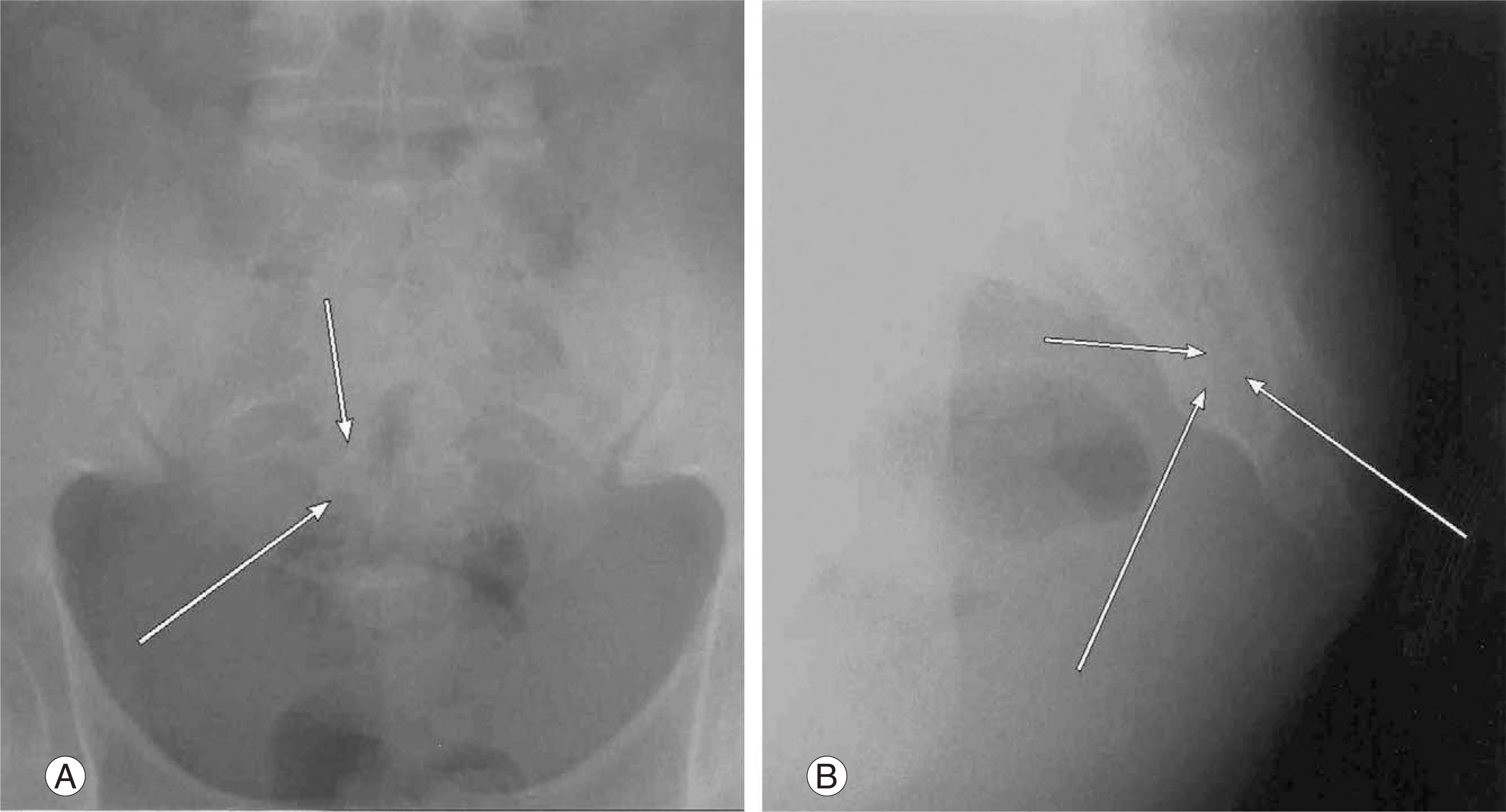
Fig. 2.
Preoperative CT scans show a low density lesion in which contains high density lesion in the 4th sacrum(arrows). (A) Axial view. (B) Sagittal view.
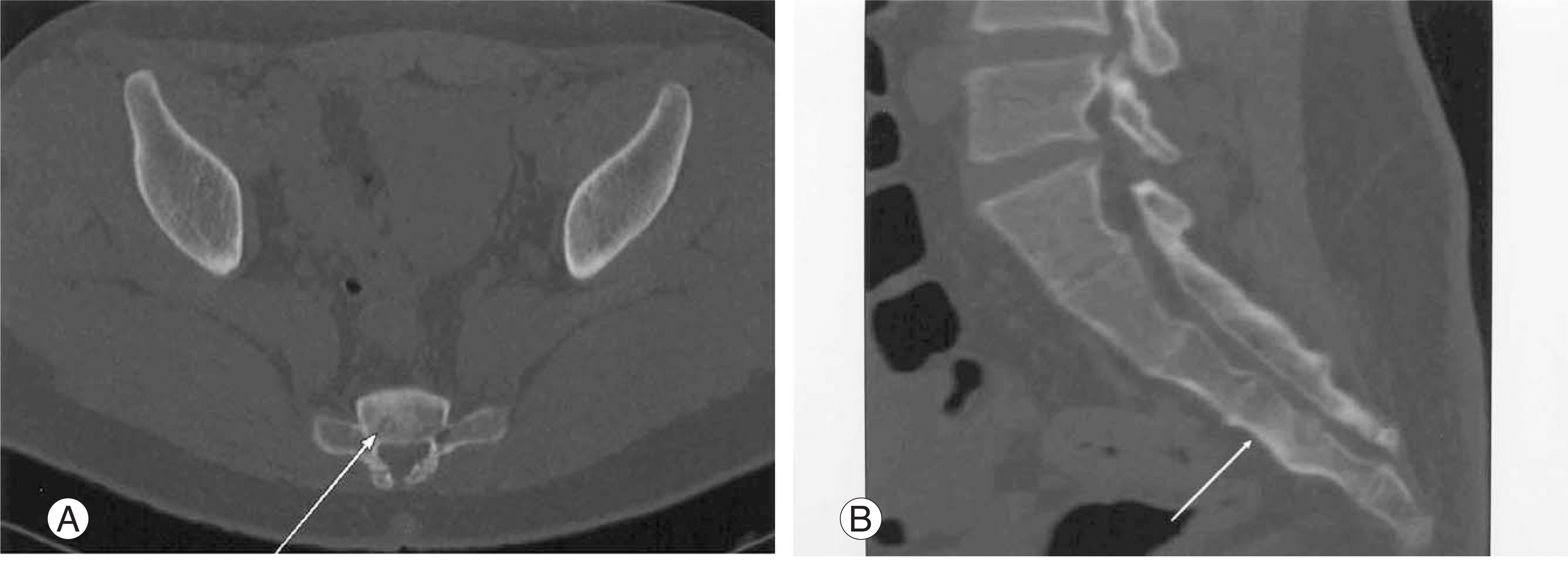
Fig. 3.
Preoperative T2-weighted MR image shows high signal intensity area in the 4th sacrum(arrow).
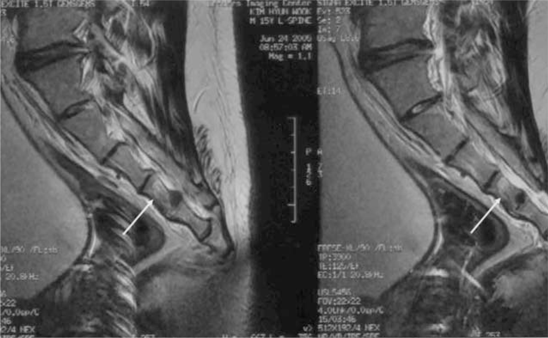
Fig. 4.
An intraoperative photogragh of the excised mass of the patient shows about 8 mm × 5 mm size.
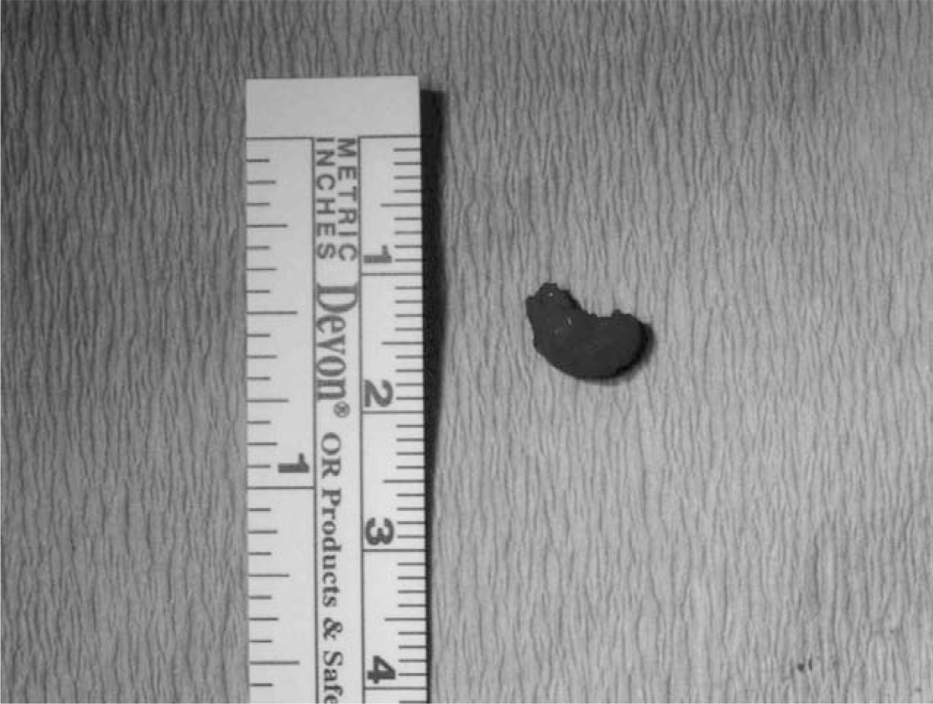
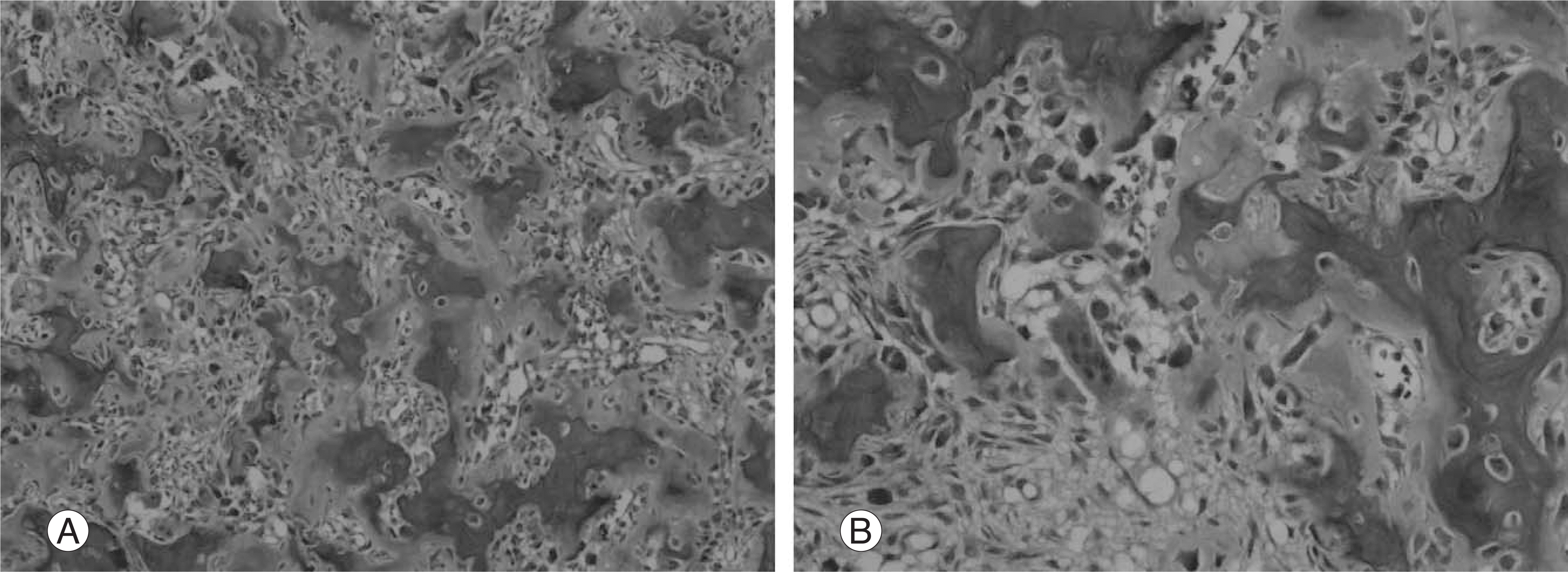
Fig. 5.
(A) The histologic examination shows irregular bone trabeculae with prominent vascularity of the intervening tissue (hematoxylin and eosin stain, ×100). (B) At high magnification, the lesional bone is covered by plump but uniform osteoblasts, and one osteoclast(hematoxylin and eosin stain, ×200).


 PDF
PDF ePub
ePub Citation
 Print
Print


 XML Download