

 PDF
PDF ePub
ePub Citation
 Print
Print


Abstract
Atlantoaxial instability with rupture of transverse atlantal ligament is mostly caused by trauma, and this can be combined with myelopathy. Although it gives rise to no neurologic deficit, it has a high possibility to quadriplegia or death by minor trauma. We experienced a rare case about atlantoaxial instability with delayed rupture of transverse atlantal ligament that was compli-cated after the treatment of posterior neck abscess.
A 44-year-old male patient had complained of posterior neck pain for 1 month. Based on a clinical examination, simple radiog-raphy, CT and MRI, his diagnosis was posterior neck abscess. He underwent an emergency operation with open drainage. One year later, he again had posterior neck pain. Atlantoaxial instability with rupture of the transverse atlantal ligament was con-firmed. Skeletal traction was applied and then C1-2 posterior fusion without wiring was performed. After the operation, antibi-otics were injected for 4 weeks and a halo-vest was put on for 6 months. Complete fusion of C1-2 was obtained without posterior neck pain at the 1 year follow-up.
Go to : 
REFERENCES
01). Bell Sir C. The nervous system of the human body, embracing papers delivered to the Royal Society on the subject of nerves. London: Longman, Rees and Orme;403. 1830.
02). Chung JY., Chung GH., Jeung JC. Pyogenic Atlatno-Axial instability Complicated after Tonsillectomy. A case report. J Kor Orthop Asscoc. 1991. 26:1338–1341.
03). Dickman CA., Sonntag VKH., Drayer BP. Magnetic res-onance imaging of the transverse atlantal ligament for the evaluation of atlantoaxial instability. J Neurosurg. 1991. 75:221–227.

04). Dickman CA., Sonntag VKH. Injuries involving the transverse atlantal ligament: classification and treatment guidelines based upon experience with 39 injuries. Neurosurgery. 1997 Apr. 40(4):886–887.
05). Fielding JW., Cochran GB., Lawsing JF III., Hohl M. Tears of the transverse ligament of the atlas. J Bone Joint Surg Am. 1974. 56:1683–1691.
06). Hunter G.A. Non-traumatic displacement of the atlantoaxial joint. A report of seven cases. J Bone Joint Surg Br. 1968. 50:44–51.
07). Papadopoulos SM., Dickman CA., Sonntag VK. Atlantoaxial stabilization in rheumatoid arthritis. J Neurosurg. 1991. 74:1–7.
08). Spence K.F.., Decker S.., Sell K.W. Bursting atlantal fracture associated with rupture of the transverse ligament. J Bone Joint Surg Am. 1970. 52:543–549.
09). Welinder NR., Hoffmann P., Hakansson S. Pathogenesis of non-traumatic atlantoaxial subluxation(Grisel's syndrome). Dur Arch Otorhinolaryngol. 1997. 254:251–254.
Go to :
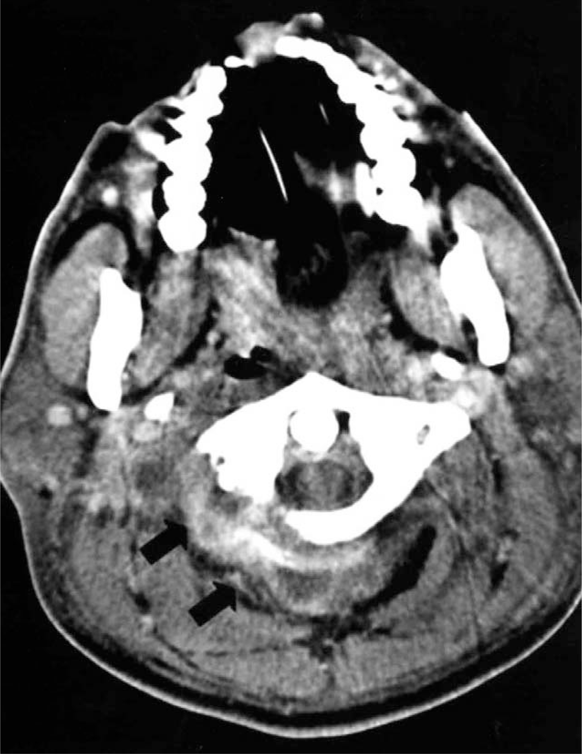
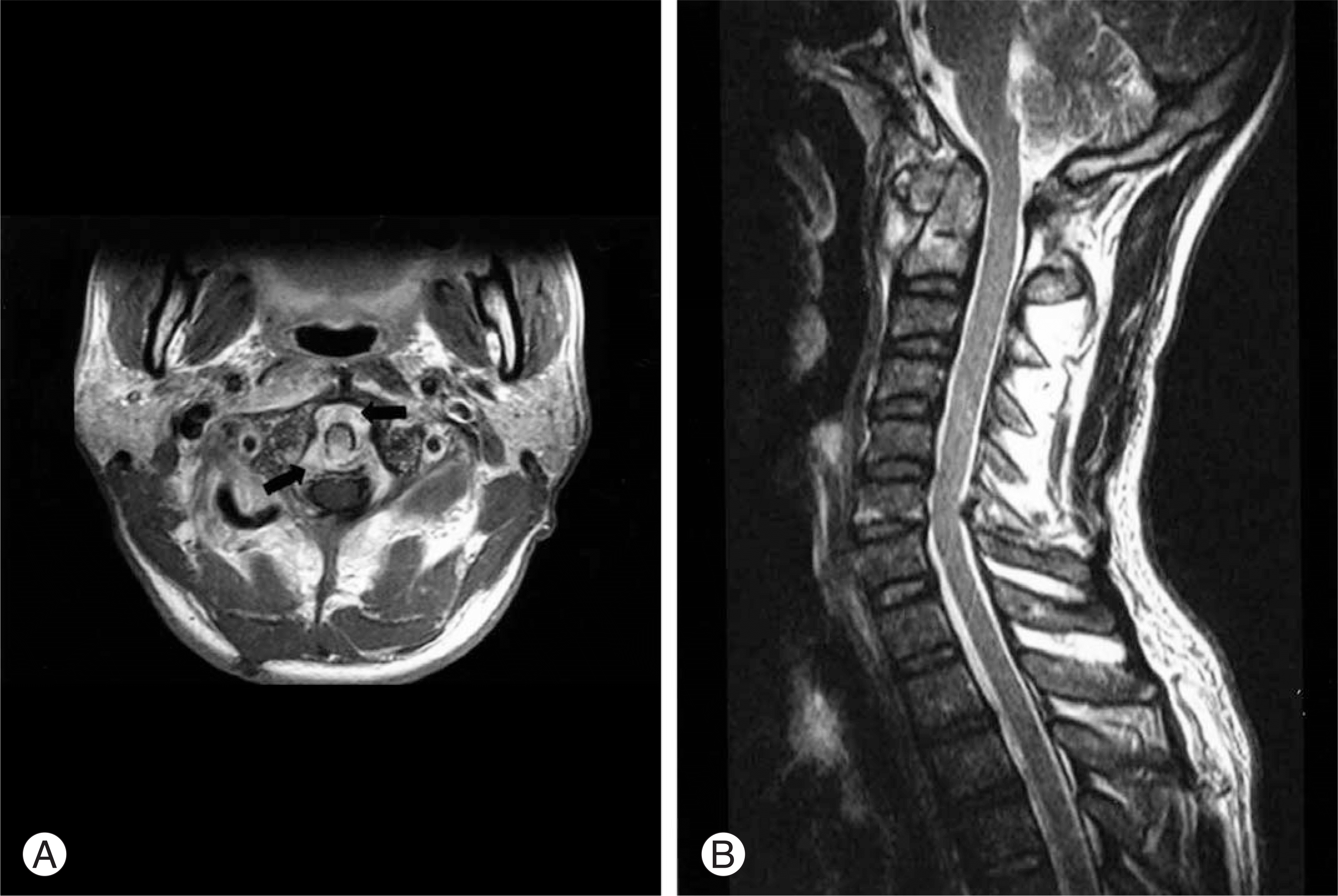
 | Fig. 2.Initial MRI (enhanced axial T1 weighted) shows paraspinal abscess around C1-2 level (arrows). Transverse atlantal ligament and C1-2 vertebrae are intact. There is no abscess in spinal canal. |
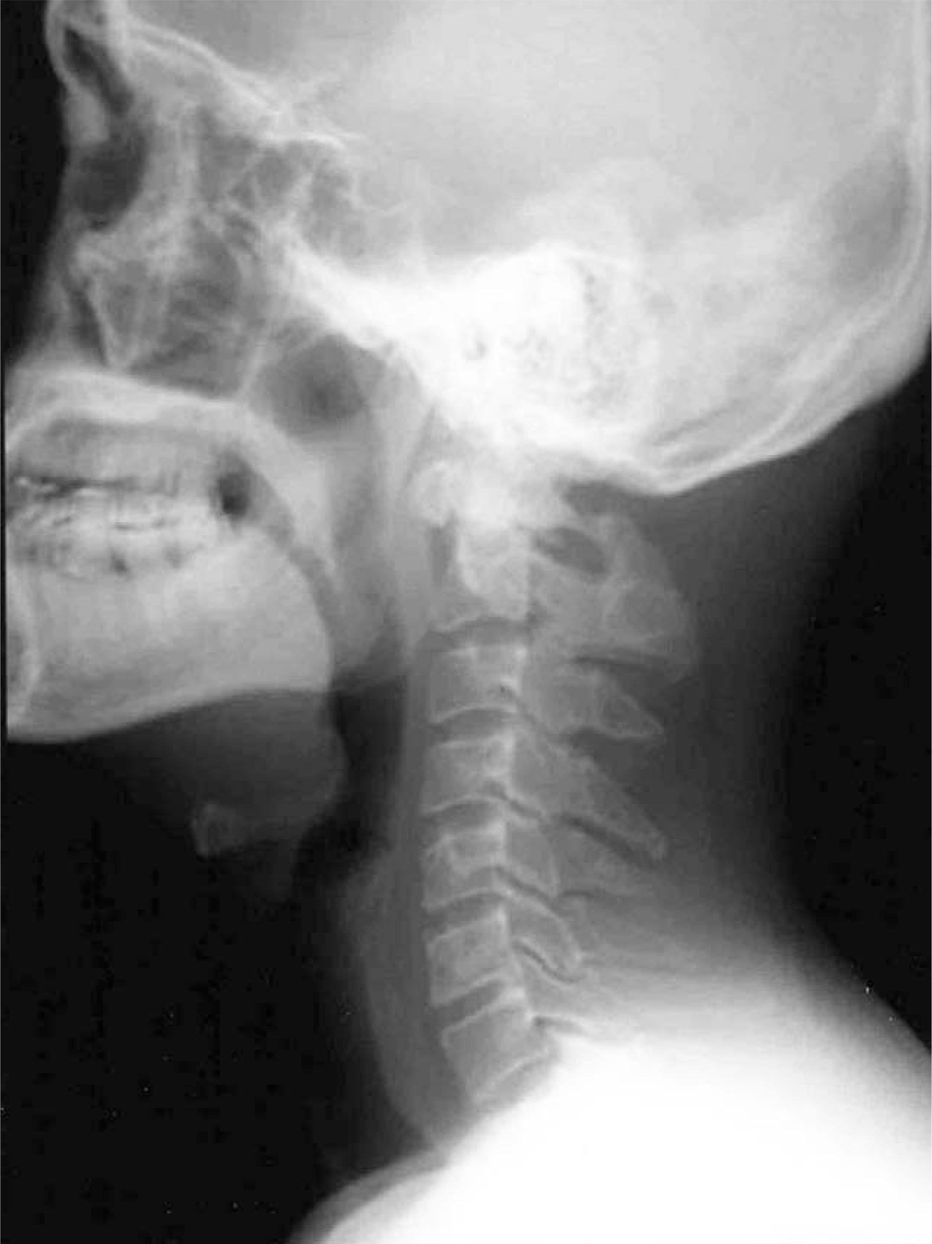
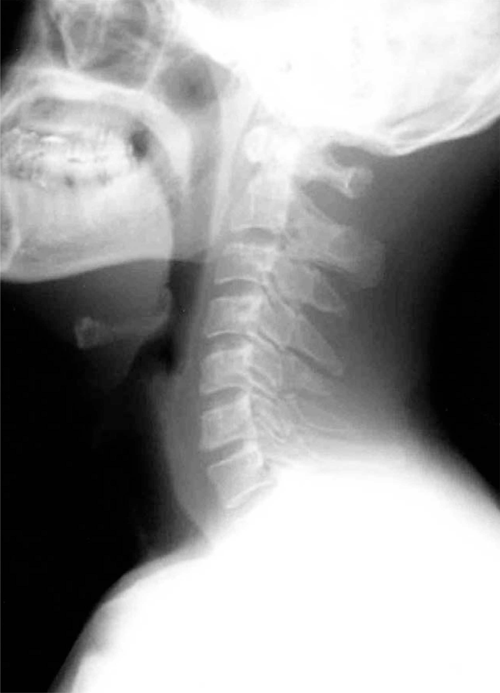 | Fig. 3.Lateral radiograph of neck taken at 3 months after open drainage shows normal bony structure. |
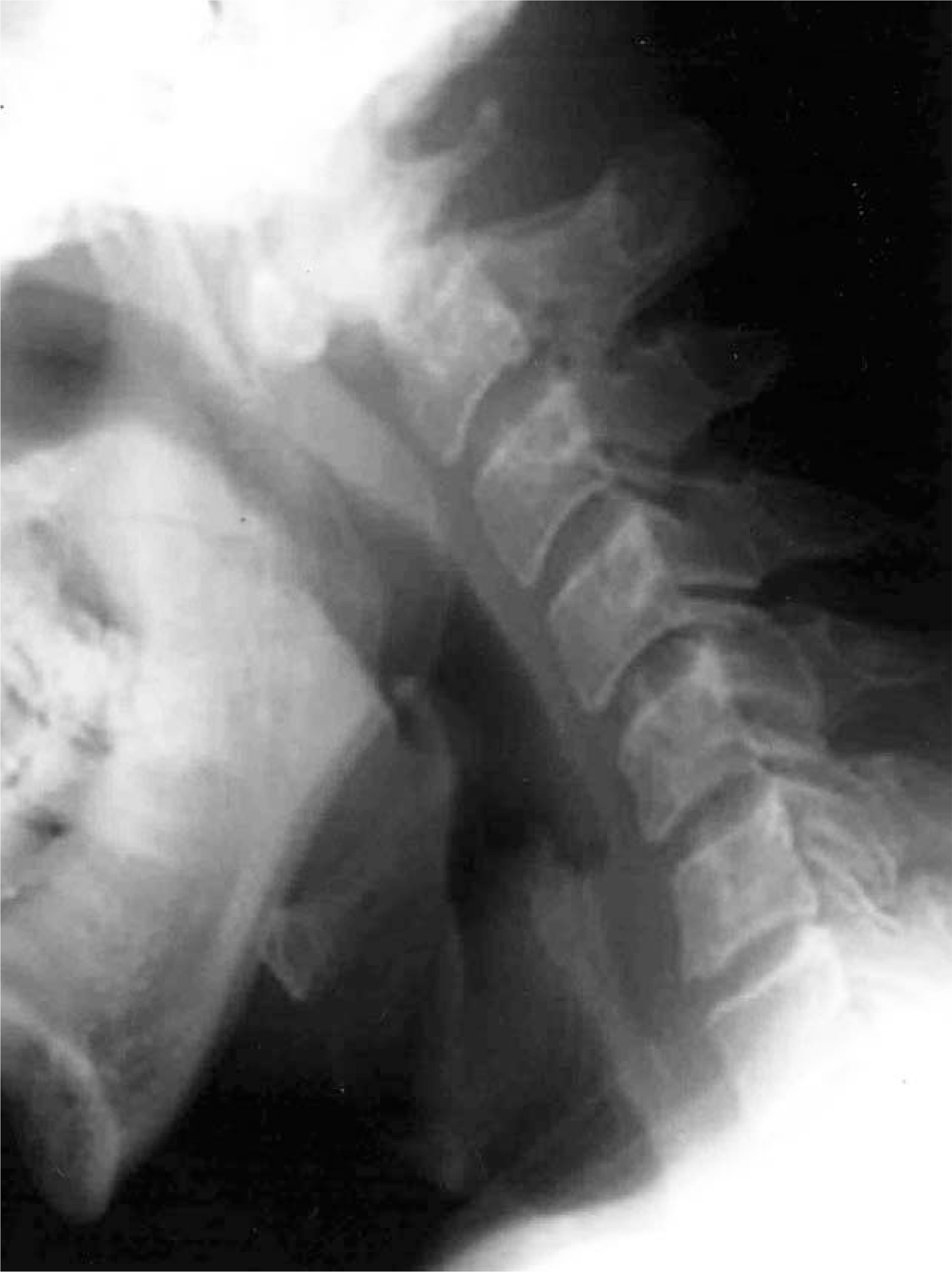 | Fig. 4.Preoperative lateral flextion radiograph of neck shows widening of the atlantoaxial interval and destruction of posterior arch of atlas (atlanto-dens interval 10 mm). |
 XML Download