Abstract
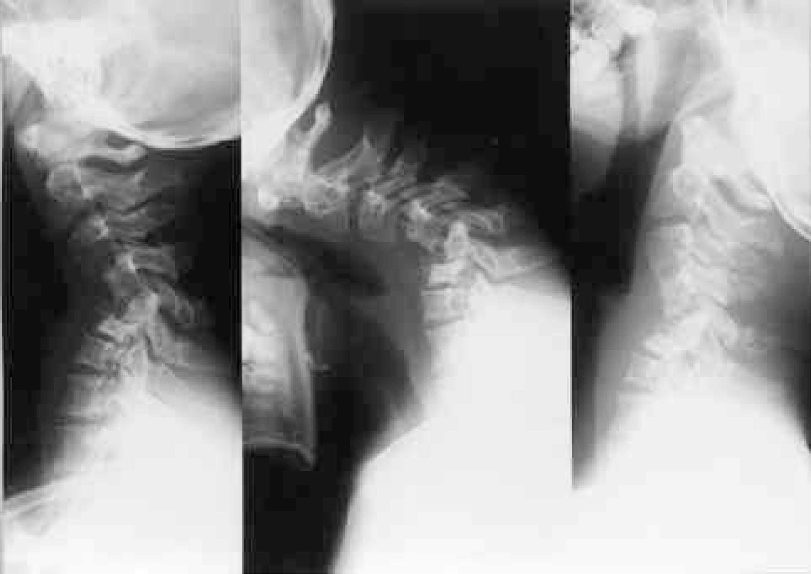
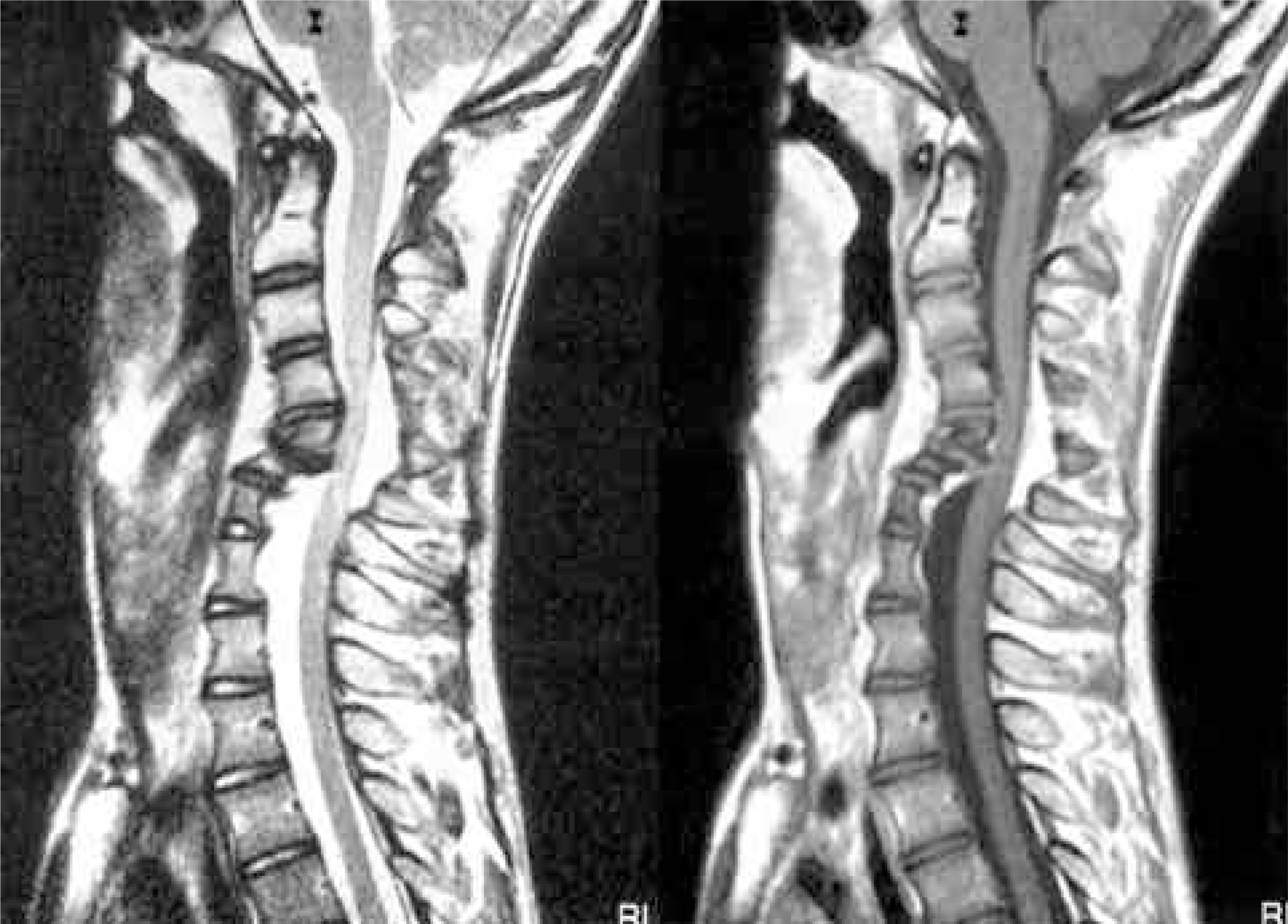
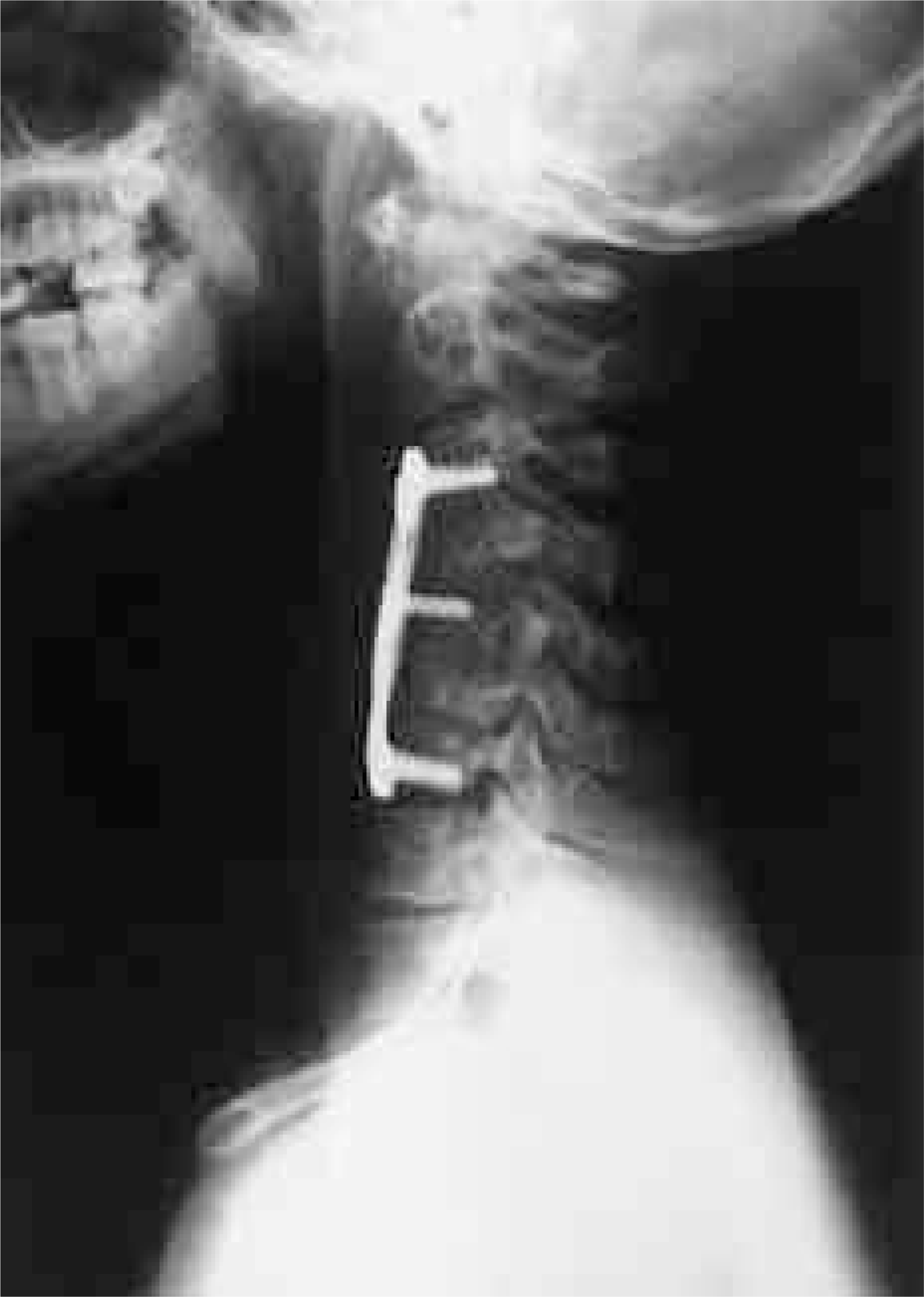
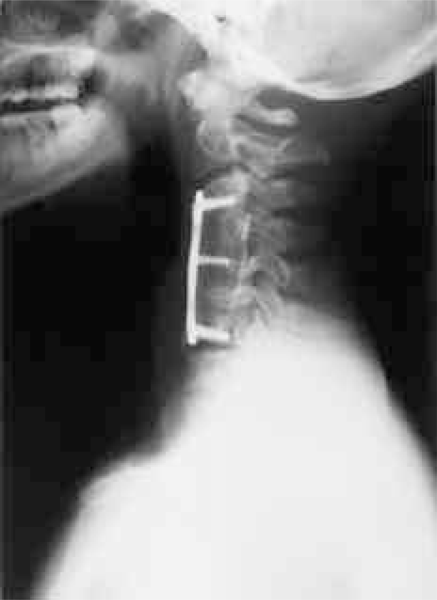
Scoliosis is the most common deformity of the spine in neurofibromatosis patient, but kyphosis of the cervical spine has rarely been reported. Most authors have reported anterior corpectomy and multilevel interbody grafting & plate osteosynthesis, combined with posterior arthrodesis, as the treatment of cervical kyphosis in neurofibromatosis. A case is presented of a 17- year-old boy with neurofibromatosis Who had 52 degrees of dystrophic kyphosis (as measured on radiographs between C3 and C7) of the cervical spine. He was treated successfully by anterior multilevel interbody grafting using an autogenous iliac bone graft. A nterior corpectomy and arthrodesis appears to provide another surgical option with a moderate degree of cervical kyphosis.
Go to : 
REFERENCES
1). Chaglassian JH, Riseborough EJ. Neurofibromatous scoliosis. J Bone joint Surg. 1976; 58-A:695–702.
2). Nijland EA, Van Royen BJ, Winters HAH, Ouwerkerk WJR. Correction of a dystrophic cervicothoracic spine deformity in Recklinghausen's disease. Clin Orthop. 1996; 349:149–155.

3). Ken YH, Kalamchi A, MacEwen GD. Cervical spine abnormalities in neurofibromatosis. J Bone Joint Surg. 1979; 61-A:695–699.
4). Haddad FS, William RL, Bentley G. The cervical spine in neurofibromatosis. Br J Hosp Med. 1995; 53:318–319.
5). Crawford AH. Pitfalls of spinal deformities associated with neurofibromatosis in children. Clin Orthop. 1989; 245:29–42.

6). Takashi A, Masaakki Y, Koichi N, Masatoshi A, Mitsu-ru O, Kyosuke F. Spinal fusion using a vascularized fIbu -lar bone graft for a patient with cervical kyphosis Due to Neurofibromatosis. J Spinal Disord. 1997; 10:537–540.
Go to : 
 | Fig. 1.Preoperative radiographs showed dysplastic C3,4,5 body. Cervical kyphosis was 52 degrees in neutral lateral view, 75 degrees in flexion view and 22 degrees in extension view at the level of C3-C7. |


 PDF
PDF ePub
ePub Citation
 Print
Print


 XML Download