

 PDF
PDF ePub
ePub Citation
 Print
Print


Abstract
Objectives
To assess the efficiency of undercorrection and transpedicular screw fixation through a posterior approach in osteoporotic spine fractures with a thoracolumbar kyphotic deformity.
Summary of Literature Review
The surgical treatment of osteoporotic spine fractures with a thoracolumbar kyphotic deformity requires extensive surgical procedures to obtain complete restoration of the sagittal alignment, but it has a few technical limitations due to insufficient mechanical stability at the bone- screw interface. A special strategy is essential for transpedicular screw fixation for osteoporotic spine fractures with a thoracolumbar kyphotic deformity.
Materials and Methods
We reviewed 14 osteoporotic spine fracture cases, with a thoracolumbar kyphotic deformity, which had undergone undercorrection and transpedicular screw fixation through a posterior approach, between March 2000 and June 2003, with an average followup period of 15.2 months. A ccording to the Jikei grade of the osteoporosis, 9 and 5 cases were grades 2 and 3, respectively. As a radiographic assessment, we measured the kyphotic angles of the fused segments on the pre-operative, postoperative and last follow up thoracolumbar lateral views on standing using Cobb's method, and also assessed the kyphotic angle correction (KA C). The clinical results were evaluated at the last followup.
Results
The kyphotic angles at the preoperative, postoperative and last followup were 33.5°± 9.3, 22.4°± 6.9 and 24.7°± 6.8, respectively. We obtained a mean KA C gain of 11.1° postoperatively (p<0.05), but a loss of 2.3° at the last followup (p>0.05).
The clinical results were analyzed as good, fair and poor in 8, 5 and 1 case, respectively. Fusions were achieved in all cases.
REFERENCES
1). Park BC, Lee SM. Correction of thoracolumbar kyhotic deformity using a posterior interbody fusion. J Kor Spine Surg. 2000; 7:639–646.
2). Cho KN, Yoon HK, Jeon HS, Jeon SJ, Kim WS. Efficiency of posterior lumbar interbody fusion in lumbar spinal stenosis with osteoporosis. J Kor Spine Surg. 1999; 6:380–387.
3). Soshi S, Shiba R, Kondo H, Murota K. An experimental study on transpedicular screw fixation in relation to osteoporosis of the lumbar spine. Spine. 1991; 16:1335–1341.

4). Kumano K, Hirabayashi S, Ogawa Y, Aota Y. Pedicle Screws and Bone Mineral Density. Spine. 1994; 19:1157–1161.
5). HU SS. Internal fixation in the osteoporotic spine. Spine. 1997; 22:958–963.
6). Coe JD, Warden KE, Herzig MA, Mcafee PC. Influence of bone mineral density on the fixation of thoracolumbar implants: A comparative study of transpedicular screws, laminar hooks, and spinous process wires. Spine. 1990; 15:902–907.
7). Hasegawa K, Takahashi HE, Uchiyama S, et al. A n experimental study of a combination method using a pedicle screw and laminar hook for the osteoporotic spine. Spine. 1997; 22:958–963.
8). Cho KN, Yoon HK, Jeon HS, et al. Effect of bone cement augmentation of pedicular screwing for osteoporotic lumbar spine. J Kor Spine Surg. 2002; 9:223–229.
9). Glassman SD, Alegre GM. Adult spinal deformity in the osteoporotic spine: options and pitfalls. Instr Course Lect. 2003; 52:579–588.
10). Simmons EH. Kyphotic deformity of the spine in ankylosing spondylitis. Clin Orthop. 1977; 128:65–77.
11). Suk SI. Kyphosis. Spinal surgery. 1997; 1:388–410.
12). Suk SI, Kim JH, Kim WJ, et al. Treatment of fixed lumbosacral kyphosis by posterior vertebral column resection. A preliminary report. J Kor Spine Surg. 1998; 5:307–313.
13). Holt RT, Dopf CA, Isaza JE, Rahn KA, Crawford MK, Kostuik JP. Adult kyphosis. (in. Frymoyer JW, editor. The adult spine, 2nd ed. Philadelpia: Lippincott-Raven;p. 1574. 1997.
14). Goel MK. Vertebral osteotomy for correction of fixed flexion deformity of the spine. J Bone Joint Surg. 1968; 50-2:287–294.
15). Winter RB, Moe JH, Wang JF. Congenital kyphosis, it's natural history and treatment as observed in a study of one hundred and thirty patients. J Bone Joint Surg. 1973; 55-2:223–256.
Figures and Tables%
Fig. 1.
65-year-old female with osteoporotic compression fracture at T11/T12/L1/L2 associated with posttraumatic kyphoscoliosis. Preoperative AP and lateral radiographs show Jikei grade III/III osteoporosis with 35° of T10 to L2 kyphosis. Fig. 1. 3 years ago, vertebroplasty performed for compression fracture T11/T12 (A). Postoperative radiographs after kyphosis correction T10 to L2, percutaneous vertebroplasty for L1/L2 and PMMA augmentation for Rt T10, L3, L4 and L5 show correcrion of T10 to L2 kyphotic angle to 30° (B). The 16 months followup radiograph shows no loss of correction and no device-related problems (C).
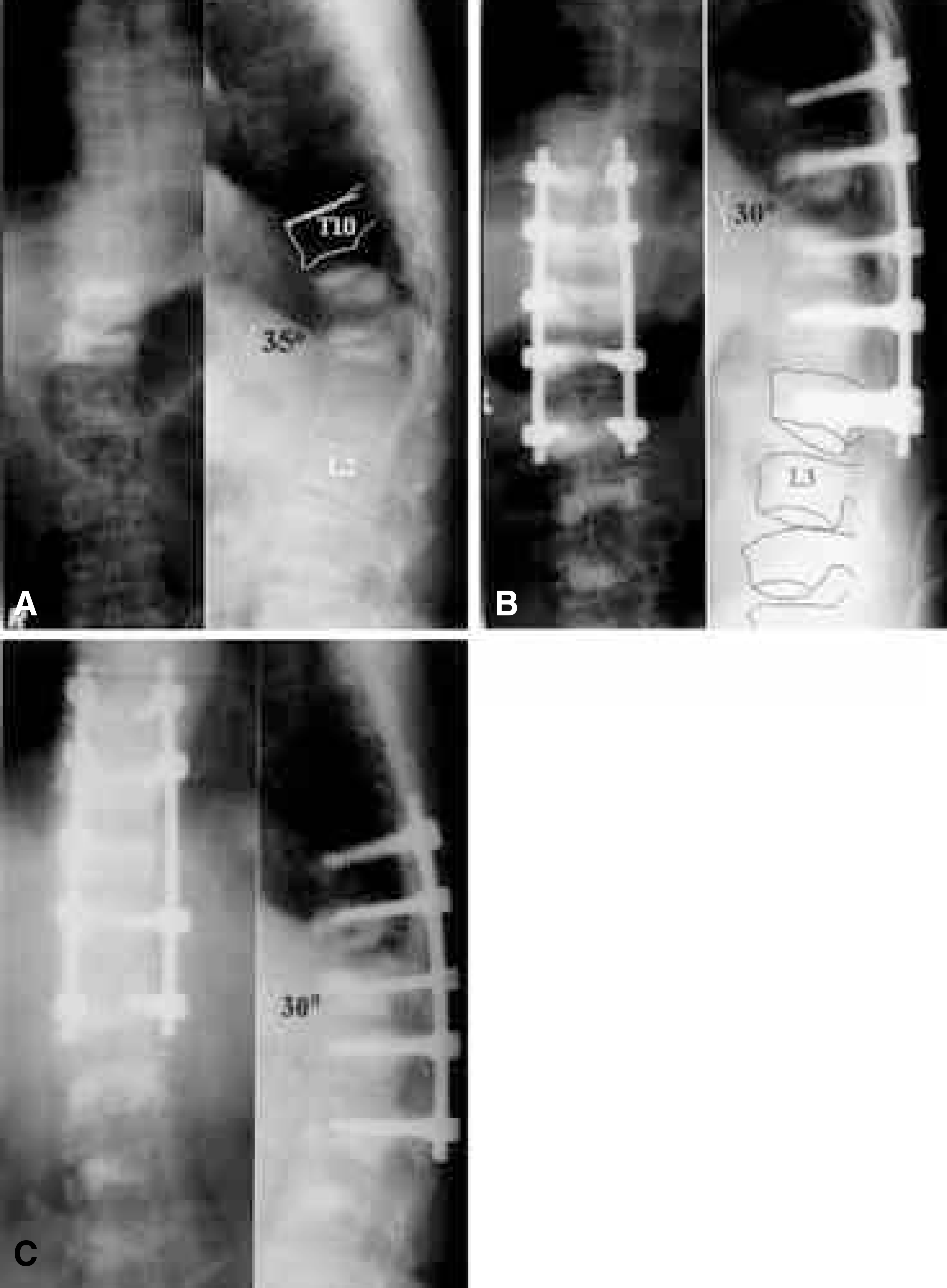
Table 1.
Analysis of Case
Table 2.
Kyphotic Angle Correction
| Pre- Op | Post-Op | Last F/U | |||
|---|---|---|---|---|---|
| Mean KA | 33.5° | 22.4° | 24.7° | ||
| KAC* Gain | (11.1°) | (-2.3°) |
 XML Download